

Physiologic hypoxia and oxygen homeostasis in the healthy intestine. A Review in the Theme: Cellular Responses to Hypoxia
- PMID: 26179603
- PMCID: PMC4572369
- DOI: 10.1152/ajpcell.00191.2015
Physiologic hypoxia and oxygen homeostasis in the healthy intestine. A Review in the Theme: Cellular Responses to Hypoxia
Abstract
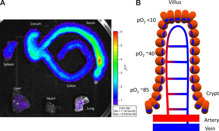
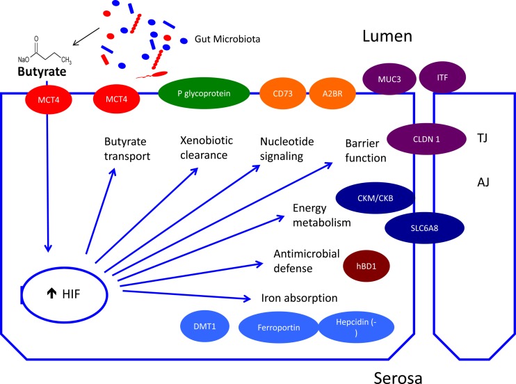
In recent years, the intestinal mucosa has proven to be an intriguing organ to study tissue oxygenation. The highly vascularized lamina propria juxtaposed to an anaerobic lumen containing trillions of metabolically active microbes results in one of the most austere tissue microenvironments in the body. Studies to date have determined that a healthy mucosa contains a steep oxygen gradient along the length of the intestine and from the lumen to the serosa. Advances in technology have allowed multiple independent measures and indicate that, in the healthy mucosa of the small and large intestine, the lumen-apposed epithelia experience Po2 conditions of <10 mmHg, so-called physiologic hypoxia. This unique physiology results from a combination of factors, including countercurrent exchange blood flow, fluctuating oxygen demands, epithelial metabolism, and oxygen diffusion into the lumen. Such conditions result in the activation of a number of hypoxia-related signaling processes, including stabilization of the transcription factor hypoxia-inducible factor. Here, we review the principles of mucosal oxygen delivery, metabolism, and end-point functional responses that result from this unique oxygenation profile.
Keywords: barrier function; colon; hypoxia; intestine; metabolism.
Copyright © 2015 the American Physiological Society.
Figures

References
-
- Albenberg L, Esipova TV, Judge CP, Bittinger K, Chen J, Laughlin A, Grunberg S, Baldassano RN, Lewis JD, Li H, Thom SR, Bushman FD, Vinogradov SA, Wu GD. Correlation between intraluminal oxygen gradient and radial partitioning of intestinal microbiota. Gastroenterology 18: 1055–1063, 2014. - PMC - PubMed
-
- Antoni L, Nuding S, Weller D, Gersemann M, Ott G, Wehkamp J, Stange EF. Human colonic mucus is a reservoir for antimicrobial peptides. J Crohns Colitis 7: e652–e664, 2013. - PubMed
-
- Arteel GE, Thurman RG, Raleigh JA. Reductive metabolism of the hypoxia marker pimonidazole is regulated by oxygen tension independent of the pyridine nucleotide redox state. Eur J Biochem 253: 743–750, 1998. - PubMed
Publication types
MeSH terms
Substances
Grants and funding
LinkOut - more resources
Full Text Sources
Other Literature Sources