. 2015 Jul 16:6:7660.
doi: 10.1038/ncomms8660.

Deactivation of excitatory neurons in the prelimbic cortex via Cdk5 promotes pain sensation and anxiety

Affiliations

Deactivation of excitatory neurons in the prelimbic cortex via Cdk5 promotes pain sensation and anxiety

Guo-Qiang Wang et al. Nat Commun. .

Erratum in

Abstract

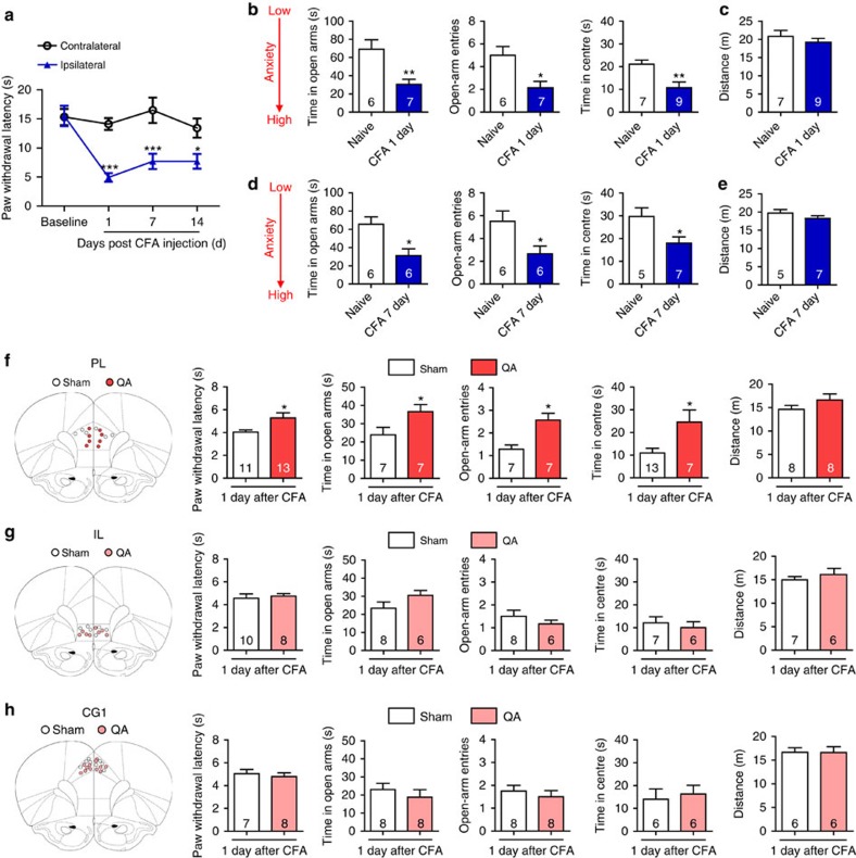
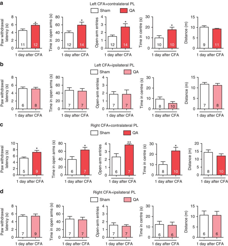
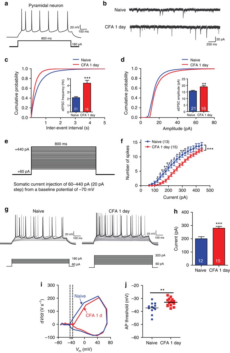
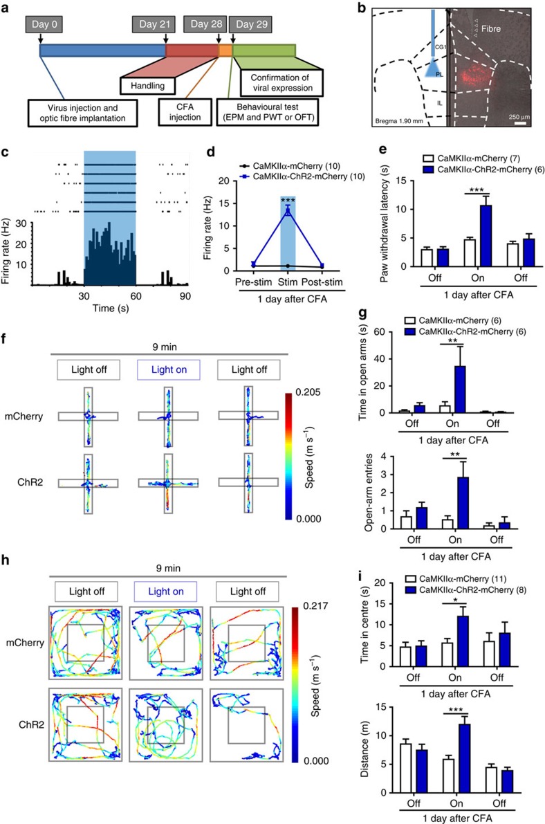
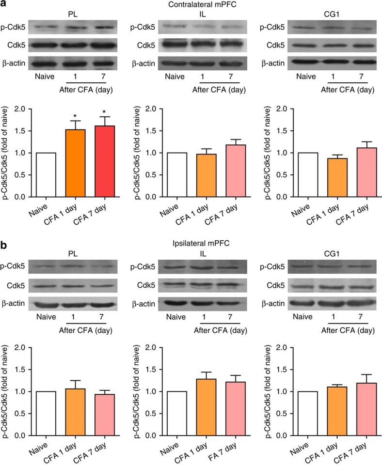
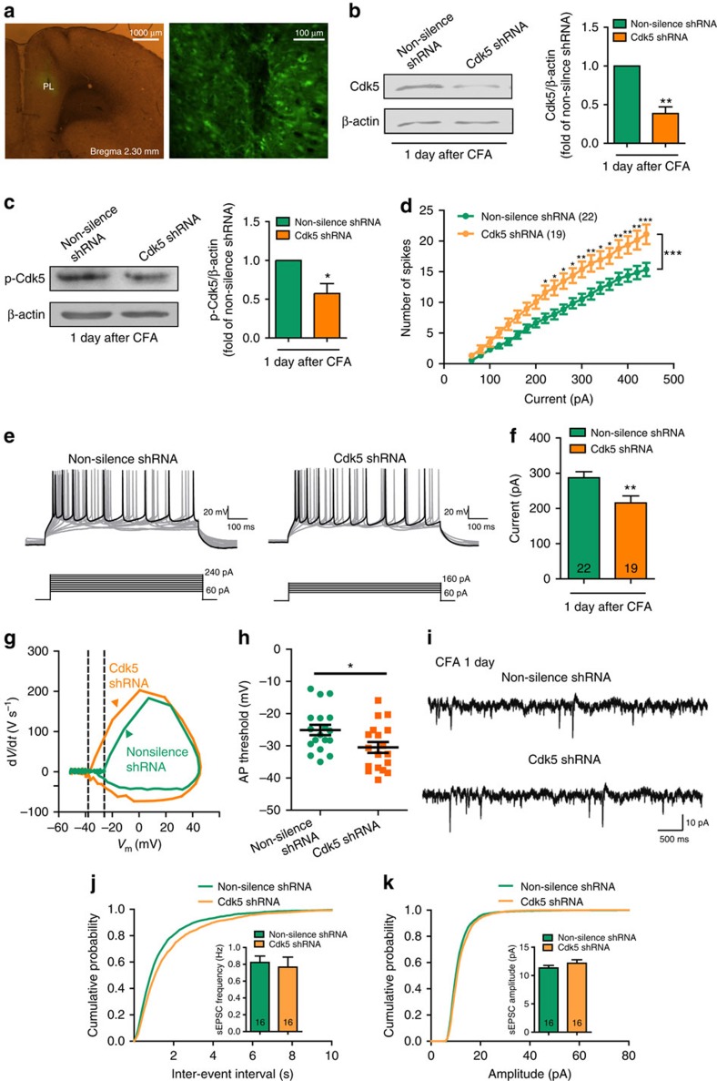
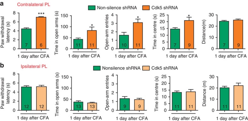
The medial prefrontal cortex (mPFC) is implicated in processing sensory-discriminative and affective pain. Nonetheless, the underlying mechanisms are poorly understood. Here we demonstrate a role for excitatory neurons in the prelimbic cortex (PL), a sub-region of mPFC, in the regulation of pain sensation and anxiety-like behaviours. Using a chronic inflammatory pain model, we show that lesion of the PL contralateral but not ipsilateral to the inflamed paw attenuates hyperalgesia and anxiety-like behaviours in rats. Optogenetic activation of contralateral PL excitatory neurons exerts analgesic and anxiolytic effects in mice subjected to chronic pain, whereas inhibition is anxiogenic in naive mice. The intrinsic excitability of contralateral PL excitatory neurons is decreased in chronic pain rats; knocking down cyclin-dependent kinase 5 reverses this deactivation and alleviates behavioural impairments. Together, our findings provide novel insights into the role of PL excitatory neurons in the regulation of sensory and affective pain.


Conflict of interest statement

The authors declare no competing financial interests.

Figures

Figure 1
Figure 1. Bilateral lesions of the PL attenuate CFA-induced heat hyperalgesia and anxiety-like behaviours.
(a) Paw withdrawal latency of the contralateral and ipsilateral hind paws after CFA injection (n=5 animals, *P<0.05, ***P<0.001, two-way ANOVA with Bonferroni post tests). (b) Elevated plus maze (left and middle, n=6, 7 animals, *P<0.05, **P<0.01, two-tailed t-test) and open field test (right, n=7, 9 animals, **P<0.01, two-tailed t-test) 1 day after CFA injection. (c) Locomotor activity 1 day after CFA injection (n=7, 9 animals, two-tailed t-test). (d) Elevated plus maze (left and middle, n=6, 6 animals, *P<0.05, two-tailed t-test) and open field test (right, n=5, 7 animals, *P<0.05, two-tailed t-test) 7 days after CFA injection. (e) Locomotor activity 7 days after CFA injection (n=5, 7 animals, two-tailed t-test). (fh) Paw withdrawal latency, elevated plus maze, open field test and locomotor activity of the CFA rats after bilateral lesion of the PL, IL or CG1 by quinolinic acid, respectively (*P<0.05, two-tailed t-test). The data are presented as mean±s.e.m.
Figure 2
Figure 2. Contralateral lesion of the PL exerts analgesic and anxiolytic effects.
(a, b) Paw withdrawal latency, elevated plus maze, open field test and locomotor activity of the rats with CFA injected in the left hind paw after contralateral or ipsilateral lesion of the PL by QA, respectively (*P<0.05, two-tailed t-test). (c, d) Paw withdrawal latency, elevated plus maze, open field test and locomotor activity of the rats with CFA injected in the right hind paw after contralateral or ipsilateral lesion of the PL by QA, respectively (*P<0.05, **P<0.01, two-tailed t-test). The data are presented as mean±s.e.m.
Figure 3
Figure 3. The intrinsic excitability of layer 2/3 neurons is decreased in the contralateral PL 1 day after CFA injection.
(a) Current-clamp recordings to identify excitatory neurons in the PL. (b) Representative sEPSC recording traces from the naive rats and the rats 1 day after CFA. Scale bars, 20 pA and 250 ms. (c, d) Cumulative probabilities (***P<0.001, Kolmogorov–Smirnov two sample test) and average sEPSC frequencies and amplitudes from the naive rats (frequency: 2.154±0.1671 Hz, amplitude: 15.67±0.5745, pA, n=21 neurons) and the rats 1 day after CFA (frequency: 4.3324±0.4222 Hz, amplitude: 18.93±0.9648, pA, n=16 neurons, **P<0.01, ***P<0.001, two-tailed t-test). (e) Recording paradigm of the excitatory neurons in the PL. (f) Number of spikes induced by injected currents in the contralateral PL excitatory neurons from the naive rats and the rats 1 day after CFA (n=13, 15 neurons, *P<0.05, **P<0.01, ***P<0.001, two-way ANOVA with Bonferroni post tests). (g) Examples of the AP responses to positive current steps recorded from pyramidal neurons from the naive rats and the rats 1 day after CFA. (h) The current required to drive APs at a frequency of 8.75 Hz from naive rats (200±15.37 pA, n=12 neurons) and the rats 1 day after CFA (280±13.66 pA, n=15 neurons; ***P<0.001, two-tailed t-test). (i) Phase plots of the APs from the naive rats and the rats 1 day after CFA evoked by brief current injections. (j) Plot diagram summary of the AP thresholds from the naive rats (−37.29±1.185 mV, n=12 neurons) and the rats 1 day after CFA (−32.95±0.7555, mV, n=15 neurons; **P<0.01, two-tailed t-test). The data are presented as mean±s.e.m.
Figure 4
Figure 4. Optogenetic activation of contralateral PL excitatory neurons attenuates CFA-induced heat hyperalgesia and anxiety-like behaviours.
(a) Schematic diagram of the experiment. One group animals were tested for EPM followed by paw withdrawal latency test. Another group animals were tested for OFT. (b) ChR2 expression in the PL excitatory neurons after viral injection. Strong staining is shown in the PL (Bregma 1.90 mm). (c, d) Multiple channel recordings of the firing rate of the contralateral PL excitatory neurons in the CFA mice infected with AAV-CaMKIIα-mCherry and AAV-CaMKIIα-ChR2-mCherry virus before, with and after 20 Hz, 6–9 mW 473-nm light photostimulation (n=10, 10 neurons, ***P<0.001, two-way ANOVA with Bonferroni post tests). (e) Paw withdrawal latency in the CFA mice of AAV-CaMKIIα-mCherry or AAV-CaMKIIα-ChR2-mCherry virus injection with 473 nm blue light off-on-off stimulation (n=7, 6 animals, ***P<0.001, two-way ANOVA with Bonferroni post tests). (f, g) Schematic and animal heat traces of the elevated plus maze with 3 min light-off, 3 min light-on and 3 min light-off (n=6, 6 animals, **P<0.01, two-way ANOVA with Bonferroni post tests). (h, i) Schematic and animal heat traces of the open field test with 3 min light-off, 3 min light-on and 3 min light-off (n=11, 8 animals, *P<0.05, ***P<0.001, two-way ANOVA with Bonferroni post tests). The data are presented as mean±s.e.m.
Figure 5
Figure 5. Optogenetic inhibition of PL excitatory neurons elicits anxiety-like behaviours.
(a, b) Multiple channel recordings of the firing rate of the PL excitatory neurons in the mice infected with AAV-CaMKIIα-mCherry and AAV-CaMKIIα-NpHR-mCherry virus before, with and after continuous 6–9 mW 593 nm light photostimulation (n=6, 7 neurons, ***P<0.001, two-way ANOVA with Bonferroni post tests). (c) Paw withdrawal latency in the naive mice of the AAV-CaMKIIα-mCherry or AAV-CaMKIIα-NpHR-mCherry virus injection with 593 nm yellow light off-on-off stimulation (n=7, 9 animals, two-way ANOVA with Bonferroni post tests). (d, e) Schematic and animal heat traces of the elevated plus maze with 3 min light-off, 3 min light-on and 3 min light-off (n=6, 9 animals, *P<0.05, two-way ANOVA with Bonferroni post tests). (f, g) Schematic and animal heat traces of the open field test with 3 min light-off, 3 min light-on and 3 min light-off (n=7, 11 animals, *P<0.05, **P<0.01, two-way ANOVA with Bonferroni post tests). The data are presented as mean±s.e.m.
Figure 6
Figure 6. Cdk5 is activated in the contralateral PL after CFA injection.
(a, b) Time course of the p-Cdk5 and Cdk5 protein levels in the contralateral or ipsilateral PL, PL or CG1 after CFA injection (*P<0.05, one-way ANOVA with Newman–Keuls post tests). The data are presented as mean±s.e.m.
Figure 7
Figure 7. Knockdown of Cdk5 in the contralateral PL reverts the deactivation of PL excitatory neurons.
(a) Expression of Cdk5 shRNA lentiviruses in the PL (Bregma 2.30 mm). (b) Cdk5 protein level after infection of lentiviruses that express Cdk5 shRNA (**P<0.05, paired t-test). (c) p-Cdk5 protein level after infection of lentiviruses that express Cdk5 shRNA (*P<0.05, paired t-test). (d) Number of spikes induced by injected currents in the contralateral PL excitatory neurons from the rats injected with lentiviruses that express non-silence shRNA or Cdk5 shRNA 1 day after CFA (n=22, 19 neurons, *P<0.05, **P<0.01, ***P<0.001, two-way ANOVA with Bonferroni post tests). (e) Examples of AP responses to positive current steps recorded from pyramidal neurons from the non-silence shRNA and Cdk5 shRNA rats 1 day after CFA. (f) The current required to drive APs at a frequency of 12.5 Hz was smaller in the Cdk5 shRNA rats (215.8±19.92 pA, n=19 neurons) compared with the non-silence shRNA rats (287.3±17.13 pA, n=22 neurons; **P<0.01, two-tailed t-test). (g) Phase plots of APs from the non-silence shRNA and Cdk5 shRNA rats 1 day after CFA evoked by brief current injections. Note the different initial point of the firing threshold. (h) Plot diagram summary of the AP thresholds from the non-silence shRNA rats (−25.09±1.590 mV, n=18 neurons) and Cdk5 shRNA rats (−30.49±1.692 mV, n=18 neurons, *P<0.05, two-tailed t-test) 1 day after CFA. (i) Representative sEPSC recording traces from the rats injected with lentiviruses that express non-silence shRNA or Cdk5 shRNA. Scale bars, 10 pA and 500 ms. (j, k) Cumulative probabilities (Frequency distribution, ***P<0.001; Amplitude distribution, **P<0.01; Kolmogorov–Smirnov two sample test) and average sEPSC frequencies and amplitudes from the non-silence shRNA group (frequency: 0.8224±0.7596 Hz, amplitude: 11.32±0.4476, pA, n=16 neurons) and the Cdk5 shRNA group (frequency: 0.7664±0.1192 Hz, amplitude: 12.16±0.6138, pA, n=16 neurons, two-tailed t-test). The data are presented as mean±s.e.m.
Figure 8
Figure 8. Knockdown of Cdk5 in the contralateral PL attenuates CFA-induced behavioural impairments.
(a, b) Paw withdrawal latency, elevated plus maze and open field test after contralateral or ipsilateral micro-infusion of lentiviruses that express the Cdk5 shRNA lentivirus or non-silence shRNA (*P<0.05, ***P<0.001, two-tailed t-test). The data are presented as mean±s.e.m.

Similar articles

Cited by

References

    1. Reid K. J. et al.. Epidemiology of chronic non-cancer pain in Europe: narrative review of prevalence, pain treatments and pain impact. Curr. Med. Res. Opin. 27, 449–462 (2011) . - PubMed
    1. Simons L. E., Elman I. & Borsook D. Psychological processing in chronic pain: a neural systems approach. Neurosci. Biobehav. Rev. 39, 61–78 (2014) . - PMC - PubMed
    1. Price D. D. Psychological and neural mechanisms of the affective dimension of pain. Science 288, 1769–1772 (2000) . - PubMed
    1. Dersh J., Polatin P. B. & Gatchel R. J. Chronic pain and psychopathology: research findings and theoretical considerations. Psychosom. Med. 64, 773–786 (2002) . - PubMed
    1. Bair M. J., Robinson R. L., Katon W. & Kroenke K. Depression and pain comorbidity: a literature review. Arch. Intern. Med. 163, 2433–2445 (2003) . - PubMed

Publication types

MeSH terms

Substances