Review
. 2015 Sep;79(3):321-46.
doi: 10.1128/MMBR.00020-15. Epub 2015 Jul 15.

Acylation of Biomolecules in Prokaryotes: a Widespread Strategy for the Control of Biological Function and Metabolic Stress

Affiliations
Review

Acylation of Biomolecules in Prokaryotes: a Widespread Strategy for the Control of Biological Function and Metabolic Stress

Kristy L Hentchel et al. Microbiol Mol Biol Rev. 2015 Sep.

Abstract

Acylation of biomolecules (e.g., proteins and small molecules) is a process that occurs in cells of all domains of life and has emerged as a critical mechanism for the control of many aspects of cellular physiology, including chromatin maintenance, transcriptional regulation, primary metabolism, cell structure, and likely other cellular processes. Although this review focuses on the use of acetyl moieties to modify a protein or small molecule, it is clear that cells can use many weak organic acids (e.g., short-, medium-, and long-chain mono- and dicarboxylic aliphatics and aromatics) to modify a large suite of targets. Acetylation of biomolecules has been studied for decades within the context of histone-dependent regulation of gene expression and antibiotic resistance. It was not until the early 2000s that the connection between metabolism, physiology, and protein acetylation was reported. This was the first instance of a metabolic enzyme (acetyl coenzyme A [acetyl-CoA] synthetase) whose activity was controlled by acetylation via a regulatory system responsive to physiological cues. The above-mentioned system was comprised of an acyltransferase and a partner deacylase. Given the reversibility of the acylation process, this system is also referred to as reversible lysine acylation (RLA). A wealth of information has been obtained since the discovery of RLA in prokaryotes, and we are just beginning to visualize the extent of the impact that this regulatory system has on cell function.


Figures

FIG 1
FIG 1
Schematic of Nε- and Nα-acetylation. Protein acetylation can occur via two methods, acetylation of the ε-amino group of internal lysine residues (Nε-acetylation) (red) (A) or acetylation of the N-terminal α-amino group (Nα-acetylation) (blue) (B). Nε-Acetylation occurs posttranslationally, can be reversible, and can alter protein structure and function. Nα-Acetylation occurs co- or posttranslationally, is typically not reversible, and alters protein stability.
FIG 2
FIG 2
RLA schematic. A protein substrate (form 1) is modified by a protein lysine acetyltransferase (Pat) (of the GNAT family), resulting in the acetylated protein (form 2). This modification is reversible, either by a NAD+-consuming class III sirtuin deacetylase, CobB, or a Zn(II)-dependent protein deacetylase. The sirtuin deacetylase uses NAD+ as a substrate and not as a coenzyme. Sirtuins modify the carboxyl group of the ribose of the NMN moiety of NAD+, simultaneously releasing nicotinamide (Nm). The resulting by-product is O-acetyl-ADP-ribose (O-AADPR).
FIG 3
FIG 3
Acyltransferase nomenclature and classification. Abbreviations: LAT/KAT, lysine (K) acetyltransferase; GNAT, Gcn5 N-acetyltransferase; HAT, histone acetyltransferase; CAT, chloramphenicol acetyltransferase; aaAT, arylamine acetyltransferase; ssAT, spermine/sperimidine acetyltransferase; Other, unclassified acetyltransferase; Nε, acetylation of the epsilon amino group of a lysine; Nα, acetylation of the alpha amino group of any N-terminal amino acid.
FIG 4
FIG 4
Acetylation mechanism of GNATs. The GNAT acetylation mechanism involves a catalytic glutamate that acts a general base, facilitating a water-mediated proton abstraction from the side chain of the substrate lysine. The ε-amino group of lysine performs a nucleophilic attack on the carbonyl carbon of the acetyl moiety of CoA, allowing direct transfer of the acetyl group to the lysine side chain.
FIG 5
FIG 5
GNAT and sirtuin structural overview. GNAT domains are comprised of a central β-sheet and contain four motifs (motifs A [gold], B [blue], C [red], and D [green]). (A and B) Examples of GNAT structures. (A) TtGcn5 is shown with an H3 11-mer peptide substrate (purple sticks). (B) SeAAC(6′) is shown in complex with its substrate kanamycin (purple sticks), and CoA is shown in black sticks. (C and D) Examples of sirtuin structures. These enzymes contain a Rossmann fold domain (blue) and a variable Zn(II) binding domain (green) [with Zn(II) shown as a gray sphere]. The binding sites for NAD+ (gold) and the acetyllysine substrate (red sticks, with lysine in black sticks) are located in a cleft between the two domains (C). The products of the reaction, nicotinamide (red sticks) and ADP-ribose (gold sticks) are shown (D). (C) Thermotoga maritima Sir2 (TmSir2) is shown with a peptide substrate (red sticks). (D) AfSir2 from the archaeon Archaeoglobus fulgidus is shown. PDB accession numbers are as follows: 1QSN (A), 2QIR (B), 2H4F (C), and 1YC2 (D).
FIG 6
FIG 6
Diversity in the domain organization of prokaryotic protein acetyltransferases. GNAT protein acetyltransferases characterized to date exhibit different domain organizations: an N-terminal domain of unknown function homologous to an ADP-forming acyl-CoA synthetase domain fused to a C-terminal GNAT domain (type I); an N-terminal GNAT domain fused to a C-terminal domain of unknown function homologous to an ADP-forming acyl-CoA synthetase domain, which contains a GPS motif, a 37-aa-long, degenerate proline-rich domain typically found in collagen (250) (type II); a GNAT domain fused to an N-terminal cAMP binding domain (type III); and a single GNAT domain (type IV).
FIG 7
FIG 7
Deacetylation mechanisms of HDACs and sirtuins. (A) HDAC-mediated catalysis is mediated by a histidinyl residue, which acts as a general base and, in conjunction with the Zn(II) ion, activates a metal-bound water molecule for nucleophilic attack of the substrate carbonyl. The products of the HDAC reaction are deacetylated protein and acetate. (B) Sirtuin-catalyzed deacetylation is initiated by binding of NAD+ to the catalytic site. The formation of an imidate intermediate occurs through one-step ADP-ribosylation and inversion of the configuration. The products of the sirtuin reaction are the deacylated protein, nicotinamide, and an O-acyl-ADP-ribose product that is derived by mono-ADP-ribosylation of the removed acyl group.
FIG 8
FIG 8
Methods for analysis of acetylomes. (Left column) Representative workflow of the methodology typically used to determine total acetylated protein from an organism. (Right column) Representative workflow of a recently described method to determine the level of acetylation of identified acetylated target proteins. LC-MS/MS, liquid chromatography-tandem mass spectrometry.
FIG 9
FIG 9
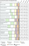
Comprehensive overview of bacterial acetylome studies. Shown are functional annotations of acetylated proteins identified from bacterial acetylome studies. ProteinAc, number of identified acetylated proteins; % TotalAc, percentage of acetylated proteins of the entire proteome; N, no; Y, yes. *, values are different than those previously reported; however, the values listed reflect the available data that were obtained from supplementary information from the cited references (28, 142, 143, 153–161). The percent scale at the top should be used to estimate the percentage of acetylated proteins in each of the categories within any given microorganism shown.
FIG 10
FIG 10
Synthesis of acyl-CoAs by AMP- and ADP-forming acyl-CoA synthetases. The AMP-forming and ADP-forming acyl-CoA synthetases convert organic acids to their CoA thioesters. AMP-forming acyl-CoA synthetases perform this reaction through two half-reactions via an adenylated intermediate (A), while the ADP-forming acyl-CoA synthetase reaction is driven by the energy of hydrolysis of the γ-phosphate of ATP (B).
FIG 11
FIG 11
Binding of cAMP induces a 40-Å structural change in M. tuberculosis PatA. In the absence of cAMP, MtPatA adopts an autoinhibited state, where a “lid” (blue) blocks the entrance of the substrate to the active site of the GNAT domain (red). In the presence of cAMP, the cAMP binding domain (gold) rotates 40° relative to the GNAT domain. This causes the lid to swing away from the GNAT domain, exposing the active-site cleft. Also shown are acetyl-CoA (black sticks), cAMP (gray spheres), and the C-terminal helix (green). The PDB accession numbers are 4AVA for the MtPatA structure and 4AVB for the MtPatA structure with cAMP.
FIG 12
FIG 12
Interactions between the T. thermophila Gcn5 protein and a peptide substrate. (A) The structure of TtGcn5 and a histone H3 11-peptide residue (PDB accession number 1QSN) demonstrates that the presence of CoA (not shown) causes structural changes that may facilitate interactions with its protein substrate (interacting residues are shown in blue). (B) Molecular interactions of TtGcn5 (blue) with the peptide substrate (green).
FIG 13
FIG 13
Molecular interactions of S. lividans PatAGNAT and S. enterica AcsCTD. (A) Crystal structure of the interactions between S. lividans PatAGNAT and S. enterica AcsCTD (PDB accession number 4U5Y). The SlPatAGNAT catalytic residue (E123) is shown in red sticks, and the acetylated lysine of SeAcsCTD (K609) is shown in blue sticks. (B) Interactions between SlPatAGNAT (surface) and SeAcsCTD (sticks). (C and D) Electrostatic potential of the SlPatAGNAT-SeAcsCTD surface interface, with negatively charged regions in red, positively charged regions in blue, and neutral residues in white. (Adapted from reference . © American Society for Biochemistry and Molecular Biology.)
FIG 14
FIG 14
Acetylation determinants outside the motif containing the acetylation site. (A) Consensus motif containing the acetylation site (indicated by the arrow) generated from the alignments of acyl-CoA synthetases acetylated by R. palustris Pat (RpPat). The letter height corresponds to the frequency of a particular amino acid residue in that position. (B) Electrostatic potential of RpMatB (methylmalonyl CoA synthetase), a protein that is not acetylated by RpPat. Negatively charged regions are shown in red, and positively charged regions are shown in blue. (C) Electrostatic potential of the RpMatB and B. xenovorans BclM (benzoate:CoA synthetase) chimera protein (RpMatB-BxBclM chimera B3), a protein that is acetylated by RpPat. Negatively charged regions are in red, and positively charged regions are in blue. (D) Overlay of the C-terminal domain of the RpMatB-BxBclM chimeras (PDB accession numbers 4GXQ for B1 and 4GXR for B3) aligned with the C-terminal domains of RpMatB (PDB accession number 4FUQ), with the BxBclM-derived residues of the B1 chimera in yellow, BxBclM-derived residues of the B3 chimera in orange, and wild-type RpMatB residues in cyan. The consensus motif containing the acetylation site (PX4GK) is shown in red in the active-site loop, with the acetylated lysine residue (K488) shown as red sticks. (Adapted from references [panel A] and [panels B to D]. © American Society for Biochemistry and Molecular Biology.)
FIG 15
FIG 15
CoA homeostasis. Shown is a schematic of the contributions of CoA and acetyl-CoA to cellular metabolism. CoASH, coenzyme A; Ac-CoA, acetyl-CoA, O-AADPR, O-acetyl-ADP-ribose; Ac-P, acetyl-phosphate; Ac-AMP, acetyl-AMP, PPi, pyrophosphate; Pr-CoA, propionyl-CoA; α-KG, alpha-ketoglutarate; OAA, oxaloacetate; Suc-CoA, succinyl-CoA; dP-CoA, dephospho-coenzyme A; AcAc-ACP, acetoacetyl-acyl carrier protein; KAS III, β-ketoacyl-(acyl-carrier-protein) synthase III; MTA, malonyl-CoA:acyl carrier protein transacylase; PDH, pyruvate dehydrogenase complex; α-KGDH, α-ketoglutarate dehydrogenase complex; SucCD, succinyl-CoA synthetase.
None
None

References

    1. Walsh CT, Garneau-Tsodikova S, Gatto GJ Jr. 2005. Protein posttranslational modifications: the chemistry of proteome diversifications. Angew Chem Int Ed Engl 44:7342–7372. doi:10.1002/anie.200501023. - DOI - PubMed
    1. Phillips DM. 1963. The presence of acetyl groups of histones. Biochem J 87:258–263. - PMC - PubMed
    1. Jarrell KF, Ding Y, Meyer BH, Albers SV, Kaminski L, Eichler J. 2014. N-linked glycosylation in archaea: a structural, functional, and genetic analysis. Microbiol Mol Biol Rev 78:304–341. doi:10.1128/MMBR.00052-13. - DOI - PMC - PubMed
    1. Zverina EA, Lamphear CL, Wright EN, Fierke CA. 2012. Recent advances in protein prenyltransferases: substrate identification, regulation, and disease interventions. Curr Opin Chem Biol 16:544–552. doi:10.1016/j.cbpa.2012.10.015. - DOI - PMC - PubMed
    1. Rivera C, Gurard-Levin ZA, Almouzni G, Loyola A. 2014. Histone lysine methylation and chromatin replication. Biochim Biophys Acta 1839:1433–1439. doi:10.1016/j.bbagrm.2014.03.009. - DOI - PubMed

Publication types

LinkOut - more resources