. 2015 Aug 15;195(4):1774-81.
doi: 10.4049/jimmunol.1500522. Epub 2015 Jul 15.

Differentiation Kinetics of Blood Monocytes and Dendritic Cells in Macaques: Insights to Understanding Human Myeloid Cell Development

Affiliations

Differentiation Kinetics of Blood Monocytes and Dendritic Cells in Macaques: Insights to Understanding Human Myeloid Cell Development

Chie Sugimoto et al. J Immunol. .

Abstract

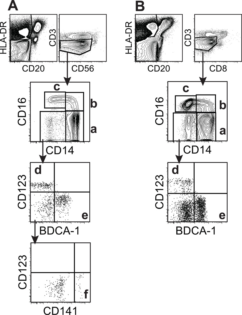
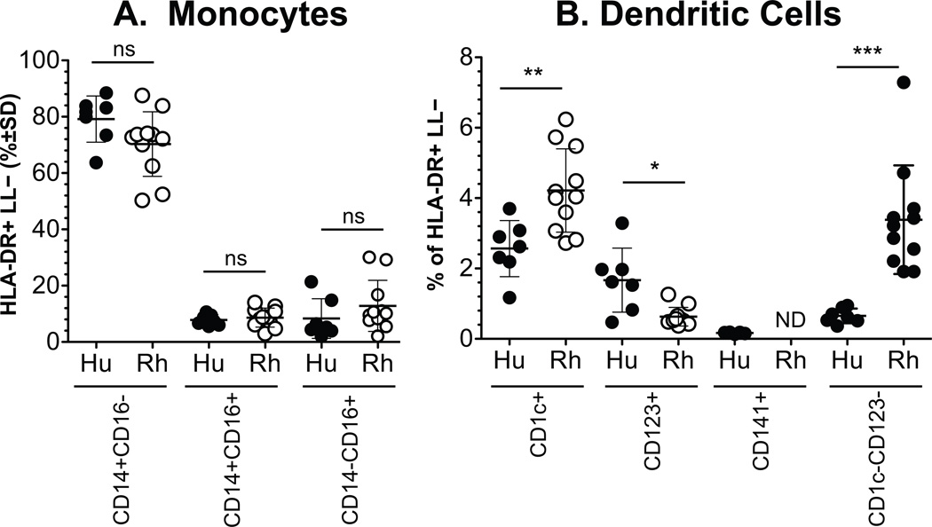
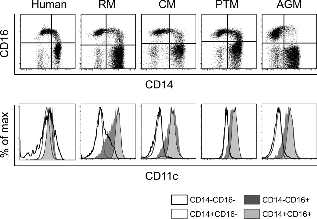
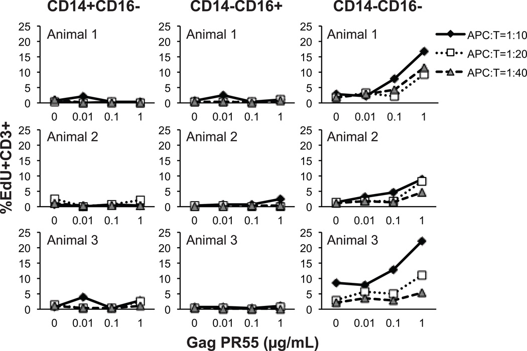
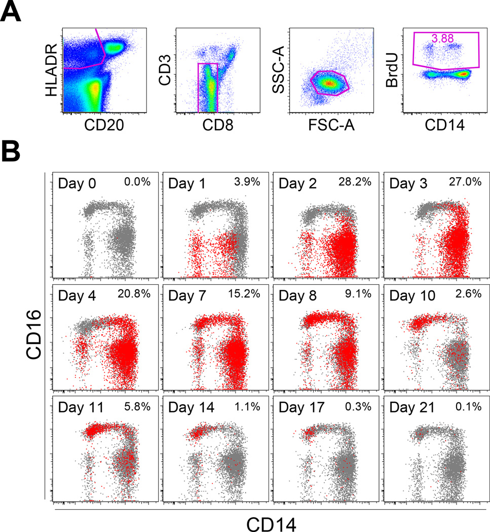
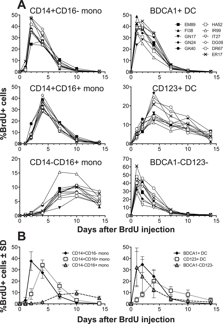
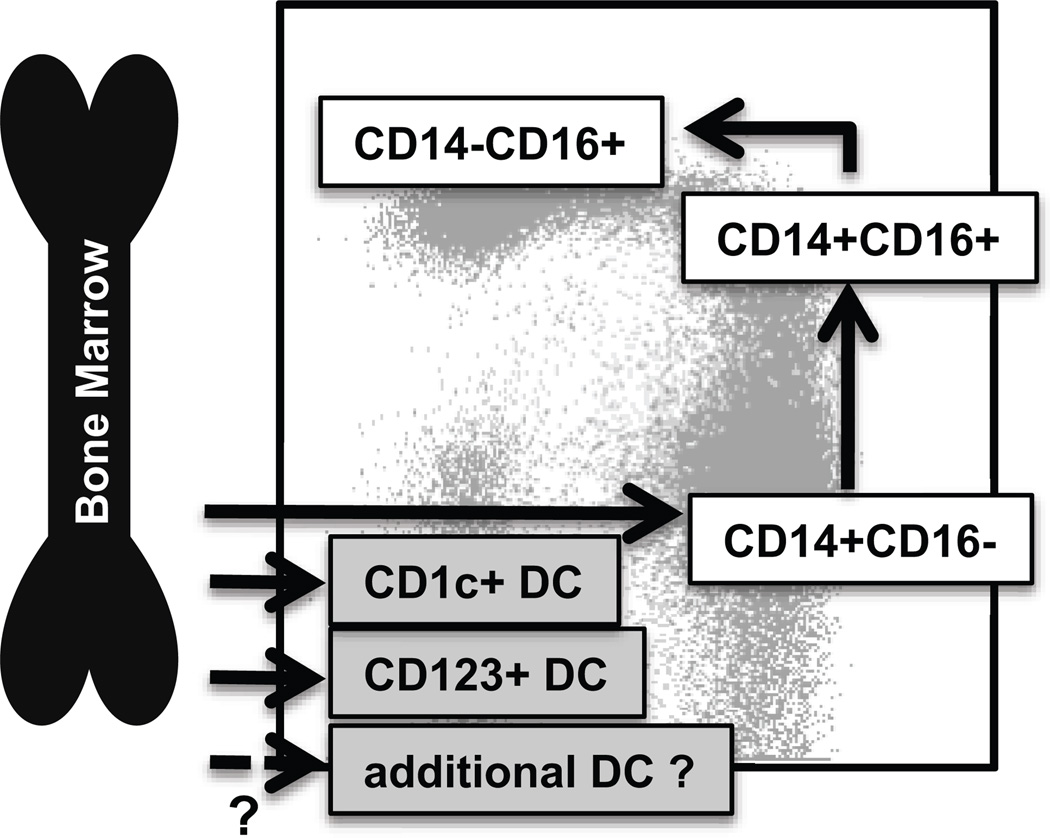
Monocyte and dendritic cell (DC) development was evaluated using in vivo BrdU pulse-chase analyses in rhesus macaques, and phenotype analyses of these cells in blood also were assessed by immunostaining and flow cytometry for comparisons among rhesus, cynomolgus, and pigtail macaques, as well as African green monkeys and humans. The nonhuman primate species and humans have three subsets of monocytes, CD14(+)CD16(-), CD14(+)CD16(+), and CD14(-)CD16(+) cells, which correspond to classical, intermediate, and nonclassical monocytes, respectively. In addition, there exist presently two subsets of DC, BDCA-1(+) myeloid DC and CD123(+) plasmacytoid DC, that were first confirmed in rhesus macaque blood. Following BrdU inoculation, labeled cells first appeared in CD14(+)CD16(-) monocytes, then in CD14(+)CD16(+) cells, and finally in CD14(-)CD16(+) cells, thus defining different stages of monocyte maturation. A fraction of the classical CD14(+)CD16(-) monocytes gradually expressed CD16(+) to become CD16(+)CD14(+) cells and subsequently matured into the nonclassical CD14(-)CD16(+) cell subset. The differentiation kinetics of BDCA-1(+) myeloid DC and CD123(+) plasmacytoid DC were distinct from the monocyte subsets, indicating differences in their myeloid cell origins. Results from studies utilizing nonhuman primates provide valuable information about the turnover, kinetics, and maturation of the different subsets of monocytes and DC using approaches that cannot readily be performed in humans and support further analyses to continue examining the unique myeloid cell origins that may be applied to address disease pathogenesis mechanisms and intervention strategies in humans.


Figures

Figure 1
Figure 1. Phenotying of blood monocyte and DC subsets in humans (A) and rhesus macaques (B)
EDTA-treated blood samples were stained with antibodies shown in Table 1 and analyzed by 11-color flow cytometry. (A) In HLA-DR+CD3CD20CD56 populations, human monocyte and DC subsets were gated and divided into 4 populations by CD14 and CD16 expression as follows; (a) CD14+CD16 monocytes, (b) CD14+CD16+ monocytes, (c) CD14CD16+ monocytes, and a CD14CD16 population that was further divided into (d) CD123+ pDC and (e) BDCA-1+ mDC. In addition, a CD141+ mDC (f) was identified. (B) To analyze rhesus monocyte and DC subsets, HLA-DR+CD3CD20CD8cell populations were similarly gated and further divided as described in panel A with the exception that antibody to human CD141 (BDCA-3) did not cross-react to, or detect this marker on rhesus macaque cells. The populations of cells identified included; (a) CD14+CD16 monocytes, (b) CD14+CD16+ monocytes, (c) CD14CD16+ monocytes, (d) CD123+ pDC, and (e) BDCA-1+ mDC.
Figure 2
Figure 2. Comparison in the proportion of blood monocyte and DC subset populations between humans and rhesus macaques
Blood from 7 humans and 11 rhesus macaques were gated and analyzed as described in the materials and methods and in Figure 1. The proportions of monocytes (A) and DC (B) subsets were calculated from the total number of HLA-DR+ lymphocyte lineage (LL) negative cells. Comparisons between cell subsets in human and rhesus macaque blood were calculated for statistically significant differences by the Mann Whitney rank test. Not detected, ND; P<0.05,*; P<0.01,**; P < 0.001,***.
Figure 3
Figure 3. Comparison of CD11c expression on monocytes and DC among primate species
Blood from human, rhesus macaque (RM), cynomolgus macaque (CM), pigtail macaque (PTM), and African green monkey (AGM) was gated for HLA-DR+ lymphocyte lineage-negative cells and divided into four populations based on CD14 and CD16 expression. CD11c staining on cell subsets CD14CD16, CD14+CD16, CD14+CD16+ and CD14CD16+ was plotted.
Figure 4
Figure 4. Cell proliferation induced by antigen presentation on rhesus monocyte and DC subsets
CD14+CD16 classical monocytes, CD14CD16+ non-classical monocytes, and the CD14CD16 fraction that includes DC subsets were sorted by flow cytometry of blood samples obtained from SIV-infected and ART-treated rhesus macaques. The subset populations were then pulsed with Gag pr55 protein at indicated concentrations for 2 hr. The antigen-pulsed effector cells were added to wells at numbers ranging from 2500 - 10000 per well and incubated with 1×10⁵ autologous CD3+ T cells per well for 4.5 days resulting in antigen presenting cell to T cell (APC:T) ratios of 1:40-1:10. The thymidine analogue, EdU, was added during the last 18 hr of incubation and EdU incorporation in CD3+ T cells was detected by immunostaining and flow cytometry. Data from three animals are shown.
Figure 5
Figure 5. Kinetics of BrdU incorporation by monocyte and DC populations
Normal healthy rhesus macaques were injected with BrdU intravenously at a dose of 60 mg/kg body weight. EDTA-treated blood was collected at varying time intervals as indicated. The data shown are representative for four animals. (A) BrdU incorporation within the HLA-DR+CD3CD20CD8 cell population was examined by immunostaining and flow cytometry. Since monocytes and DC displayed slightly distinct scatter plots, the cell population first was gated on HLA-DR, CD20, CD3, and CD8, and then the scatter plot was confirmed. (B) BrdU-staining cells, as identified in Panel A, were indicated here in red with the percent of HLA-DR+CD3CD20CD8 cells noted on the top right corner of each time point after BrdU injection, and results were overlaid onto plots of cells stained for CD14 and CD16 expression shown in gray.
Figure 6
Figure 6. Kinetics of BrdU incorporation by each monocyte and DC population in rhesus macaque blood
(A) Eleven healthy normal adult rhesus macaques were administrated BrdU intravenously and blood was drawn and analyzed at indicated time points. The percent of BrdU+ cells within each of the monocyte subsets (CD14+CD16, CD14+CD16+, and CD14CD16+ monocytes) and two DC subsets (BDCA-1+ mDC, and CD123+ pDC), as well as the non-BDCA-1 and CD123 populations identified in Figure 1B were plotted as a percent of the HLA-DR+ lymphocyte lineage-negative cells. (B) Mean values (%±SD) of BrdU incorporation by the monocyte (left panel) and DC plus BDCA-1 and CD123+ populations (right panel) were plotted from the 11 animals.
Figure 7
Figure 7. Summary of blood monocyte and DC differentiation in rhesus macaques
Myeloid precursor cells in the bone marrow migrate into blood and give rise to CD14+CD16 classical monocytes. A large proportion of these classical monocytes rapidly disappeared from the circulation to become tissue macrophages. A fraction of the classical monocytes differentiated into CD14+CD16+ intermediate monocytes and then into CD14CD16+ non-classical monocytes in the circulation. The populations of mDC, pDC and perhaps other unidentified DC appeared to differentiate from various DC precursors directly in the bone marrow prior entering the blood circulation.

References

    1. Chow A, Brown BD, Merad M. Studying the mononuclear phagocyte system in the molecular age. Nat Rev Immunol. 2011;11:788–798. - PubMed
    1. Auffray C, Sieweke MH, Geissmann F. Blood monocytes: development, heterogeneity, and relationship with dendritic cells. Annu Rev Immunol. 2009;27:669–692. - PubMed
    1. Ziegler-Heitbrock L, Ancuta P, Crowe S, Dalod M, Grau V, Hart DN, Leenen PJM, Liu Y-J, MacPherson G, Randolph GJ, Scherberich J, Schmitz J, Shortman K, Sozzani S, Strobl H, Zembala M, Austyn JM, Lutz MB. Nomenclature of monocytes and dendritic cells in blood. Blood. 2010;116:e74–e80. - PubMed
    1. Kim W-K, Sun Y, Do H, Autissier P, Halpern EF, Piatak M, Lifson JD, Burdo TH, McGrath MS, Williams K. Monocyte heterogeneity underlying phenotypic changes in monocytes according to SIV disease stage. J Leukoc Biol. 2010;87:557–567. - PMC - PubMed
    1. Moniuszko M, Bodzenta-Lukaszyk A, Kowal K, Lenczewska D, Dabrowska M. Enhanced frequencies of CD14++CD16+, but not CD14+CD16+, peripheral blood monocytes in severe asthmatic patients. Clin Immunol. 2009;130:338–346. - PubMed

Publication types