. 2015 Sep;138(Pt 9):2521-36.
doi: 10.1093/brain/awv204. Epub 2015 Jul 15.

Myelin-associated glycoprotein gene mutation causes Pelizaeus-Merzbacher disease-like disorder

Affiliations

Myelin-associated glycoprotein gene mutation causes Pelizaeus-Merzbacher disease-like disorder

Alexander Lossos et al. Brain. 2015 Sep.

Abstract

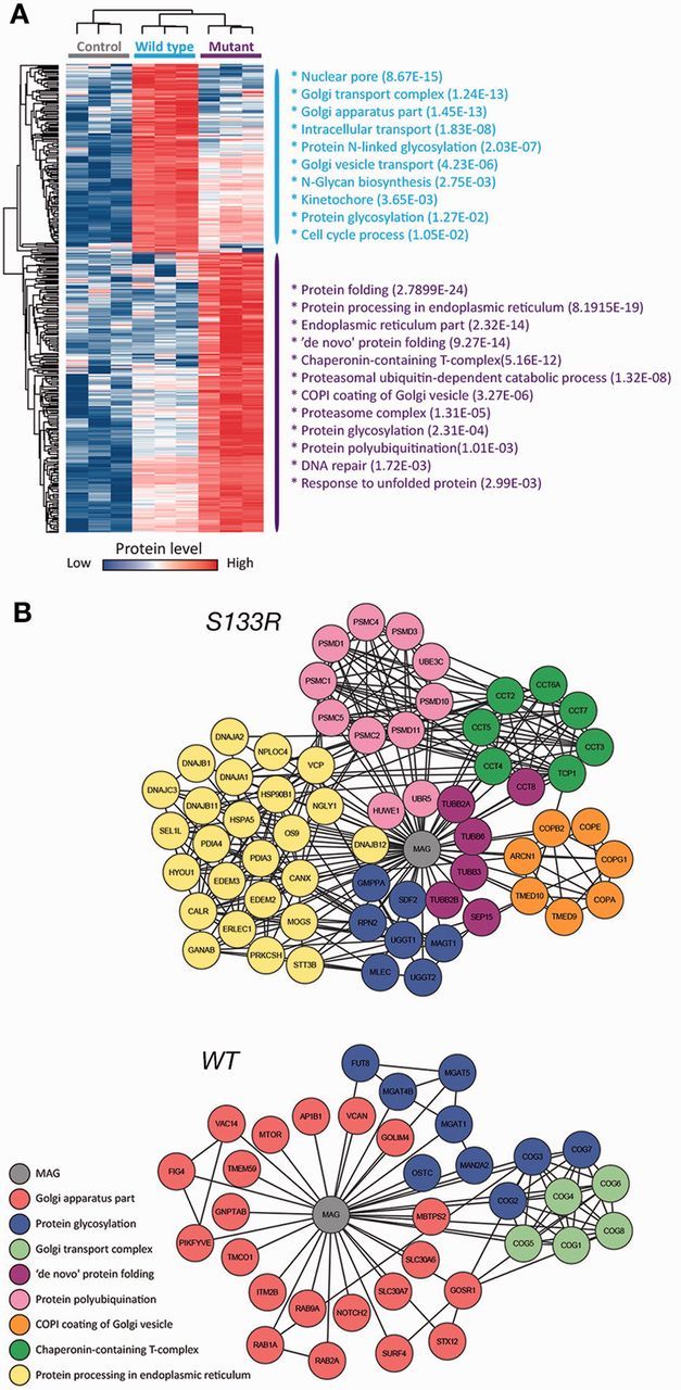
Pelizaeus-Merzbacher disease is an X-linked hypomyelinating leukodystrophy caused by mutations or rearrangements in PLP1. It presents in infancy with nystagmus, jerky head movements, hypotonia and developmental delay evolving into spastic tetraplegia with optic atrophy and variable movement disorders. A clinically similar phenotype caused by recessive mutations in GJC2 is known as Pelizaeus-Merzbacher-like disease. Both genes encode proteins associated with myelin. We describe three siblings of a consanguineous family manifesting the typical infantile-onset Pelizaeus-Merzbacher disease-like phenotype slowly evolving into a form of complicated hereditary spastic paraplegia with mental retardation, dysarthria, optic atrophy and peripheral neuropathy in adulthood. Magnetic resonance imaging and spectroscopy were consistent with a demyelinating leukodystrophy. Using genetic linkage and exome sequencing, we identified a homozygous missense c.399C>G; p.S133R mutation in MAG. This gene, previously associated with hereditary spastic paraplegia, encodes myelin-associated glycoprotein, which is involved in myelin maintenance and glia-axon interaction. This mutation is predicted to destabilize the protein and affect its tertiary structure. Examination of the sural nerve biopsy sample obtained in childhood in the oldest sibling revealed complete absence of myelin-associated glycoprotein accompanied by ill-formed onion-bulb structures and a relatively thin myelin sheath of the affected axons. Immunofluorescence, cell surface labelling, biochemical analysis and mass spectrometry-based proteomics studies in a variety of cell types demonstrated a devastating effect of the mutation on post-translational processing, steady state expression and subcellular localization of myelin-associated glycoprotein. In contrast to the wild-type protein, the p.S133R mutant was retained in the endoplasmic reticulum and was subjected to endoplasmic reticulum-associated protein degradation by the proteasome. Our findings identify involvement of myelin-associated glycoprotein in this family with a disorder affecting the central and peripheral nervous system, and suggest that loss of the protein function is responsible for the unique clinical phenotype.

Keywords: MAG; Pelizaeus-Merzbacher-like disease; hereditary spastic paraplegia.


Figures

None
Pelizaeus-Merzbacher disease is an X-linked hypomyelinating leukodystrophy. Lossos et al. describe a family with an early-onset Pelizaeus-Merzbacher disease-like phenotype that slowly evolves into complicated hereditary spastic paraplegia, affecting both the CNS and PNS. Exome sequencing reveals a causative homozygous missense mutation in MAG, which encodes myelin associated glycoprotein.
Figure 1
Figure 1
Homozygous c.399C>G mutation in MAG in the autosomal recessive PMD-like disorder. (A) Pedigree of the family. Circles represent females and squares males. Filled symbols indicate affected individuals and double lines consanguinity by descent. (B) DNA sequencing identified a c.399C>G, p.S133R mutation in MAG. Upper panel shows wild-type sequence (WT) and lower panel shows affected homozygous Patient III-2. (C) Restriction-based analysis of individuals from the extended family. PCR products were digested with AvaII resulting in a wild-type (183 + 65 bp) and a mutant allele (141 + 65 + 42 bp). M = marker; UC = uncut; HTZ = heterozygote; HMZ = homozygote, with the letters designating position in the pedigree. (D) Modelling of the p.S133R mutation effect on MAG stability and structure. Structural model of the first Ig-like domain (residues 22–139) suggests that S133 points into the core of this domain to form hydrogen bonds with a neighbouring proline residue. Mutation to arginine will create strong clashes leading to unfolding and destabilization of the protein. (I) Overall model of the first Ig-like domain of MAG. S133 (in red) is located in the c-terminal strand of the first Ig-like domain (in green), adjacent to the n-terminal strand of this (in blue). Interaction between these two strands holds the domain together. (II) Close-up of the S133 position (green central residue in sticks representation) and its surrounding showing a potential hydrogen bond formed with a neighboring backbone, P27 at the N-terminal of the Ig-like domain (dashed lines). (III) Mutation of serine to arginine (in white) at position 133 leads to strong clashes (in red) and may cause opening of the sheet to its unfolding and misfolding. (E) Representative imaging findings. Cerebral MRI in Patient III-2 at age 7 (I) and 25 (II–V and VII) years and in Patient III-3 at age 24 (VI) showing mild progressive atrophy of the optic chiasm, corpus callosum and cerebellum on T1-weighted images (I and II), a periventricular rim of the cerebral white matter hyperintensity on FLAIR (III) and T2-weighted (IV) images, which appears hypointense on T1(+Gd)-weighted images (V, arrow), and scattered foci of hyperintensity on FLAIR-weighted images (VI). Areas of the unaffected cerebral white matter appear of a normal signal. (VII) Cerebral 1H-magnetic resonance spectroscopy (echo time 135) spectrum at the centrum semiovale showing an increase in choline peak.
Figure 2
Figure 2
Sural nerve biopsy findings in Patient III-2. Paraffin embedded sections stained with haematoxylin and eosin (A), Luxol Fast blue–periodic acid–Schiff (B), and with immunohistochemical stains for neurofilament (C), MBP (D), S-100 protein (E) and MAG (G), as well as electron-microscopy (F and H) did not reveal remarkable alterations, except for rare ill-formed onion-bulb structures, noted only on S-100 stained section (E, inset, arrow) and electron-microscopy (F, H, encircled). Myelin sheath of the affected nerve appears relatively thin (F, black arrow) as opposed to myelin sheath of most axons (F, white arrow). Possible minimal loss of axon-glia interaction (H, asterisks) was noted in rare myelinated axons, but traction/tearing artefact cannot be excluded. There was complete absence of myelin sheath staining for MAG (G), compared to a normal control (G, inset). SC = Schwann cell nucleus; ax = axon.
Figure 3
Figure 3
Characterization of the MAG-S133R mutant. (A) Expression of the wild-type (WT) and mutant (S133R) human MAG (hMAG) and of these proteins fused to GFP (MAG-GFP) in HEK-293 cells. Western blot analysis was done on total cell lysates using antibodies to MAG or GFP. The location of molecular weight markers is shown in kDa on the right. (B) MAG-S133R mutant lacks complex glycans. Immunocomplexes of wild-type (WT) and mutant (S133R) MAG-GFP expressed in HEK-293 cells were left untreated or treated with endoglycosidase H (EndoH), which cleaves asparagine-linked mannose rich oligosaccharides but not highly processed complex oligosaccharides from glycoproteins. Western blot analysis was done using an antibody to GFP. Arrowheads mark the location of MAG containing high complex oligosaccharides (a), mannose rich oligosaccharides (b) and the deglycosylated protein (containing only the core N-acetylglucosamines) (c). The location of molecular weight markers is shown in kDa on the right. (C) Differential recognition of the MAG-S133R mutant by specific antibodies. Fixed and permeabilized COS7 cells expressing the wild-type (WT) and mutant (S133R) forms of MAG were immunolabelled using the indicated antibodies. Note that MAb 513 does not recognize the mutant protein. Scale bars = 20 μm.
Figure 4
Figure 4
MAG-S133R does not reach the cell surface. (A) Cell surface expression of the wild-type (WT) and MAG-S133R (S133R) proteins in transfected COS7 cells. Cells were stained with the D3A2G5 antibody, which recognize the extracellular region of MAG, either without (-Tx100) or with (+Tx100) fixation and permeabilization. The cellular nuclei were labelled with DAPI (blue). (B) Distribution of MAG-GFP or MAG-S133R-GFP in transfected rat Schwann cells. (C) Distribution of MAG-MYC or MAG-S133-MYC co-infected with plasma membrane marker farnesylated GFP (FarGFP) in rat Schwann cells. Scale bars: A–C = 20 μm. (D) Cell surface expression of MAG-GFP and MAG-S133R-GFP proteins in MAG-GFP or MAG-S133R-GFP expressing HEK 293T cells. Surface proteins where labelled using impermeable NHS-sulfo biotin. Lysates were precipitated with streptavidin-conjugated agarose beads followed western blot analysis using antibodies to GFP. The location of molecular weight markers is shown in kDa on the right.
Figure 5
Figure 5
S133R MAG accumulates in the endoplasmic reticulum. (A and B) Hela cells expressing the wild-type (WT) or MAG-S133R (S133R) proteins were immunolabelled using an antibody to MAG and to calnexin (A), or to p115 (B). (C and D) COS7 cells expressing the wild-type (WT) or MAG-S133R (S133R) proteins were immunolabelled using an antibody to MAG and to the endoplasmic reticulum marker proteins VAPB (C) or Caspr (D). (E) Rat oligodendrocytes expressing MAG-GFP (WT) or MAG-S133R-GFP (S133R) were immunolabelled using an antibody to VAPB. (F) Rat Schwann cells expressing MAG-GFP (WT) or MAG-S133R-GFP (S133R) were immunolabelled using an antibody to VAPB. The GFP fluorescence is shown in green. Note that MAG-S133R is associated with endoplasmic reticulum markers. Scale bars = 20 μm. (G) MAG-S133R co-imunoprecipitation with endoplasmic reticulum proteins calnexin and BiP. HEK 293T expressing MAG-GFP and MAG-S133R-GFP where imunopercipitated using GFP antibody-conjugated beads and subjected to western blot analysis using antibodies to calnexin and BiP. The location of molecular weight markers is shown in kDa on the right.
Figure 6
Figure 6
MAG-S133R undergoes proteasome dependent degradation and associates with ERAD machinery. (A and B) MAG-S133R degradation rate is higher than wild-type MAG. HEK293T expressing wild-type MAG-MYC and MAG-S133-MYC subjected to cyclohexamide chase, at the indicated time points, total protein lysates were subjected to western blot analysis using MYC antibody. (B) Degradation rate plot analysis, MAG intensity relative to actin (image-lab software) from three independent experiments. (C) MAG-S133R degradation is proteasome dependent. HEK293T expressing wild-type MAG-MYC and MAG-S133-MYC subjected to cyclohexamide chase with or without bortezomib at the indicated time points, total protein lysates were subjected to western blot analysis using MYC antibody. (D and E) MAG-S133R associates with ERAD protein P97 and is polyubiquitinated. HEK293T expressing MAG-GFP and MAG-S133R-GFP where immunoprecipitation using GFP antibody conjugated beads and subjected to western blot analysis using P97 and FK-2 antibodies. The location of molecular weight markers is shown in kDa on the right.
Figure 7
Figure 7
Mass spectrometry-based interaction proteomics. MAG and MAG-S133R associate with distinct protein interaction networks. (A) Hierarchical clustering of 423 significantly changing proteins between wild-type and mutant (Student t-test; FDR = 0.05; S0 = 1.5). Selected enriched categories (GOCC, GOBP and KEGG) based on Fisher exact test are shown on the right, followed by their FDR corrected q-value. (B) Interaction network of potential interaction partners of MAG wild-type (lower map) and MAG-S133R (upper map). The protein network was built using the string database. Colours are based on selected significantly enriched categories. MAG is at the centre of each map.

Similar articles

Cited by

References

    1. Altier C, Garcia-Caballero A, Simms B, You H, Chen L, Walcher J, et al. The Cavbeta subunit prevents RFP2-mediated ubiquitination and proteasomal degradation of L-type channels. Nat Neurosci 2011; 14: 173–80. - PubMed
    1. Bizzi A, Castelli G, Bugiani M, Barker PB, Herskovits EH, Danesi U, et al. Classification of childhood white matter disorders using proton MR spectroscopic imaging. AJNR Am J Neuroradiol 2008; 29: 1270–5. - PMC - PubMed
    1. Burger D, Simon M, Perruisseau G, Steck AJ. The epitope(s) recognized by HNK-1 antibody and IgM paraprotein in neuropathy is present on several N-linked oligosaccharide structure on human P0 and myelin-associated glycoprotein. J Neurochem 1990; 54: 1569–75. - PubMed
    1. Christianson JC, Shaler TA, Tyler RE, Kopito RR. OS-9 and GRP94 deliver mutant alpha 1-antitrypsin to the Hrd1-SEL1L ubiquitin ligase for ERAD. Nat Cell Biol 2008; 10: 272–82. - PMC - PubMed
    1. Cox J, Neuhauser N, Michalski A, Scheltema RA, Olsen JV, Mann M. Andromeda: a peptide search engine integrated into the MaxQuant environment. J Proteome Res 2011; 10: 1794–805. - PubMed

Publication types

MeSH terms