. 2015 Jul 15;35(28):10188-201.
doi: 10.1523/JNEUROSCI.1096-15.2015.

Zebrafish Models for the Mechanosensory Hair Cell Dysfunction in Usher Syndrome 3 Reveal That Clarin-1 Is an Essential Hair Bundle Protein

Affiliations

Zebrafish Models for the Mechanosensory Hair Cell Dysfunction in Usher Syndrome 3 Reveal That Clarin-1 Is an Essential Hair Bundle Protein

Suhasini R Gopal et al. J Neurosci. .

Abstract

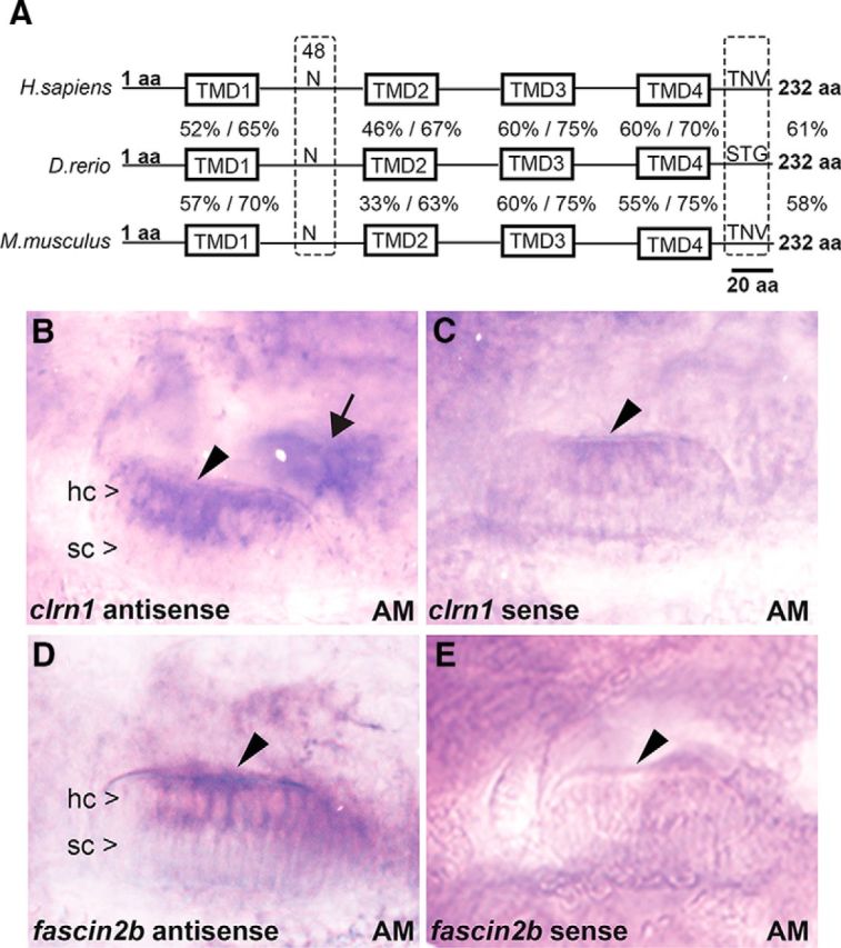
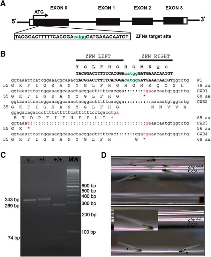
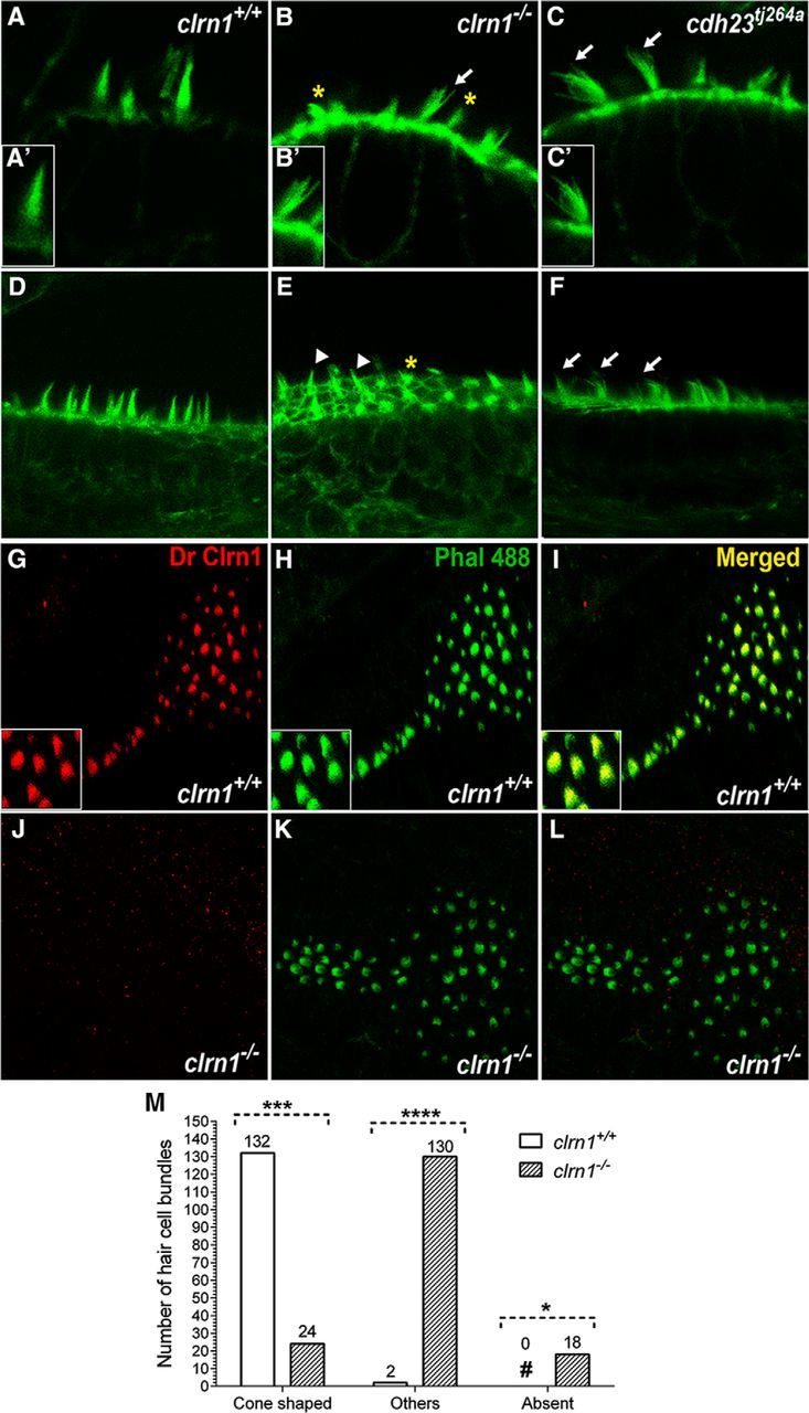
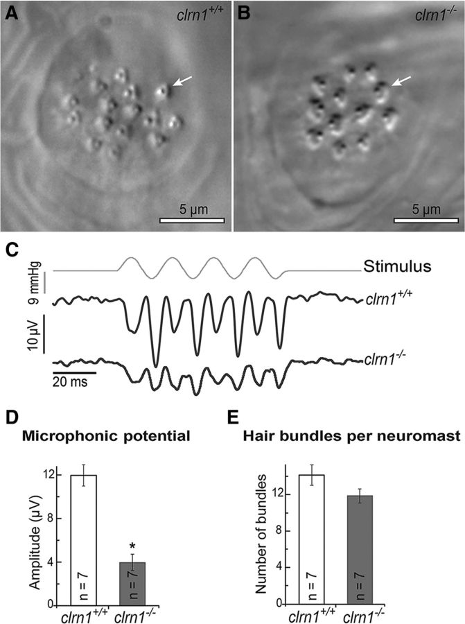
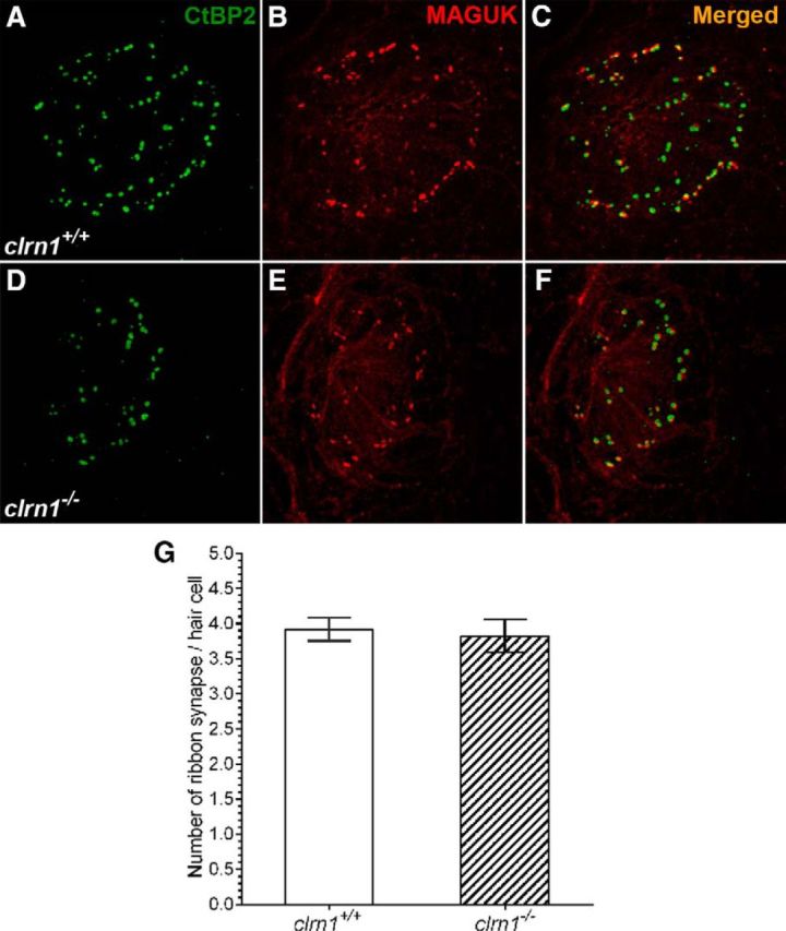
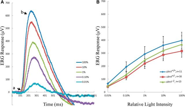
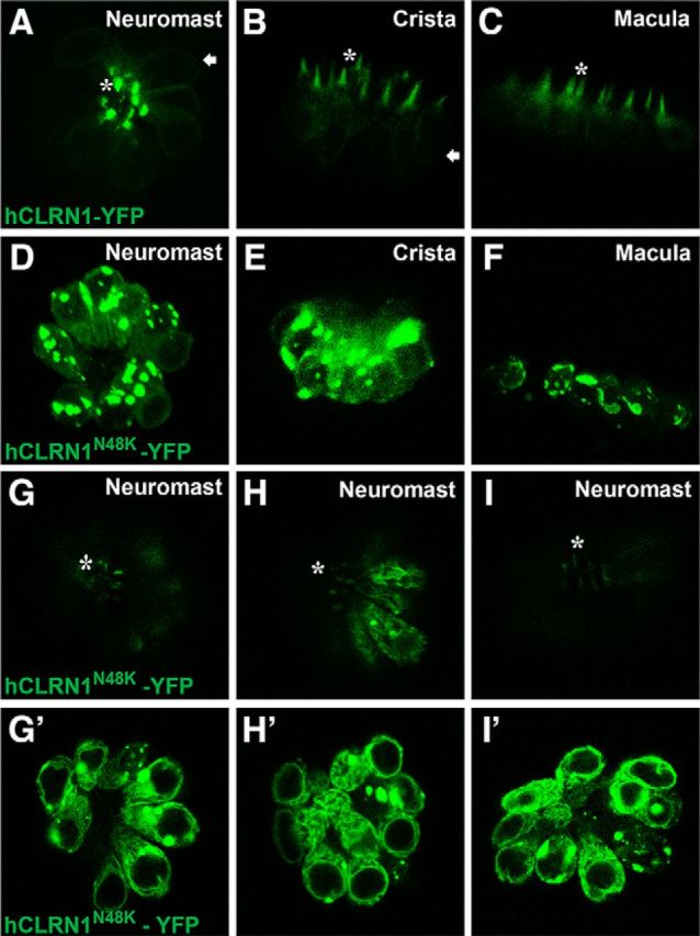
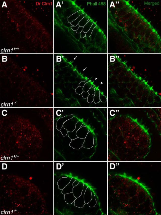
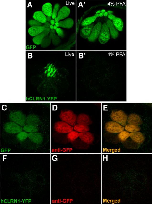
Usher syndrome type III (USH3) is characterized by progressive loss of hearing and vision, and varying degrees of vestibular dysfunction. It is caused by mutations that affect the human clarin-1 protein (hCLRN1), a member of the tetraspanin protein family. The missense mutation CLRN1(N48K), which affects a conserved N-glycosylation site in hCLRN1, is a common causative USH3 mutation among Ashkenazi Jews. The affected individuals hear at birth but lose that function over time. Here, we developed an animal model system using zebrafish transgenesis and gene targeting to provide an explanation for this phenotype. Immunolabeling demonstrated that Clrn1 localized to the hair cell bundles (hair bundles). The clrn1 mutants generated by zinc finger nucleases displayed aberrant hair bundle morphology with diminished function. Two transgenic zebrafish that express either hCLRN1 or hCLRN1(N48K) in hair cells were produced to examine the subcellular localization patterns of wild-type and mutant human proteins. hCLRN1 localized to the hair bundles similarly to zebrafish Clrn1; in contrast, hCLRN1(N48K) largely mislocalized to the cell body with a small amount reaching the hair bundle. We propose that this small amount of hCLRN1(N48K) in the hair bundle provides clarin-1-mediated function during the early stages of life; however, the presence of hCLRN1(N48K) in the hair bundle diminishes over time because of intracellular degradation of the mutant protein, leading to progressive loss of hair bundle integrity and hair cell function. These findings and genetic tools provide an understanding and path forward to identify therapies to mitigate hearing loss linked to the CLRN1 mutation.

Significance statement: Mutations in the clarin-1 gene affect eye and ear function in humans. Individuals with the CLRN1(N48K) mutation are born able to hear but lose that function over time. Here, we develop an animal model system using zebrafish transgenesis and gene targeting to provide an explanation for this phenotype. This approach illuminates the role of clarin-1 and the molecular mechanism linked to the CLRN1(N48K) mutation in sensory hair cells of the inner ear. Additionally, the investigation provided an in vivo model to guide future drug discovery to rescue the hCLRN1(N48K) in hair cells.

Keywords: clarin-1; hair cells; hearing.


Conflict of interest statement

The authors declare no competing financial interests.

Figures

Figure 1.
Figure 1.
An amino acid sequence comparison and mRNA expression analysis of zebrafish clarin-1. A, The zebrafish (D. rerio) amino acid sequence is 61% identical to the human (Homo sapiens) and 58% identical to the mouse (Mus musculus) clarin-1 sequences. The TMDs (TMD1–4), asparagine “N” at position 48 (N-glycosylation site), and putative PDZ class I-binding domain (TNV, STG) at the C terminus are indicated. The percentage of identity/similarity of the amino acid sequence within each TMD in zebrafish Clrn1 is compared with the corresponding domain in human (above) and mouse (below) in the line diagram. B–E, In situ hybridization (5 dpf) shows clrn1 mRNA expression in hair cells (hc) but not in supporting cells (sc) of the anterior macula (AM; arrowhead), similar to that of the control, fscn2b. clrn1 expression is also detected in hair cells of the posterior macula (arrow). Magnification, 40×.
Figure 2.
Figure 2.
Generation of clrn1 null alleles and balance phenotype in clrn1−/− zebrafish. A, ZFN target (upper case letters) and spacer (lower case, green letters) sequences in exon 0 of clrn1. B, clrn1 mutant alleles harbor a premature stop codon in exon 0. C, For genotyping, PCR was followed by restriction digestion with MslI to distinguish wild-type, heterozygous, and homozygous mutant alleles of clrn1. D, Balance/orientation in the clrn1+/+ and clrn1−/− mutant at 6 dpf is shown. Still frames captured during swim episodes show the trajectory of clrn1+/+ larvae toward the surface or parallel to it, with a normal body orientation. clrn1−/− mutants settle to the bottom of the Petri dish in a “head down with tail tipped toward the surface” position, with their dorsal side facing the surface (arrowhead). The arrow indicates a mutant in a head-down position that is also upside down; the inset shows an enlarged image of this larva.
Figure 3.
Figure 3.
Clrn1 is an essential hair bundle protein. A–F, The inner ear hair bundle phenotype of the clrn1−/− mutant larvae at 5 dpf. F-actin-rich hair bundles were visualized using fluorescent phalloidin. The clrn1+/+ hair cells with cone-shaped bundles in the crista (A) and anterior macula (D). clrn1−/− hair cells with splayed bundles (B) similar to those of cdh23tj264a (C, arrows). Digital enlargement of a single hair bundle of A′–C′ shows more clearly the cone shape and splayed hair bundle phenotype in wild-type and mutants, respectively. Many clrn1−/− hair cells display short or fractured bundles in the crista and macula (B and E; asterisks) compared with those of cdh23tj264a bundles (C, F). Three of 19 (∼15%) clrn1−/− hair cells in this image (arrowheads, E) appear cone shaped. Magnification, 63×. Immunolabeling: Clrn1 staining of clrn1+/+ (G) or clrn1−/− (J) larvae posterior macula with a DrClrn1 antibody and F-actin with fluorescent phalloidin (H, K). Yellow bundle (I, L) in the merged image of G and H showing Clrn1 expression in the hair bundle. Insets in G–I show magnified views of a portion of the macula in each part. Magnification, 40×. M, Quantification of different hair bundle morphology types in the clrn1−/− mutants. Hair cell bundles of inner ear from the clrn1+/+ (n = 5) and clrn1−/− (n = 5) larvae at 5 dpf were classified into one of three categories based on whether they were cone shaped, splayed, or missing (degenerated). A total of 134 (132 + 2) hair cell bundles from clrn1+/+ and 172 (24 + 130 + 18) hair cell bundles from clrn1−/− larvae were evaluated. For each genotype, the number in each category is expressed as a percentage of the total number of hair cell bundles evaluated for that genotype. Approximately 2% of the wild-type hair bundles were disrupted or splayed, possibly from spontaneous defects or to the effects of mechanical force used during mounting/handling for microscopy. ****p ≤ 0.0001; ***p ≤ 0.001; *p ≤ 0.05.
Figure 4.
Figure 4.
Microphonic potentials are reduced in the neuromasts of the clrn1−/− larvae. Oblique illumination images of the neuromasts in clrn1+/+ (A) and clrn1−/− (B) zebrafish larvae at 6 dpf. Arrows indicate single hair cell bundles. C, Stimulus (top trace) and microphonic potentials in neuromasts of clrn1+/+ (middle trace) and clrn1−/− (bottom trace) larvae. The top trace shows pressure applied to the stimulating puff pipette. D, Summary of microphonic potential peak-to-peak amplitudes at twice the stimulus frequency. Average values of microphonic potentials obtained from lateral line neuromasts of clrn1+/+ and clrn1−/− larvae. E, Number of hair cells per neuromast from clrn1+/+ and clrn1−/− larvae. Data represent the mean ± SEM. Analysis of the same larvae (n = 7) is shown in D and E. Asterisks indicate statistical significance, Student's t test, p < 0.001.
Figure 5.
Figure 5.
Evaluation of ribbon synapse in hair cells. A–F, Show ribbon synapse staining in the neuromast from clrn1+/+ (A–C) and clrn1−/− (B–D) zebrafish larvae at 6 dpf. A, D, Presynaptic (CtBP2). B, E, Postsynaptic (MAGUK) marker. C, F, Merged images. A–F, Maximum intensity projection of z-stack images (each section: 0.5 μm). Magnification, 63×. G, Quantification of ribbon synapse in the hair cells of clrn1+/+ and clrn1−/− zebrafish larvae at 6 dpf. The number and location of the ribbon synapse were evaluated from clrn1+/+ hair cells (n = 64) from the neuromast of three larvae and from clrn1−/− hair cells (n = 44) from the neuromast of three larvae. Data represent the mean ± SEM. The difference in ribbon synapse count between clrn1+/+ and clrn1−/− larvae is statistically not significant. Student's t test, p > 0.5.
Figure 6.
Figure 6.
Seven dpf clrn1−/− mutants show no defects in the ERG response. A, A typical ERG response recorded from a clrn1−/− mutant. The flash duration was 100 ms and the interval was 10 s. Flashes were delivered 50 ms after the start of recording. The stimulation intensity increased from 0.01 to 100% of the maximum value. Each curve represents the average of two responses. B, Mean b-wave amplitudes were plotted against the corresponding light intensities. Data represent the mean ± SEM.
Figure 7.
Figure 7.
A–C, Human CLRN1 is a hair bundle protein. Live confocal images of transgenic zebrafish (5 dpf) stably expressing hCLRN1-YFP or hCLRN1N48K-YFP in hair cells show that hCLRN1-YFP localizes predominantly to the hair bundle (asterisk), with a small amount at the plasma membrane (arrows). The pathogenic mutant, hCLRN1N48K-YFP, is predominantly retained within the cell body (D–F), although a small amount of protein reaches the hair bundle (G–I, asterisks). A–F, Maximum intensity projections that retain the brightest pixel value along each projection ray. In D–F, it is hard to visualize the small amount of hCLRN1N48K-YFP localized to the hair bundle. G, G′, Images showing top and middle (optical) sections of the same neuromast; these reveal that a small amount of hCLRN1N48K protein is translocated to the hair bundle (G, asterisk), and the remaining is retained in the cell body (G′); similar confocal images from two additional hCLRN1N48K-YFP-expressing larvae are shown to demonstrate that dual localization of hCLRN1N48K-YFP in hair cells is reproducible (H, H′, I, I′). Magnification, 40×.
Figure 8.
Figure 8.
Clrn1 is sensitive to PFA. A–D, Confocal images of the DrClrn1 antibody labeling the anterior macula (A, B) and cristae (C, D) of clrn1+/+ (A, C) and clrn1−/− (B, D) zebrafish larvae at 5 dpf. Splayed (arrow) and cone-shaped (arrowhead) hair bundles within the macula (B′) confirm the genotype of larvae; crista from the same larva was imaged (D′). To distinguish hair cells from supporting cells, borders of some of the hair cells are outlined in the sections of the middle column (A′–D′). Similar scattered fluorescent dots across the tissue (hair cells, supporting cells, and beyond) are seen in both wild-type and knock-out tissue. Magnification, 63×.
Figure 9.
Figure 9.
Human CLRN1 is sensitive to PFA. Confocal images of hair cells in neuromasts expressing GFP in the cell body (A) or hCLRN1-YFP in the hair bundle (B). After fixation, GFP (A′) but not hCLRN1 (B′) was detected in hair cells. In PFA-fixed larvae, anti-GFP antibodies detected GFP (C–E) but not hCLRN1-YFP (F–H) in hair cells. Magnification, 40×.

References

    1. Adato A, Vreugde S, Joensuu T, Avidan N, Hamalainen R, Belenkiy O, Olender T, Bonne-Tamir B, Ben-Asher E, Espinos C, Millán JM, Lehesjoki AE, Flannery JG, Avraham KB, Pietrokovski S, Sankila EM, Beckmann JS, Lancet D. USH3A transcripts encode clarin-1, a four-transmembrane-domain protein with a possible role in sensory synapses. Eur J Hum Genet. 2002;10:339–350. doi: 10.1038/sj.ejhg.5200831. - DOI - PubMed
    1. Adato A, Lefèvre G, Delprat B, Michel V, Michalski N, Chardenoux S, Weil D, El-Amraoui A, Petit C. Usherin, the defective protein in Usher syndrome type IIA, is likely to be a component of interstereocilia ankle links in the inner ear sensory cells. Hum Mol Genet. 2005;14:3921–3932. doi: 10.1093/hmg/ddi416. - DOI - PubMed
    1. Balciunas D, Wangensteen KJ, Wilber A, Bell J, Geurts A, Sivasubbu S, Wang X, Hackett PB, Largaespada DA, McIvor RS, Ekker SC. Harnessing a high cargo-capacity transposon for genetic applications in vertebrates. PLoS Genet. 2006;2:e169. doi: 10.1371/journal.pgen.0020169. - DOI - PMC - PubMed
    1. Blanco-Sanchez B, Clément A, Fierro J, Jr, Washbourne P, Westerfield M. Complexes of Usher proteins preassemble at the endoplasmic reticulum and are required for trafficking and ER homeostasis. Dis Model Mech. 2014;7:547–559. doi: 10.1242/dmm.014068. - DOI - PMC - PubMed
    1. Boëda B, El-Amraoui A, Bahloul A, Goodyear R, Daviet L, Blanchard S, Perfettini I, Fath KR, Shorte S, Reiners J, Houdusse A, Legrain P, Wolfrum U, Richardson G, Petit C. Myosin VIIa, harmonin and cadherin 23, three Usher I gene products that cooperate to shape the sensory hair cell bundle. EMBO J. 2002;21:6689–6699. doi: 10.1093/emboj/cdf689. - DOI - PMC - PubMed

Publication types

MeSH terms