

Cytological Kinetics of Periodontal Ligament in an Experimental Occlusal Trauma Model
- PMID: 26180510
- PMCID: PMC4502058
- DOI: 10.7150/ijms.12217
Cytological Kinetics of Periodontal Ligament in an Experimental Occlusal Trauma Model
Abstract
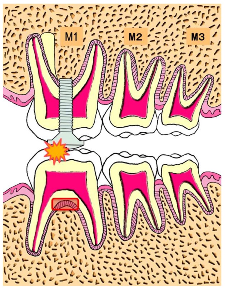
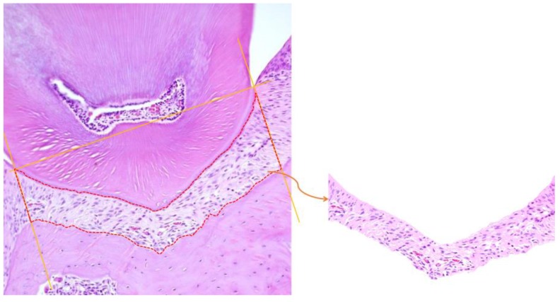
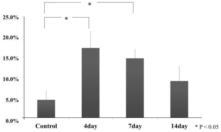
Using a model of experimental occlusal trauma in mice, we investigated cytological kinetics of periodontal ligament by means of histopathological, immunohistochemical, and photographical analysis methods. Periodontal ligament cells at furcation areas of molar teeth in the experimental group on day 4 showed a proliferation tendency of periodontal ligament cells. The cells with a round-shaped nucleus deeply stained the hematoxylin and increased within the day 4 specimens. Ki67 positive nuclei showed a prominent increase in the group on days 4 and 7. Green Fluorescent Protein (GFP) positivity also revealed cell movement but was slightly slow compared to Ki67. It indicated that restoration of mechanism seemed conspicuous by osteoclasts and macrophages from bone-marrow-derived cells for the periodontal ligament at the furcation area. It was suggested that the remodeling of periodontal ligament with cell acceleration was evoked from the experiment for the group on day 4 and after day 7. Periodontal ligament at the furcation area of the molar teeth in this experimental model recovered using the cells in situ and the bone-marrow-derived cells.
Keywords: Green fluorescent protein (GFP); Ki67; Mouse; Occlusal trauma; Periodontal tissue.
Conflict of interest statement
Competing Interests: The authors have declared that no competing interest exists.
Figures


References
-
- Svanberg G. Influence of trauma from occlusion on the periodontium of dogs with normal or inflamed gingivae. Odontol Revy. 1974;25:165–178. - PubMed
-
- Stahl SS. Accommodation of the periodontium to occlusal trauma and inflammatory periodontal disease. Dent Clin North Am. 1975;19:531–542. - PubMed
-
- Lindhe J, Ericsson I. The influence of trauma from occlusion on reduced but healthy periodontal tissues in dogs. J Clin Periodontol. 1976;3:110–122. - PubMed
-
- Biancu S, Ericsson I, Lindhe J. Periodontal ligament tissue reactions to trauma and gingival inflammation. An experimental study in the beagle dog. J Clin Periodontol. 1995;22:772–779. - PubMed
-
- Glickman I, Smulow JB. Effect of excessive occlusal forces upon the pathway of gingival inflammation in humans. J Periodontol. 1965;36:141–147. - PubMed
Publication types
MeSH terms
LinkOut - more resources
Full Text Sources
Other Literature Sources
