. 2015 Apr 28;35(3):e00220.
doi: 10.1042/BSR20150004.

SNAP23 is selectively expressed in airway secretory cells and mediates baseline and stimulated mucin secretion

Affiliations

SNAP23 is selectively expressed in airway secretory cells and mediates baseline and stimulated mucin secretion

Binhui Ren et al. Biosci Rep. .

Abstract

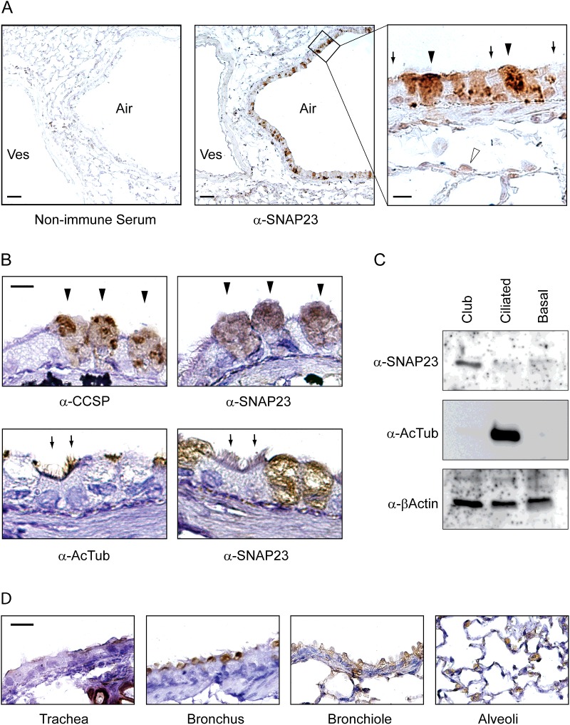
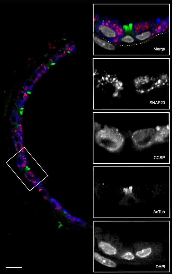
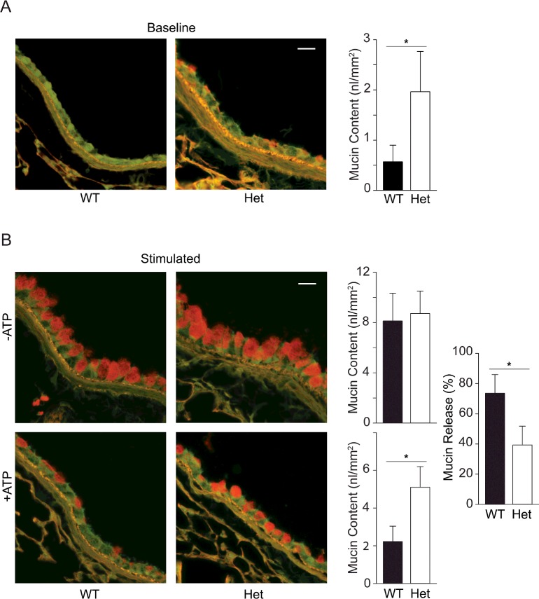
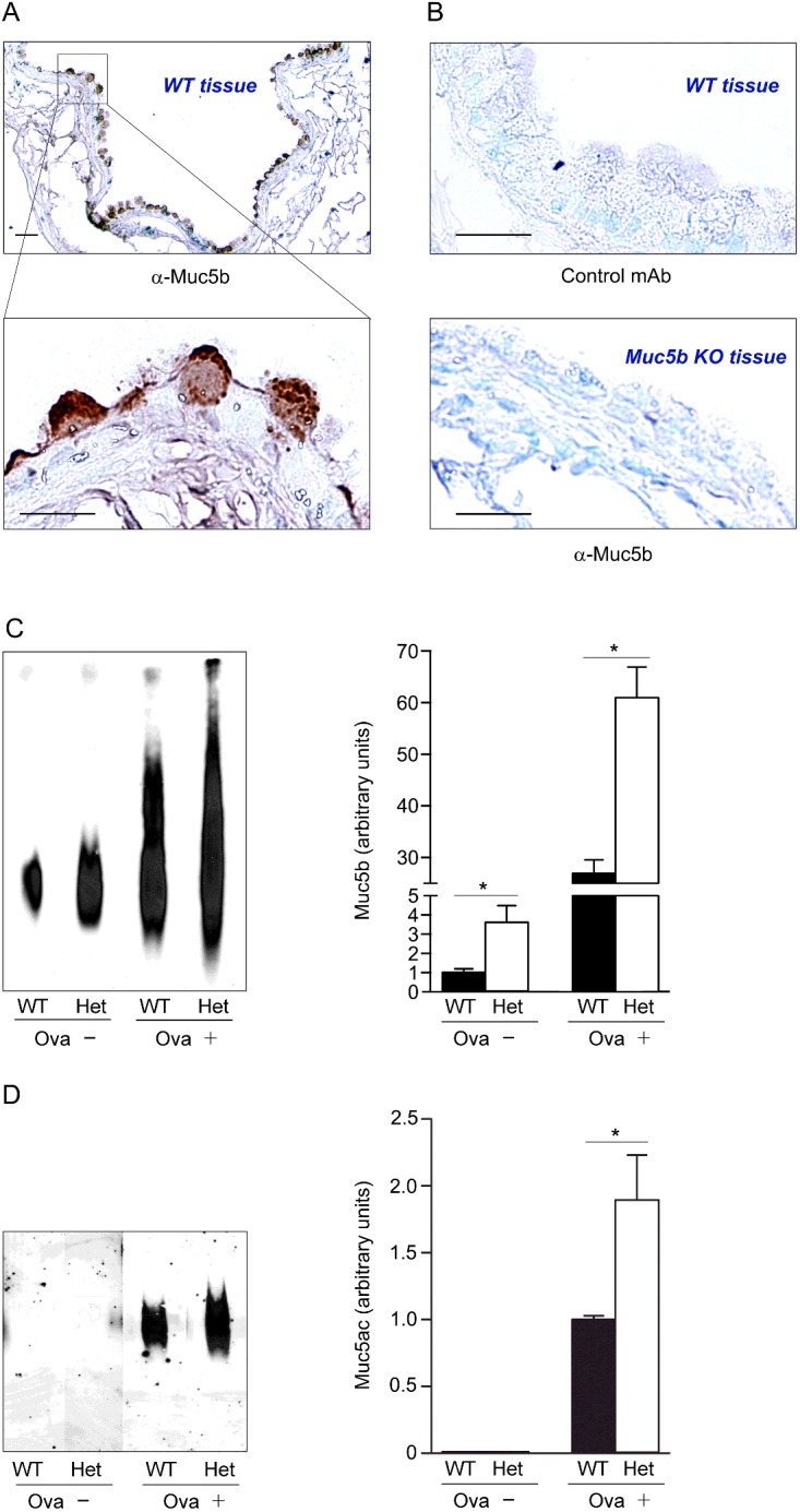
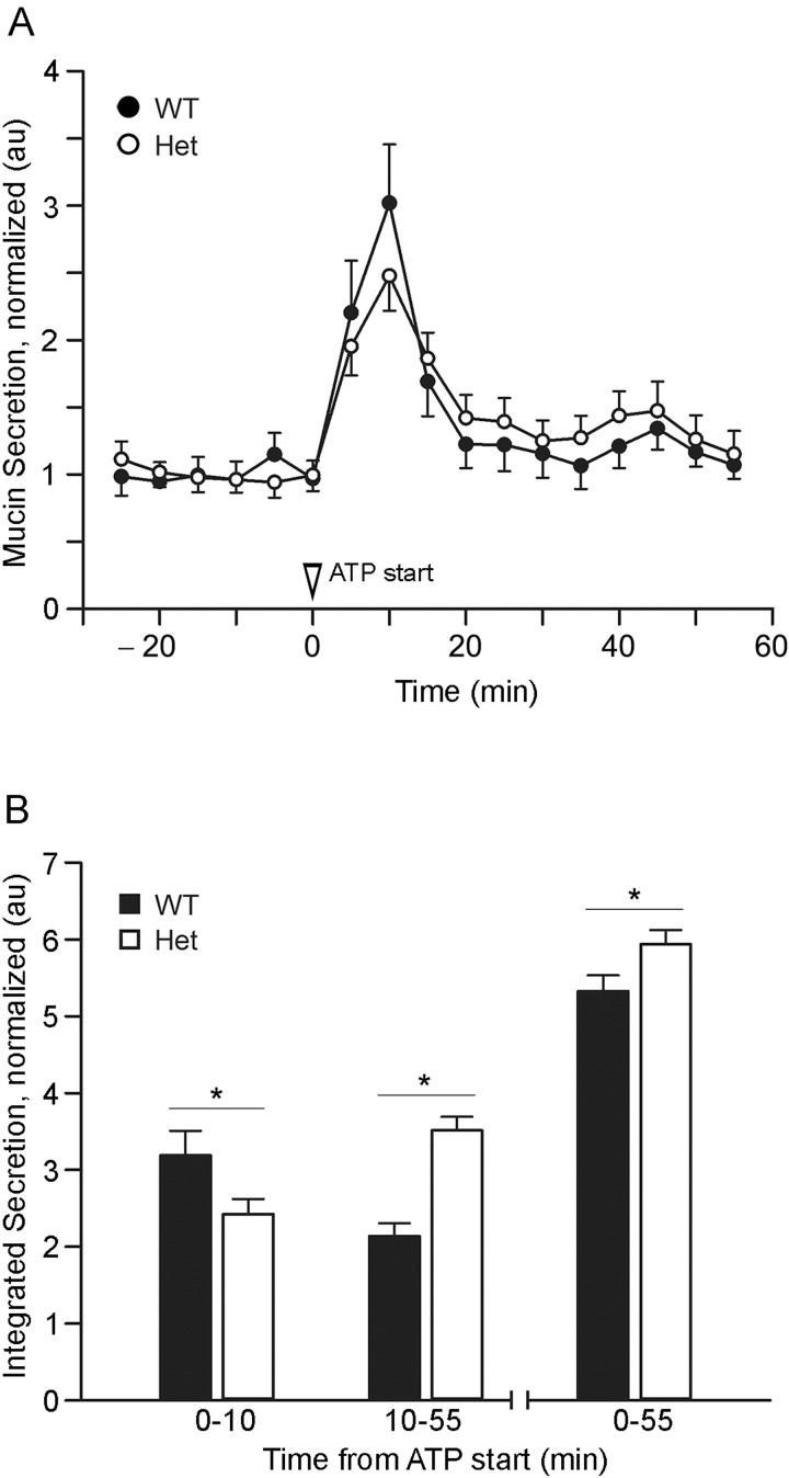
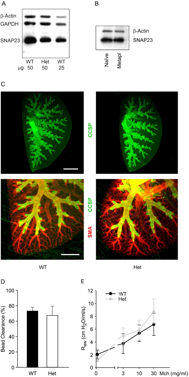
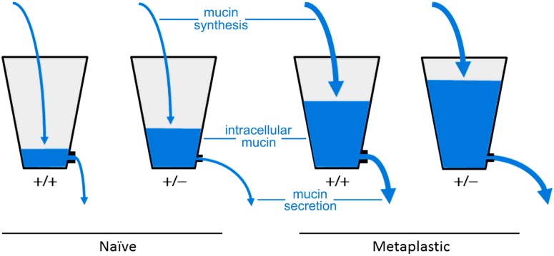
Airway mucin secretion is important pathophysiologically and as a model of polarized epithelial regulated exocytosis. We find the trafficking protein, SNAP23 (23-kDa paralogue of synaptosome-associated protein of 25 kDa), selectively expressed in secretory cells compared with ciliated and basal cells of airway epithelium by immunohistochemistry and FACS, suggesting that SNAP23 functions in regulated but not constitutive epithelial secretion. Heterozygous SNAP23 deletant mutant mice show spontaneous accumulation of intracellular mucin, indicating a defect in baseline secretion. However mucins are released from perfused tracheas of mutant and wild-type (WT) mice at the same rate, suggesting that increased intracellular stores balance reduced release efficiency to yield a fully compensated baseline steady state. In contrast, acute stimulated release of intracellular mucin from mutant mice is impaired whether measured by a static imaging assay 5 min after exposure to the secretagogue ATP or by kinetic analysis of mucins released from perfused tracheas during the first 10 min of ATP exposure. Together, these data indicate that increased intracellular stores cannot fully compensate for the defect in release efficiency during intense stimulation. The lungs of mutant mice develop normally and clear bacteria and instilled polystyrene beads comparable to WT mice, consistent with these functions depending on baseline secretion that is fully compensated.

Keywords: 23-kDa paralogue of synaptosome-associated protein of 25 kDa (SNAP23); exocytosis; mucin; mucus; secretion.


Figures

Figure 1
Figure 1. Expression of SNAP23 in the lungs of mice
(A) Sections of mouse lung were incubated with non-immune rabbit serum (left) or rabbit serum reactive to SNAP23 (centre), then labelled with HRP-conjugated secondary antibodies. Lumens of airways (Air) and blood vessels (Ves) are indicated. A high magnification image of a portion of the centre image is shown on the right, with closed arrowheads pointing to stained club cells, arrows pointing to unstained ciliated cells and an open arrowhead pointing to a type 2 alveolar epithelial cell. Scale bar in the left and centre images=100 μm and in the right image=10 μm. (B) Adjacent thin sections of mouse airways were labelled with antibodies against CCSP or SNAP23 (top row) and arrowheads point to the same three club cells stained in each image. Other adjacent sections were labelled with antibodies against acetlylated α-tubulin (AcTub) or SNAP23 (bottom row) and arrows heads point to the same two ciliated cells stained with acetylated tubulin but not with SNAP23 antibodies. Scale bar=10 μm. (C) Mouse tracheal epithelial cells were separated by FACS using lineage-specific surface markers, then equal numbers of club, ciliated and basal cells were Western blotted and probed with antibodies to SNAP23, acetylated α-tubulin as a lineage probe and β-actin as a loading control. (D) Sections of airways at multiple levels from the trachea to alveoli were probed with antibodies against SNAP23. Scale bar=20 μm.
Figure 2
Figure 2. Co-localization of SNAP23 with airway epithelial lineage markers
A section of mouse lung was probed with primary antibodies from different species and then labelled with species-specific fluorescent secondary antibodies. The low-magnification image (left) shows the epithelium of a small airway with SNAP23 stained red, CCSP stained blue and AcTub stained green. The boxed area is shown at higher magnification with DAPI-labelled nuclei stained grey in the merged image and with each fluorochrome shown separately in grey scale below. The sub-epithelial basement membrane was identified by auto-fluorescence in the green channel and its position is indicated by the yellow dotted line. The nucleus in the submucosa probably belongs to a smooth muscle cell based on its elongated shape and proximity to the epithelium. Scale bar for low magnification image=20 μm and for high magnification images=10 μm.
Figure 3
Figure 3. Steady state mucin secretory function in WT and SNAP23 Het mice
(A) Baseline secretory function was examined by PAFS staining of bronchial airways of untreated WT and Het mice. Spontaneously accumulated intracellular mucin stains red and is quantified in the bar graph by analysis of images from two airway sections each from ≥ seven mice per group. *P<0.05. Scale bar=20 μm. (B) Stimulated secretory function was examined by PAFS staining of bronchial airways of mice with mucous metaplasia induced by ovalbumin sensitization and challenge, then not further treated (top row) or treated with an aerosol of 100 mM ATP to induce acute mucin secretion (bottom row). Intracellular mucin content is quantified in the adjacent bar graphs and the mucin content in mice treated with ATP as a percentage of that in untreated mice is shown at far right. Results are the weighted means ± S.E.M. of ≥ two separate experiments with a total of ≥ 12 mice per group, *P<0.05.
Figure 4
Figure 4. Measurement of Muc5ac and Muc5b in lungs of WT and SNAP23 Het mice
(A) Monoclonal antibody MDA-3E1 generated against a peptide in Muc5b stains granules in naive airway secretory cells. Scale bar=20 μm in low magnification image and 10 μm in high magnification image. (B) There is no brown staining with an isotype control monoclonal antibody (top) or with MDA-3E1 in Muc5b KO mice (bottom). (C) Immunoblot of lung lysates from WT and SNAP23 Het mice, without airway inflammation (Ova−) or with allergic airway inflammation (Ova+), probed with antibody MDA-3E1 to detect Muc5b. Densitometric analysis of three similar blots are plotted at right showing means ± S.E.M., *P<0.05. (D) Western blot of lung lysates from WT and SNAP23 Het mice, with or without airway inflammation as in (C), probed with UEA1 lectin to detect Muc5ac. Densitometric analysis of three similar blots is plotted at right, as for (C).
Figure 5
Figure 5. Kinetic mucin secretory function in WT and SNAP23 Het mice
(A) Mucous metaplasia was induced by intrapharyngeal instillation of interleukin (IL)-13, then tracheas were excised and perfused with isotonic cell culture medium; secreted mucins in the perfusate were measured every 5 min. At time 0, 100 μM ATPγS was added to the perfusate. Results are the weighted means ± S.E.M. of ≥ two separate experiments with a total of ≥ eight mice per group. (B) Total mucins secreted during the early (0–10 min), late (10–55 min) or entire (0–55 min) periods of stimulation with ATPγS are compared between WT and Het mice, *P<0.05.
Figure 6
Figure 6. Structure and clearance function of the lungs of WT and SNAP23 Het mice
(A) Western blots of lung homogenates from adult WT and Het mice were probed with antibodies against SNAP23. Antibodies against β-actin and GAPDH were used as loading controls. (B) Western blots of lung homogenates from WT mice, with or without mucous metaplasia, were probed with antibodies against SNAP23. Antibodies against β-actin were used as a loading control. (C) The branching structure of airways from 3-week-old WT and Het mice was revealed by whole mount immunostaining using antibodies against CCSP to mark airway secretory cells and against smooth muscle actin (SMA) to mark arterioles. Scale bar in top image=2 mm and in bottom image=1 mm. (D) Fluorescent 2 μm polystyrene microspheres were instilled into the lungs of adult WT and Het mice and the percentage of microspheres cleared from the lungs in 15 min was measured as a percentage of those instilled. Results are the weighted means ± S.E.M. of ≥ two separate experiments with a total of ≥ four mice per group. (E) Total respiratory resistance during mechanical ventilation with MCh aerosol challenge. Results are the weighted means ± S.E.M. of ≥ two separate experiments with a total of ≥ four mice per group.
Figure 7
Figure 7. Hydrostatic steady state model of the effects of SNAP23 mutation on mucin accumulation and baseline secretion
The naive uninflamed condition with low mucin synthesis is illustrated with thin arrows of water flowing into the two left buckets, whereas the metaplastic inflamed condition with high mucin synthesis is illustrated with thick arrows flowing into the two right buckets. Buckets representing WT (+/+) mice have large outlets so that little water accumulates in the naive condition and only a moderate amount in the metaplastic condition, whereas buckets representing SNAP23 Het (+/−) mice have small outlets so that some water accumulates even in the naive condition and a large amount in the metaplastic condition. In all scenarios, hydrostatic pressure increases until steady state outflow matches inflow.

References

    1. Fahy J.V., Dickey B.F. Airway mucus function and dysfunction. N. Engl. J. Med. 2010;363:2233–2247. doi: 10.1056/NEJMra0910061. - DOI - PMC - PubMed
    1. Knowles M.R., Boucher R.C. Mucus clearance as a primary innate defense mechanism for mammalian airways. J. Clin. Invest. 2002;109:571–577. doi: 10.1172/JCI0215217. - DOI - PMC - PubMed
    1. Roy M.G., Livraghi-Butrico A., Fletcher A.A., McElwee M.M., Evans S.E., Boerner R.M., Alexander S.N., Bellinghausen L.K., Song A.S., Petrova Y.M., et al. Muc5b is required for airway defence. Nature. 2014;505:412–416. doi: 10.1038/nature12807. - DOI - PMC - PubMed
    1. Adler K.B., Tuvim M.J., Dickey B.F. Regulated mucin secretion from airway epithelial cells. Front. Endocrinol. 2013;4:129. doi: 10.3389/fendo.2013.00129. - DOI - PMC - PubMed
    1. Davis C.W., Dickey B.F. Regulated airway goblet cell mucin secretion. Annu. Rev. Physiol. 2008;70:487–512. doi: 10.1146/annurev.physiol.70.113006.100638. - DOI - PubMed

Publication types