. 2015 Aug 4;112(31):E4197-205.
doi: 10.1073/pnas.1422981112. Epub 2015 Jul 16.

Structural basis for methyl-donor-dependent and sequence-specific binding to tRNA substrates by knotted methyltransferase TrmD

Affiliations

Structural basis for methyl-donor-dependent and sequence-specific binding to tRNA substrates by knotted methyltransferase TrmD

Takuhiro Ito et al. Proc Natl Acad Sci U S A. .

Abstract

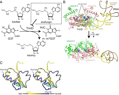
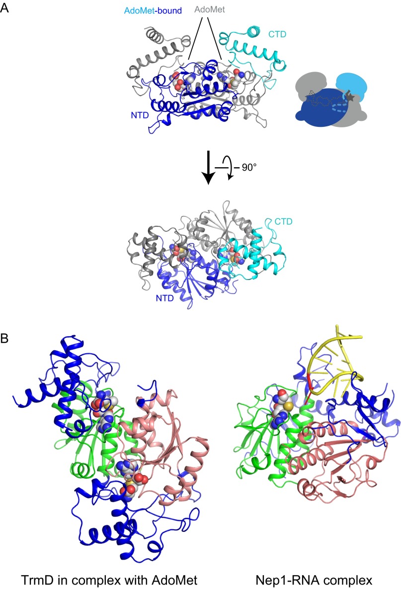
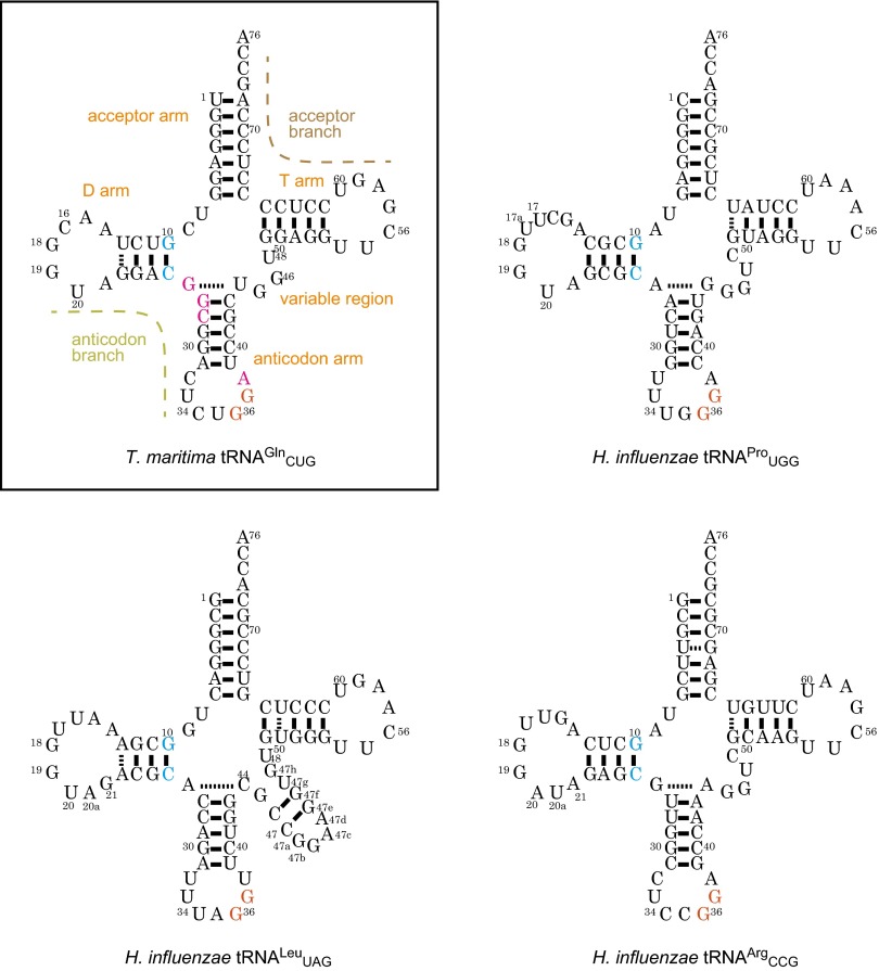
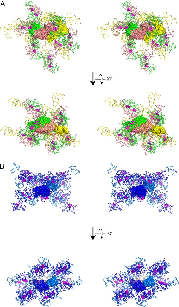
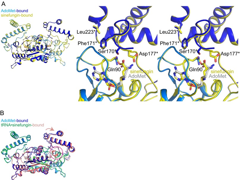
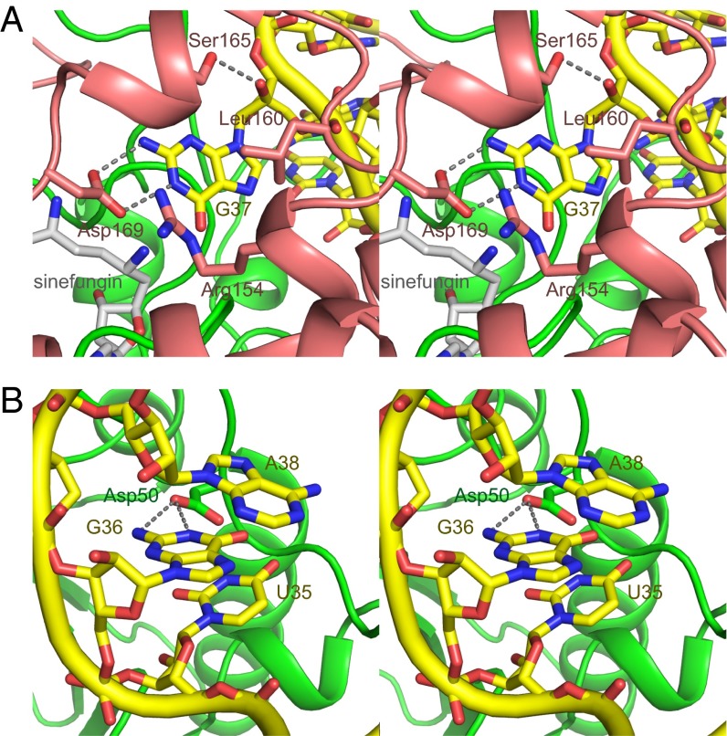
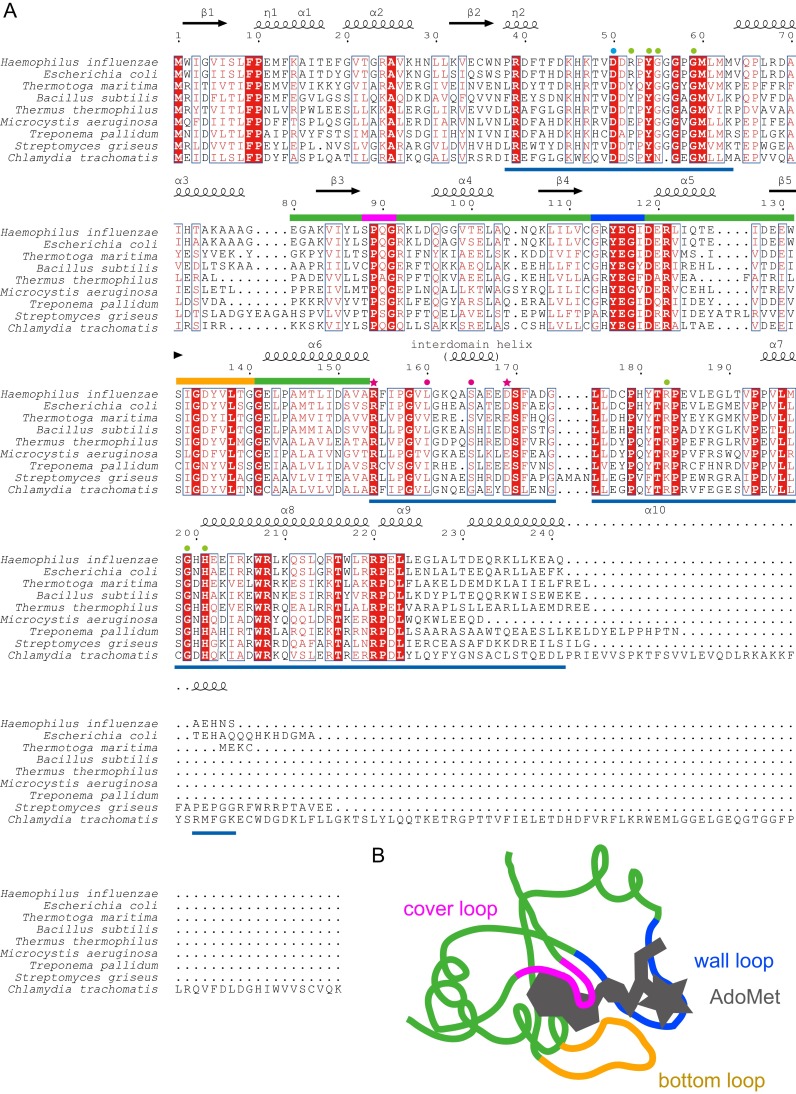
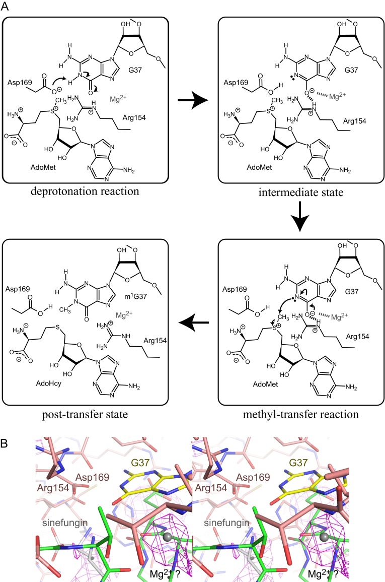
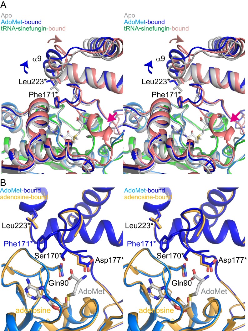
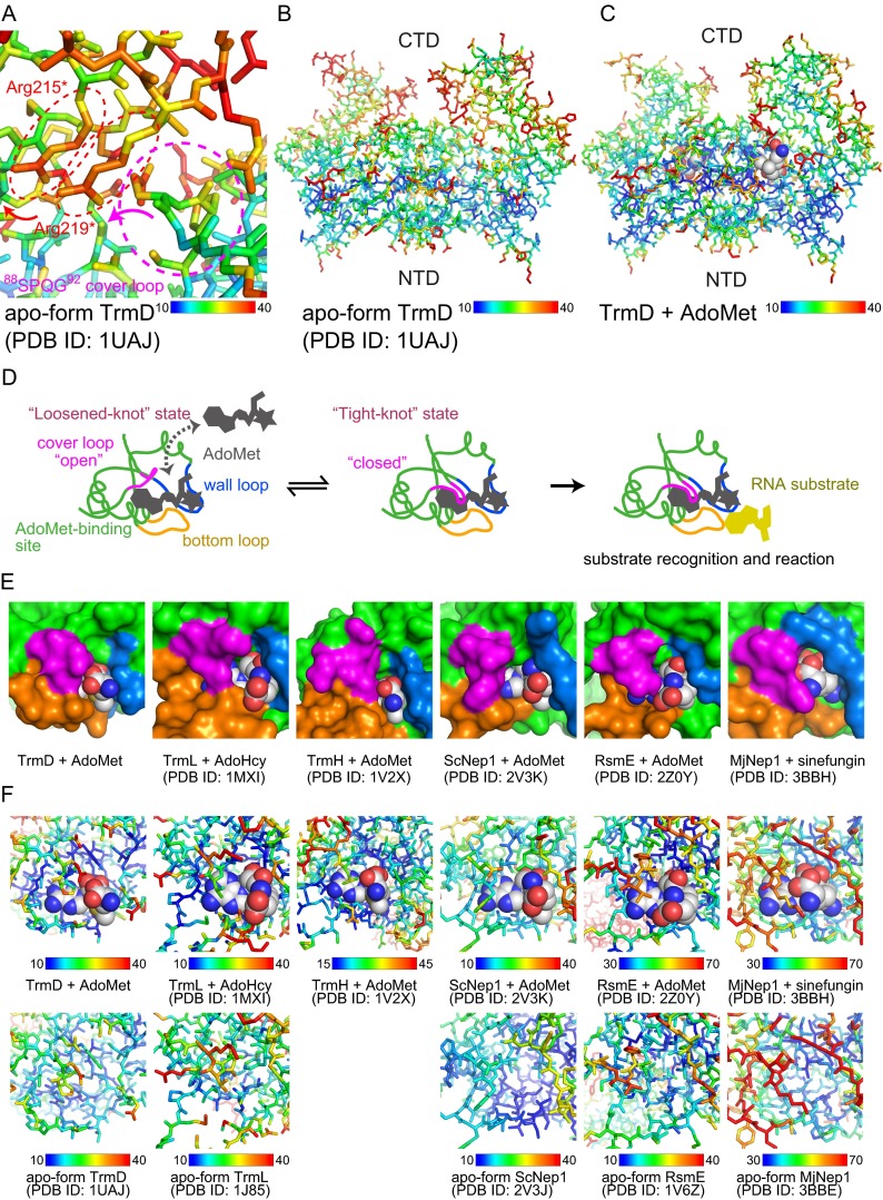
The deep trefoil knot architecture is unique to the SpoU and tRNA methyltransferase D (TrmD) (SPOUT) family of methyltransferases (MTases) in all three domains of life. In bacteria, TrmD catalyzes the N(1)-methylguanosine (m(1)G) modification at position 37 in transfer RNAs (tRNAs) with the (36)GG(37) sequence, using S-adenosyl-l-methionine (AdoMet) as the methyl donor. The m(1)G37-modified tRNA functions properly to prevent +1 frameshift errors on the ribosome. Here we report the crystal structure of the TrmD homodimer in complex with a substrate tRNA and an AdoMet analog. Our structural analysis revealed the mechanism by which TrmD binds the substrate tRNA in an AdoMet-dependent manner. The trefoil-knot center, which is structurally conserved among SPOUT MTases, accommodates the adenosine moiety of AdoMet by loosening/retightening of the knot. The TrmD-specific regions surrounding the trefoil knot recognize the methionine moiety of AdoMet, and thereby establish the entire TrmD structure for global interactions with tRNA and sequential and specific accommodations of G37 and G36, resulting in the synthesis of m(1)G37-tRNA.

Keywords: RNA modification; SPOUT methyltransferase; TrmD; X-ray crystallography.


Conflict of interest statement

The authors declare no conflict of interest.

Figures

Fig. 1.
Fig. 1.
Crystal structure of the TrmD•sinefungin•tRNA complex. (A) Schematic representation of the synthesis of m1G37-tRNA catalyzed by TrmD. The chemical structures of AdoMet, AdoHcy, and sinefungin are also shown. (B) Ribbon representation of the crystal structure of the ternary complex of TrmD, sinefungin, and tRNA. The TrmD subunits A and B are colored green and salmon, respectively. The acceptor and anticodon branches of tRNA are colored brown and yellow, respectively. G36 and G37 in the tRNA are represented by stick models. The sinefungin molecules in the AdoMet-binding sites of the TrmD subunits A and B are represented by CPK models, colored light green and light salmon, respectively. (C) The backbone wire model of the deep trefoil knot structure of subunit A in the tRNA•sinefungin-bound form of TrmD. The backbone model of the residues from Glu80 to Phe155 is presented in a stereoview, from the same angle as the lower panel of B. The color of the residues from Glu80 to Ala153 gradually changes from yellow to blue, to clarify the relationship between the positions in the primary structure and the knotted part in the tertiary structure, as in the bar indicated below. The bound sinefungin is represented by a stick model, and its simulated-annealing omit map, contoured at 3σ, is illustrated on the model.
Fig. S1.
Fig. S1.
Comparison of the TrmD and Nep1 structures. (A) Ribbon representation of the crystal structure of the complex between TrmD and S-adenosyl-l-methionine (AdoMet): the tRNA-free AdoMet-bound form of TrmD. One subunit of the dimeric TrmD is colored blue and light blue for the NTD and CTD, respectively, and the other symmetric subunit is colored gray. The bound AdoMet molecule is represented by a CPK model. (B) Ribbon representations of TrmD in complex with AdoMet and Nep1 in complex with an RNA substrate are shown, and the SPOUT core domains of subunit A of TrmD and Nep1 are presented from the same viewing angle. The Nep1-bound RNA molecule is colored yellow, and the methylation residue is highlighted in red. The characteristic SPOUT domain structures are colored green and salmon for subunits A and B, respectively, and the specific structures of TrmD or Nep1 are colored blue. The TrmD-bound AdoMet and Nep1-bound AdoHcy are depicted by CPK models.
Fig. S2.
Fig. S2.
Sequences of tRNA transcripts used in this study. The sequences of the transcripts of Thermotoga maritima tRNACUGGln, Haemophilus influenzae tRNAUGGPro, H. influenzae tRNAUAGLeu, and H. influenzae tRNAArgCCG are shown as cloverleaf models. The conserved G10:25C pair and 36GG37 sequence are colored cyan and orange, respectively. In the boxed panel of T. maritima tRNAGlnCUG, the residues with phosphate groups that are recognized by TrmD are colored magenta. In addition, the names of the five arms and the two branches of tRNA are indicated.
Fig. S3.
Fig. S3.
Crystal packing of the TrmD•tRNA•sinefungin ternary and TrmD•AdoMet binary complexes. (A) The packing manner in the crystal of the TrmD•tRNA•sinefungin ternary complex is represented in a stereoview from two orthogonal directions. Subunits A and B of TrmD, tRNA, and sinefungin are colored green, salmon, yellow, and magenta, respectively. A central ternary complex, constituting an asymmetric unit, is represented by a surface model, and the interacting symmetric molecules are represented by wire models, except that sinefungin is represented by the CPK model. (B) The packing manner in the crystal of the TrmD•AdoMet binary complex is represented in a similar manner to A. The central asymmetric unit of the TrmD monomer and AdoMet is represented by a surface model, and the other symmetric TrmD and AdoMet are represented by wire and CPK models, respectively. The N-terminal and C-terminal domains of TrmD and AdoMet are colored blue, light blue, and magenta, respectively, except that the TrmD dimerizing with the surface model is colored gray.
Fig. S4.
Fig. S4.
Structural comparison between the different forms of TrmD. (A) Structural comparisons between the binary complexes of the AdoMet-bound and sinefungin-bound TrmDs. The structures are overlaid according to the backbone atoms in the NTD. The overall structures and the close-up views of the AdoMet-binding site are presented on the left and right, respectively. Subunits A and B of the AdoMet-bound form are colored blue and light blue, respectively, and the sinefungin-bound form is colored yellow. The models on the right lack the NTD of subunit B, to facilitate visualization (stereoview). (B) Ribbon representations of the AdoMet-bound TrmD and the tRNA•sinefungin-bound TrmD, overlaid according to the backbone atoms in the NTD. The AdoMet-bound binary complex of TrmD is colored as in A, and the tRNA•sinefungin-bound ternary complex of TrmD is colored as in Fig. 2. The structural change of the CTD is indicated by the salmon colored arrow, and the interdomain helix induced by tRNA binding is indicated by the dotted red circle.
Fig. 2.
Fig. 2.
TrmD interactions with G37 and G36 in the target tRNA. (A) Close-up views of the G37-binding pocket (stereoview). The molecules are colored as in Fig. 1B, except that sinefungin is colored gray. The interacting residues of TrmD and sinefungin are indicated by stick models. The hydrogen bonds between TrmD and tRNA are indicated by gray dotted lines. (B) Close-up views of the G36-binding pocket (stereoview). The molecules are depicted as in A.
Fig. S5.
Fig. S5.
Sequence alignment of TrmDs. (A) Amino acid sequences of TrmD enzymes from nine bacteria (H. influenzae, Escherichia coli, T. maritima, Bacillus subtilis, Thermus thermophilus, Microcystis aeruginosa, Treponema pallidum, Streptomyces griseus, and Chlamydia trachomatis) were aligned by the program ClustalW (41), and visualized by the program ESPript (42). The fully conserved residues are highlighted in red boxes, and the moderately conserved residues are indicated by red characters. The secondary structural elements are indicated above the sequences. The G36-interacting residues, the G37-interacting residues, and the anticodon branch-interacting residues are indicated by cyan, magenta, and green circles, respectively. The G37-interacting catalytic residues of Arg154 and Asp169 are indicated by magenta stars. The deep trefoil-knot region is indicated by green bars, except that the cover, wall and bottom loops are indicated by magenta, blue, and orange bars, respectively. The TrmD-specific structures are indicated by blue lines under the sequences. (B) The schematic deep trefoil-knot structure is indicated with the bound AdoMet, which is also shown in Fig. S9 D and E. The cover, wall and bottom loops are colored as in A.
Fig. S6.
Fig. S6.
Proposed mechanism of the catalytic reaction. (A) The proposed mechanism of the catalytic reaction is indicated in four panels: the deprotonation reaction, the intermediate state, the methyl-transfer reaction, and the posttransfer state. In the intermediate state, the electrostatic interaction between G37 and Arg154 is indicated by bold dotted lines. The Mg2+ ion, which stabilizes the developing negative charge on O6 of G37, and its interaction with O6 of G37 are indicated by a gray character and a dotted line, respectively. (B) Close-up views of the presumed Mg2+ ion-binding site in the TrmD•tRNA•sinefungin ternary complex (stereoview). The possible position of the Mg2+ ion is indicated by the gray ball, and the other molecules are indicated by line models, colored as in Fig. 2A. The mFo-DFc map, contoured at 3σ, is illustrated on the model by a magenta mesh.
Fig. S7.
Fig. S7.
Binding experiments between sinefungin-bound TrmD and tRNA variants. The tRNA-binding was monitored by the quenching of the intrinsic tryptophan fluorescence of TrmD. For the wild-type and G36A tRNAs, the determined dissociation constants for the first strong tRNA binding (Kd1) are indicated next to the fitting lines. The calculated slopes for the weak binding are also indicated.
Fig. 3.
Fig. 3.
The structural changes induced by the G36 interaction. (A) Ribbon representation of the ternary complex of TrmD, sinefungin, and the G36U variant tRNA (Left), together with the schematic representation (Right Upper), and the stick model colored with B-factors (Right Lower). The key residues are depicted by stick models and labeled. The peptide bond between Gly59 and Pro58 is depicted by a stick model, and the hydrogen bond from the NH group of Gly59 is indicated by the gray dotted line. The viewpoint of the left panel is indicated by the black arrow in the schematic representation. The red dotted square corresponds to the region represented in the B-factor-colored model in the right lower panel. The colors of the molecules are fainter than those of the wild-type, as in Fig. 2. (B) Representations of the ternary complex of TrmD, sinefungin, and the wild-type tRNA, in the same manner as in A.
Fig. 4.
Fig. 4.
The interaction between the anticodon branch and TrmD to find position 37. (A) The loop between helices α7 and α8 recognizes the minor groove next to the G10:C25 pair (stereoview). The key residues in TrmD are depicted by stick models. The identified electrostatic and hydrogen bonds are indicated by the dotted gray lines. (B) The recognition of the phosphate groups of G26, G27, C28, and A38 by TrmD is indicated in the same manner as in A.
Fig. 5.
Fig. 5.
A model of the enzymatic cycle of TrmD. (A) The AdoMet-binding site of the tRNA•sinefungin-bound TrmD (stereoview). The residues recognizing the methionine-mimic moiety of sinefungin are depicted by stick models, and the identified hydrogen bonds are indicated by the dotted gray lines. The model lacks the NTD of subunit B to facilitate visualization, and is colored as in Fig. 2, except that the 88SPQG91 cover loop is colored magenta. (B) Based on the crystal structures, the kinetic experiments and the structural analyses, the recognition order of AdoMet and the elements in the 36GG37-containing tRNA, and the subsequent methyl transfer reaction by TrmD are presented. The states in the AdoMet-binding stage (states I to V) are illustrated as close-up views of the AdoMet-binding site with schematic trefoil-knot structures, those in the tRNA-binding stage (states VI to X) are illustrated around the tRNA-binding site, and the methyltransfer stage (state XI) is illustrated schematically. The TrmD, AdoMet, and tRNA in the graphical panels are depicted by surface models, CPK models, and stick models with ribbons, respectively. The figures are colored as in Fig. 2, except that the nucleotides at positions 36 and 37 are colored cyan, the 88SPQG91 cover loop is colored magenta, and the ordered loop in the CTD and the interdomain helix are colored purple. The right panel of state I, the panels of states II and III, and the left panel of state IV lack the CTD of subunit B, to facilitate visualization.
Fig. S8.
Fig. S8.
Structural differences among the various forms of TrmD around the AdoMet-binding site. (A) Ribbon representations of the apo, AdoMet-bound, and tRNA•sinefungin-bound TrmDs are overlaid, according to the backbone atoms in the NTD of subunit A (stereoview). The key residues are depicted by stick models. The structural change from the apo to AdoMet-bound form is indicated by the blue arrows, and that from the AdoMet-bound to tRNA•sinefungin-bound form is shown by the salmon arrows. The interdomain helix in the tRNA•sinefungin-bound form is indicated by the magenta arrow. (B) Structural comparison between the AdoMet-bound and adenosine-bound TrmDs (stereoview). The structures are overlaid according to the backbone atoms in the NTD. The AdoMet-bound form is colored as in Fig. S4A, and the adenosine-bound form is colored yellow. The models lack the NTD of subunit B, to facilitate visualization. The specific orientation of Phe171* is observed only in the AdoMet-bound and tRNA•sinefungin-bound TrmDs.
Fig. S9.
Fig. S9.
Structural analysis of AdoMet accommodation. (A) Expanded view of the B-factor mapping around the 88SPQG92 cover loop of the apo-form TrmD. The cover loop and the two key arginine residues are encircled by the dashed light magenta and red ellipses, respectively. The B-factors in each model are mapped onto the stick model, and the B-factor color spectrum is indicated below. (B and C) The B-factor maps of the (B) apo and (C) AdoMet-bound TrmDs. The stick models are represented from the same angle as in the top panel of Fig. S1A. These panels show that the B-factors of the CTD are generally higher than those of the NTD. (D) Schematic representation of the function of the deep trefoil knot structure of the SPOUT MTases in the accommodation of AdoMet and the following target interaction. The cover, wall, and bottom loops from the deep trefoil knot are colored magenta, blue, and orange, respectively. (E) Structural comparison of the AdoMet-binding sites in the SPOUT MTases. Six SPOUT domains from TrmD (3), TrmL (4), TrmH (43), ScNep1 (44), RsmE, and MjNep1 (45) are represented by green surface models. All figures represent the monomer to facilitate visualization. The cover, wall, and bottom loops from the deep trefoil knot are colored as in D, and the bound AdoMet, AdoHcy, or sinefungin is indicated by a CPK model. The reference PDB ID codes are indicated under each panel. (F) The B-factor mapping of the AdoMet-binding sites from the six SPOUT MTases indicated in E. The AdoMet/AdoHcy/sinefungin-bound and apo SPOUT MTases are shown at the top and the bottom, respectively, together with the color spectrum of the B-factors, the enzyme name, and the PDB ID code.

Similar articles

Cited by

References

    1. Michel G, et al. The structure of the RlmB 23S rRNA methyltransferase reveals a new methyltransferase fold with a unique knot. Structure. 2002;10(10):1303–1315. - PubMed
    1. Nureki O, et al. An enzyme with a deep trefoil knot for the active-site architecture. Acta Crystallogr D Biol Crystallogr. 2002;58(Pt 7):1129–1137. - PubMed
    1. Ahn HJ, et al. Crystal structure of tRNA(m1G37)methyltransferase: insights into tRNA recognition. EMBO J. 2003;22(11):2593–2603. - PMC - PubMed
    1. Lim K, et al. Structure of the YibK methyltransferase from Haemophilus influenzae (HI0766): A cofactor bound at a site formed by a knot. Proteins. 2003;51(1):56–67. - PubMed
    1. Elkins PA, et al. Insights into catalysis by a knotted TrmD tRNA methyltransferase. J Mol Biol. 2003;333(5):931–949. - PubMed

Publication types

MeSH terms

LinkOut - more resources