. 2015 Sep;35(9):1945-53.
doi: 10.1161/ATVBAHA.115.305597. Epub 2015 Jul 16.

Local MicroRNA Modulation Using a Novel Anti-miR-21-Eluting Stent Effectively Prevents Experimental In-Stent Restenosis

Affiliations

Local MicroRNA Modulation Using a Novel Anti-miR-21-Eluting Stent Effectively Prevents Experimental In-Stent Restenosis

Dong Wang et al. Arterioscler Thromb Vasc Biol. 2015 Sep.

Erratum in

  • Correction.
    [No authors listed] [No authors listed] Arterioscler Thromb Vasc Biol. 2015 Oct;35(10):e51. doi: 10.1161/ATV.0000000000000024. Arterioscler Thromb Vasc Biol. 2015. PMID: 26399923 No abstract available.

Abstract

Objective: Despite advances in stent technology for vascular interventions, in-stent restenosis (ISR) because of myointimal hyperplasia remains a major complication.

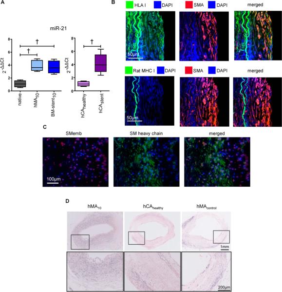
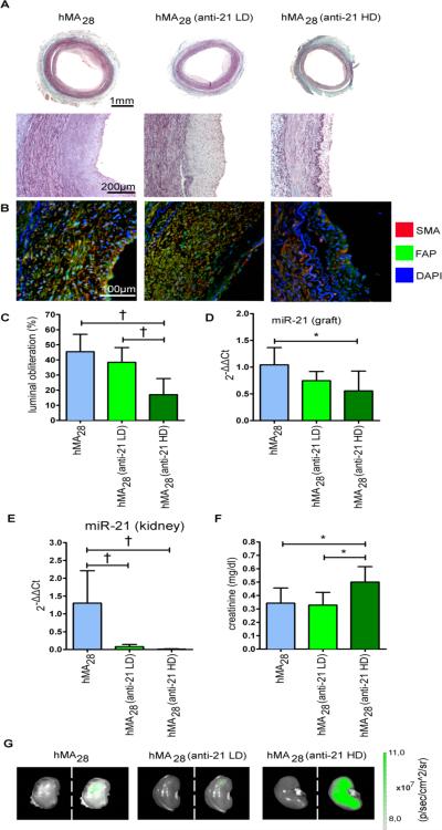
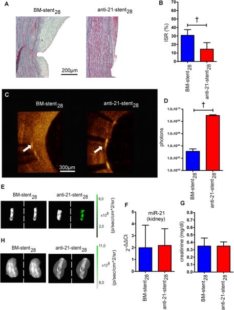
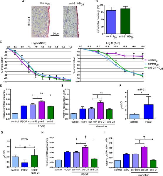
Approach and results: We investigated the regulatory role of microRNAs in myointimal hyperplasia/ISR, using a humanized animal model in which balloon-injured human internal mammary arteries with or without stenting were transplanted into Rowett nude rats, followed by microRNA profiling. miR-21 was the only significantly upregulated candidate. In addition, miR-21 expression was increased in human tissue samples from patients with ISR compared with coronary artery disease specimen. We systemically repressed miR-21 via intravenous fluorescein-tagged-locked nucleic acid-anti-miR-21 (anti-21) in our humanized myointimal hyperplasia model. As expected, suppression of vascular miR-21 correlated dose dependently with reduced luminal obliteration. Furthermore, anti-21 did not impede reendothelialization. However, systemic anti-miR-21 had substantial off-target effects, lowering miR-21 expression in liver, heart, lung, and kidney with concomitant increase in serum creatinine levels. We therefore assessed the feasibility of local miR-21 suppression using anti-21-coated stents. Compared with bare-metal stents, anti-21-coated stents effectively reduced ISR, whereas no significant off-target effects could be observed.

Conclusion: This study demonstrates the efficacy of an anti-miR-coated stent for the reduction of ISR.

Keywords: coronary restenosis; hyperplasia; microRNAs; rats; stents.


Figures

Figure 1
Figure 1. MiR-21 is involved in human myointimal hyperplasia
(A) Human IMAs underwent either balloon denudation (hMA), or denudation and stenting (BM-stent) and were implanted into the abdominal aortic position of RNU rats to await myointima development. After 10 days, qRT-PCR measurement of miR-21 revealed significant overexpression in both hMA and BM-stent groups as compared to native IMA (n=10 (native), n=6 (hMA), n=6 (BM-stent), ANOVA with Bonferroni's post-hoc test). Likewise, miR-21 levels in diseased, stented human coronary artery (hCAstent n=5;) were similarly altered vs. normal vessels (hCAhealthy n=4; unpaired t-test). (B) Human origin of myointimal cells in the hMA model was confirmed by double-positive immunofluorescence staining for SMA and human leukocyte antigen class I (HLA I) and the lack of rat major histocompatibility complex class I (rat MHC I). (C) Smooth muscle cells (SMC) within the myointima of hMA28 showed expression of SMemb and/or SM heavy chain. Synthetic, SMemb positive cells were distributed more in neo-myointimal regions and SM-heavy chain positive, contractile SMCs were found near the elastic laminae. (D) Presence of miR-21 in hMA10 confirmed by in situ hybridization in comparison to un-denuded hMA (hMAcontrol) and uninjured healthy human coronary arteries (hCAhealthy; purple chromagen). † p<0.01.
Figure 2
Figure 2. Systemic modulation of miR-21 inhibits myointima development, but causes off-target effects
(A) To assess the effect of miR-21 modulation on myointimal hyperplasia, 1mg/kg (LD), 5mg/kg (HD) anti-miR-21 (anti-21) or vehicle were injected intravenously one day post-IMA-implantation. After 28 days, hMA-vessels were excised and stained with Masson's Trichrome. (B) Double immunofluorescence staining against SMA and FAP demonstrates similar myointimal composition in all groups. (C) Analysis of hMA vessels revealed marked luminal obliteration in the non-treated group, while anti-21-treated vessels showed less obliteration in a dose dependent manner (mean ± s.d., n=7 animals per group, ANOVA with Bonferroni's post-hoc test). (D) Sufficient inhibition of miR-21 in hMA vessel through anti-21 was confirmed by qRT-PCR. (mean ± s.d., n=7 (hMA), n=6 (anti-21 LD), n=6 (anti-21 HD), ANOVA with Bonferroni's post-hoc test). (E) Systemic miR-21 inhibition caused marked reduction of miR-21 expression in kidneys (mean ± s.d., n=6 (hMA), n=7 (anti-21 LD), n=7 (anti-21 HD), ANOVA with Bonferroni's post-hoc test). (F) This correlated with an increase in creatinine (mean ± s.d., n=7 animals per group, ANOVA with Bonferroni's post-hoc test). (G) Injected anti-21 was tracked via fluorescence imaging. High fluorescence signal was detected in HD-treated rat kidneys, indicating an accumulation of anti-21 (left: photo picture; right: overlay picture with fluorescence signal). * p<0.05, † p<0.01.
Figure 3
Figure 3. Local inhibition of miR-21 prevents myointima development without exerting off-target effects
(A) For local miR-21 inhibition, anti-21-coated stents were expanded into denuded IMAs, which were then implanted into RNU rats. Histological sections of stented vessel stained with Masson's Trichrome are shown 28 days after implantation. (B) Anti-21-stent coating resulted in significantly less luminal obliteration compared to the BM-stent group (mean± s.d., n=8 (BM-stent), n=6 (anti-21-stent), unpaired t-test). (C) OCT images revealed considerably less myointima formation in the anti-21-stent group (white arrow marks a stent strut, luminal side is to the right). (D) Fluorescent signal intensity measurements confirm anti-21 uptake in the anti-21-stent group (mean± s.d., n=5 per group, unpaired t-test). (E) Corresponding representative fluorescence images to signal intensity measurements shown in D. (F) miR-21 levels in kidneys were not affected by anti-21-coated stent delivery (mean± s.d., n=6 per group, unpaired t-test) and (G) serum creatinine showed no significant difference between the BM-stent and anti-21-stent groups (mean± s.d., n=8 (BM-stent), n=6 (anti-21-stent), unpaired t-test). (H) In fluorescence imaging, similar signals were detected in kidneys of both groups (left: photo picture; right: overlay picture with fluorescence signal). † p<0.01.
Figure 4
Figure 4. Anti-21 treatment does not impair re-endothelialization and abolishes stimulatory proliferative effects on SMCs in vitro
(A) To assess the effect of anti-21 on vessel re-endothelialization, rat aortas underwent endothelial denudation via balloon injury. Animals in the treatment group received 5mg/kg anti-21 intravenously one day post-denudation. After 28 days, vessels were recovered and stained for endothelial cells. (B) There were no relevant differences in endothelial coverage between control28 and anti-2128 (mean± s.d., n=5 per group, unpaired t-test). (C)Endothelial function was assessed in organ chamber experiments (mean± SEM, n=6 (control28), n=6 (anti-21 HD28), n=3 (control3)). All three groups showed similar endothelial-independent relaxation characteristics, when stimulated with nitroglycerine. In contrast, stimulation with acetylcholine had no effect 3 days after vessel injury, while both control28 and anti-21 HD28 groups showed similar physiologic endothelial-dependent relaxation. (D) Proliferation assays with hCAECs after PDGF or 48h serum starvation (E) treatment revealed a negligible effect of miR-21 inhibition on EC proliferation (mean± s.d., quadruplicates of 3 independent experiments, unpaired t-test). (F) To determine the effect of PDGF on miR-21 expression in vessels, fresh pieces of IMA were stimulated with PDGF. 24h post-stimulation, miR-21 expression was significantly elevated (mean± s.d., n=6 (control), n=10 (PDGF), unpaired t-test) (G), while PTEN expression was suppressed. Concurrent transfection with anti-21 abolished this effect (mean± s.d., n=5 (control), n= 5 (PDGF), n=4 (PDGF+anti-21), ANOVA with LSD post-hoc test). (H) Inhibition of miR-21 with anti-21 caused diminished proliferation in PDGF-stimulated hCASMCs and (I) after 48h of serum starvation (mean± s.d., quadruplicates of 3 independent experiments, unpaired t-test). * p<0.05, † p<0.01.

Comment in

References

    1. Owens GK, Kumar MS, Wamhoff BR. Molecular regulation of vascular smooth muscle cell differentiation in development and disease. Physiological reviews. 2004;84:767–801. - PubMed
    1. Owens GK. Regulation of differentiation of vascular smooth muscle cells. Physiological reviews. 1995;75:487–517. - PubMed
    1. Aikawa M, Sakomura Y, Ueda M, Kimura K, Manabe I, Ishiwata S, et al. Redifferentiation of smooth muscle cells after coronary angioplasty determined via myosin heavy chain expression. Circulation. 1997;96:82–90. - PubMed
    1. Yoshida T, Owens GK. Molecular determinants of vascular smooth muscle cell diversity. Circulation research. 2005;96:280–291. - PubMed
    1. Deuse T, Hua X, Wang D, Maegdefessel L, Heeren J, Scheja L, et al. Dichloroacetate prevents restenosis in preclinical animal models of vessel injury. Nature. 2014;509:641–644. - PMC - PubMed

Publication types

MeSH terms