. 2015 Nov;29(11):4523-31.
doi: 10.1096/fj.15-274829. Epub 2015 Jul 16.

Enhanced enteroviral infectivity via viral protease-mediated cleavage of Grb2-associated binder 1

Affiliations

Enhanced enteroviral infectivity via viral protease-mediated cleavage of Grb2-associated binder 1

Haoyu Deng et al. FASEB J. 2015 Nov.

Abstract

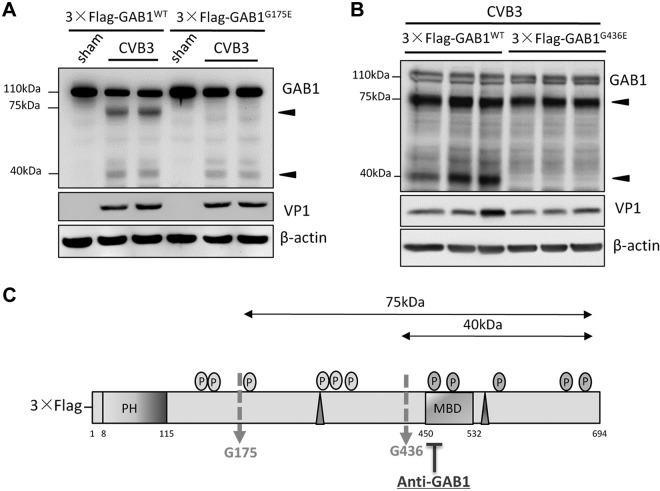
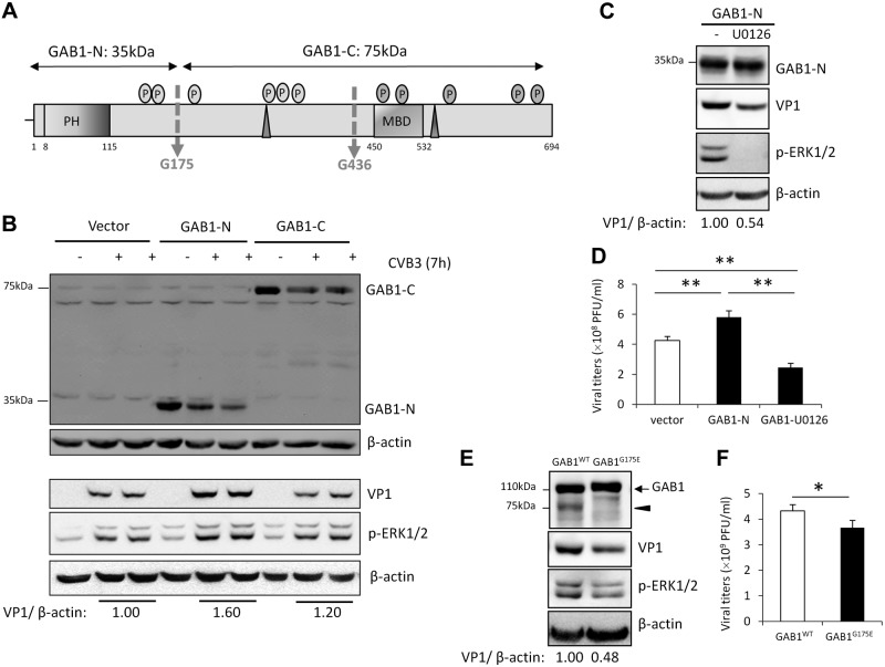
Coxsackievirus B3 (CVB3), an important human causative pathogen for viral myocarditis, pancreatitis, and meningitis, has evolved different strategies to manipulate the host signaling machinery to ensure successful viral infection. We previously revealed a crucial role for the ERK1/2 signaling pathway in regulating viral infectivity. However, the detail mechanism remains largely unknown. Grb2-associated binder 1 (GAB1) is an important docking protein responsible for intracellular signaling assembly and transduction. In this study, we demonstrated that GAB1 was proteolytically cleaved after CVB3 infection at G175 and G436 by virus-encoded protease 2A(pro), independent of caspase activation. Knockdown of GAB1 resulted in a significant reduction of viral protein expression and virus titers. Moreover, we showed that virus-induced cleavage of GAB1 is beneficial to viral growth as the N-terminal proteolytic product of GAB1 (GAB1-N1-174) further enhances ERK1/2 activation and promotes viral replication. Our results collectively suggest that CVB3 targets host GAB1 to generate a GAB1-N1-174 fragment that enhances viral infectivity, at least in part, via activation of the ERK pathway. The findings in this study suggest a novel mechanism that CVB3 employs to subvert the host signaling and facilitate consequent viral replication.

Keywords: ERK1/2; coxsackievirus B3; signal transduction; viral protease 2Apro.


Figures

Figure 1.
Figure 1.
GAB1 is cleaved during CVB3 infection. A) Cleavage of endogenous GAB1 following CVB3 infection. HeLa cells were sham- or CVB3-infected at multiplicity of infections 10 for various time points as indicated. Cell lysates were collected and processed for Western blotting for detection of viral capsid protein VP1 and GAB1 protein expression (using an anti-GAB1 antibody targeting residues surrounding Tyr472 of human GAB1). The protein level of β-actin was examined as a loading control. B) Cleavage of exogenous GAB1 following CVB3 infection. HeLa cells were transiently transfected with a plasmid expressing N-terminal Flag-tagged GAB1 (3×Flag-GAB1) for 24 h, followed by CVB3 infection for different time points as indicated. Western blotting was performed to assess the protein levels of exogenous GAB1 (using anti-Flag antibody), VP1, and β-actin. Arrowheads indicate CVB3-induced GAB1 cleavage fragments. C) Schematic diagram of full-length GAB1 with various functional domains, the resulting cleavage fragments, and the regions that individual antibodies detected. Red arrows indicate 2 potential cleavage sites. pi, post infection.
Figure 2.
Figure 2.
GAB1 is cleaved by viral protease 2Apro. A) CVB3-induced cleavage of GAB1 is dependent on viral protein production. HeLa cells infected with either WT CVB3 or UV-CVB3 were harvested at 7 h postinfection (pi). Cell lysates were processed for Western blotting to determine the protein expression levels of GAB1 (using anti-GAB1 antibody), VP1, and β-actin. B) Cleavage of GAB1 is mediated by viral protease 2Apro. Cell lysates (50 μg) from HeLa cells transfected with Flag-GAB1 were incubated with either purified viral protease 2Apro (0.1 or 0.4 µg) or catalytically inactive mutant 2Apro (0.4 µg) overnight and in vitro cleavage assay was conducted as described in the Materials and Methods. Protein levels of GAB1 (using anti-GAB1 antibody), VP1, and β-actin were examined by Western blotting. CVB3-infected HeLa cell lysates (7 h pi) were loaded (right lane) as a positive control for GAB1 cleavage. C) Cleavage of GAB1 following CVB3 infection is independent of caspase activation. HeLa cells were infected with CVB3 in the presence or absence of pan-caspase inhibitor, z-VAD-fmk (50 µM), for 7 h. Protein levels of GAB1 (using anti-GAB1 antibody), VP1, cleaved caspase-3, and β-actin were examined by Western blotting. Arrowheads indicate CVB3-induced GAB1 cleavage fragments.
Figure 3.
Figure 3.
GAB1 is cleaved at G175 and G436 during CVB3 infection. A) GAB1G175E mutant blocks the generation of the 75 kDa cleavage product triggered by CVB3 infection. HeLa cells were transiently transfected with either 3×Flag-GAB1WT or 3×Flag- GAB1G175E for 24 h, followed by sham or CVB3-infection for 7 h. Cell lysates were harvested for Western blot analysis to detect the cleaved fragments of GAB1 using anti-GAB1 antibody. VP1 and β-actin were determined as an infection and a loading control, respectively. B) GAB1G436E mutant inhibits the production of the 40 kDa product. The same protocol was performed as described above using either plasmid of 3×Flag-GAB1WT or 3×Flag-GAB1G436E. Arrowheads indicate CVB3-induced GAB1 cleavage fragments detected by anti-GAB1 antibody. C) Schematic diagram of full-length GAB1 with 2 cleavage sites at amino acid G175 and G436, respectively.
Figure 4.
Figure 4.
Knockdown of GAB1 inhibits CVB3-induced ERK phosphorylation and viral replication. A) HeLa cells were treated with either control siRNA (siCon) or GAB1-targeting siRNA (siGAB1) for 48 h, followed by CVB3 infection for 7 h. Cell lysates were harvested to examine the protein levels of GAB1 (using anti-GAB1 antibody), VP1, p-ERK1/2, and β-actin by Western blotting. Protein levels of VP1 were quantitated by densitometric analysis using ImageJ (U.S. National Institutes of Health, Bethesda, MD, USA), normalized to β-actin, and presented underneath as fold changes compared with control group, which was arbitrarily set a value of 1. B) HeLa cells were transiently transfected with 3×Flag-GAB1WT, or corresponding vector control for 24 h, followed by CVB3 infection for 7 h. Western blotting was performed and analyzed as described above. C, D) Supernatants were collected from (A) and (B) for plaque assay and the results are presented as means ± sd (n = 4). **P < 0.001.
Figure 5.
Figure 5.
The N-terminal cleavage fragment of GAB1 promotes CVB3 replication by further enhancing ERK1/2 phosphorylation. A) Schematic diagram of the N- and C-terminal cleaved fragments of GAB1 used in this figure and Fig. 6 hereafter. B) HeLa cells were transiently transfected with 3×Flag-GAB1-N1–174, 3×Flag-GAB1-C175–694, or corresponding empty vector (3×Flag) for 24 h, followed by CVB3 infection for 7 h. Cell lysates were harvested to determine protein levels of VP1, GAB1-N1–174, or GAB1-C175–694 (using anti-Flag antibody), p-ERK1/2, and β-actin. Densitometric analysis was conducted as in Fig. 4. C, D) Inhibition of ERK1/2 activation attenuates CVB3 replication induced by GAB1-N. HeLa cells were transiently transfected with 3×Flag-GAB1-N1–174 for 24 h, followed by CVB3 infection in the presence or absence of MEK inhibitor U0126 (20 µM). C) Protein levels of GAB1-N1–174 (using anti-Flag antibody), VP1, and β-actin were examined by Western blotting. D) Supernatants were collected for plaque assay (mean ± sd, n = 4). **P < 0.001. E, F) HeLa cells were transiently transfected with 3×Flag-GAB1WT or 3×Flag-GAB1G175E, followed by viral infection as described above. Protein expression of GAB1 (using anti-GAB1 antibody) and VP1 was assessed by Western blotting (E) and plaque assay was conducted to determine virus titer in supernatants (F) (mean ± sd; n = 4). *P < 0.05.
Figure 6.
Figure 6.
The N-terminal cleavage fragment of GAB1 is constitutively localized in the cell plasma membrane. A, B) HeLa cells were transiently transfected with 3×Flag-GAB1WT, 3×Flag-GAB1-N1–174, or 3×Flag-GAB1-C175–694 for 24 h, followed by sham (A, left panel), or CVB3 infection (A, right panel), or treatment with 10% fetal bovine serum (B). Cells were collected for cell fractionation. Plasma membrane and cytoplasm fractions were subjected to Western blot analysis of protein levels of WT GAB1 and GAB1-C175–694 (using anti-GAB1 antibody), GAB1-N1–174 (using anti-Flag antibody), VP1, LRP6, and β-actin. C) Cells were transfected and infected as above and then immunocytochemical staining was conducted using anti-Flag (red) and anti-VP1 antibody (green). Nuclei were counterstained by DAPI (blue). Scale bars, 10 μM.

Similar articles

Cited by

References

    1. Esfandiarei M., McManus B. M. (2008) Molecular biology and pathogenesis of viral myocarditis. Annu. Rev. Pathol. 3, 127–155 - PubMed
    1. Jensen K. J., Garmaroudi F. S., Zhang J., Lin J., Boroomand S., Zhang M., Luo Z., Yang D., Luo H., McManus B. M., Janes K. A. (2013) An ERK-p38 subnetwork coordinates host cell apoptosis and necrosis during coxsackievirus B3 infection. Cell Host Microbe 13, 67–76 - PMC - PubMed
    1. Luo H., Wong J., Wong B. (2010) Protein degradation systems in viral myocarditis leading to dilated cardiomyopathy. Cardiovasc. Res. 85, 347–356 - PMC - PubMed
    1. Marchant D., Si X., Luo H., McManus B., Yang D. (2008) The impact of CVB3 infection on host cell biology. Curr. Top. Microbiol. Immunol. 323, 177–198 - PubMed
    1. Luo H., Yanagawa B., Zhang J., Luo Z., Zhang M., Esfandiarei M., Carthy C., Wilson J. E., Yang D., McManus B. M. (2002) Coxsackievirus B3 replication is reduced by inhibition of the extracellular signal-regulated kinase (ERK) signaling pathway. J. Virol. 76, 3365–3373 - PMC - PubMed

Publication types

MeSH terms

LinkOut - more resources