

Effects of the orientation of anti-BMP2 monoclonal antibody immobilized on scaffold in antibody-mediated osseous regeneration
- PMID: 26184354
- PMCID: PMC4670793
- DOI: 10.1177/0885328215594704
Effects of the orientation of anti-BMP2 monoclonal antibody immobilized on scaffold in antibody-mediated osseous regeneration
Abstract
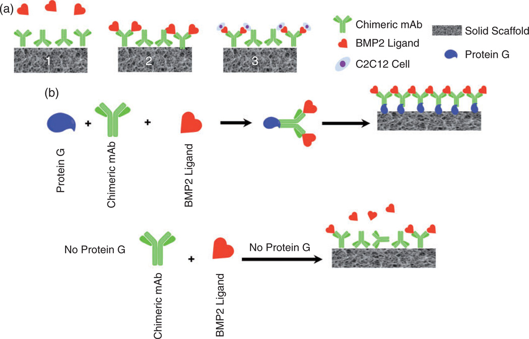
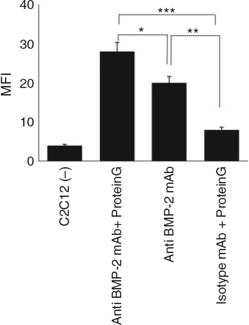
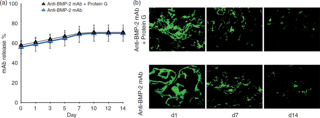
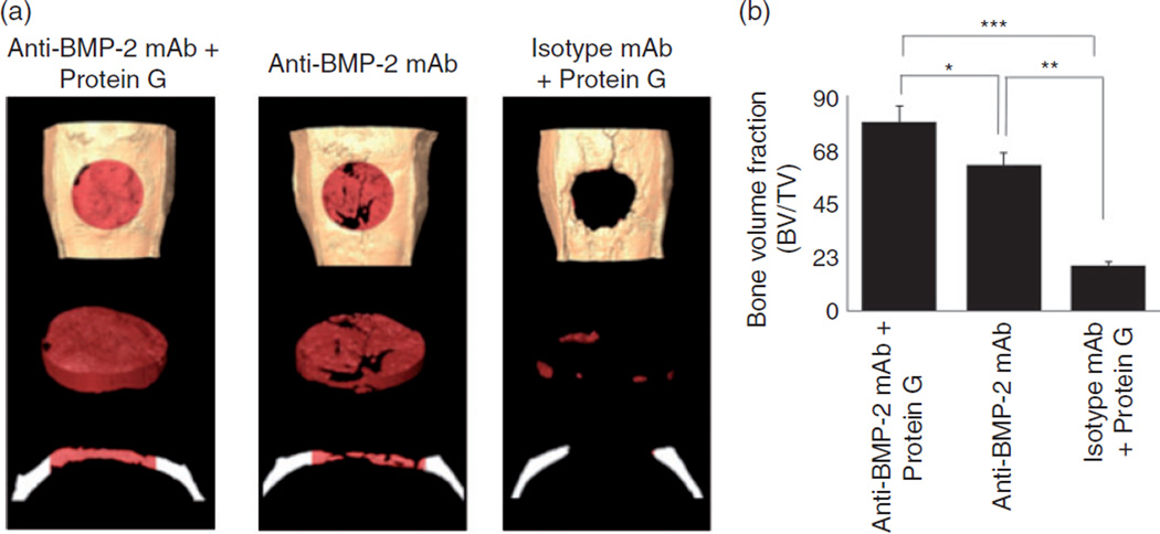
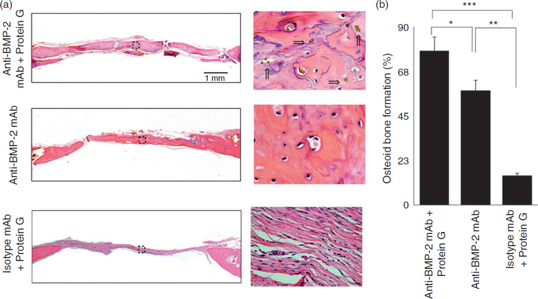
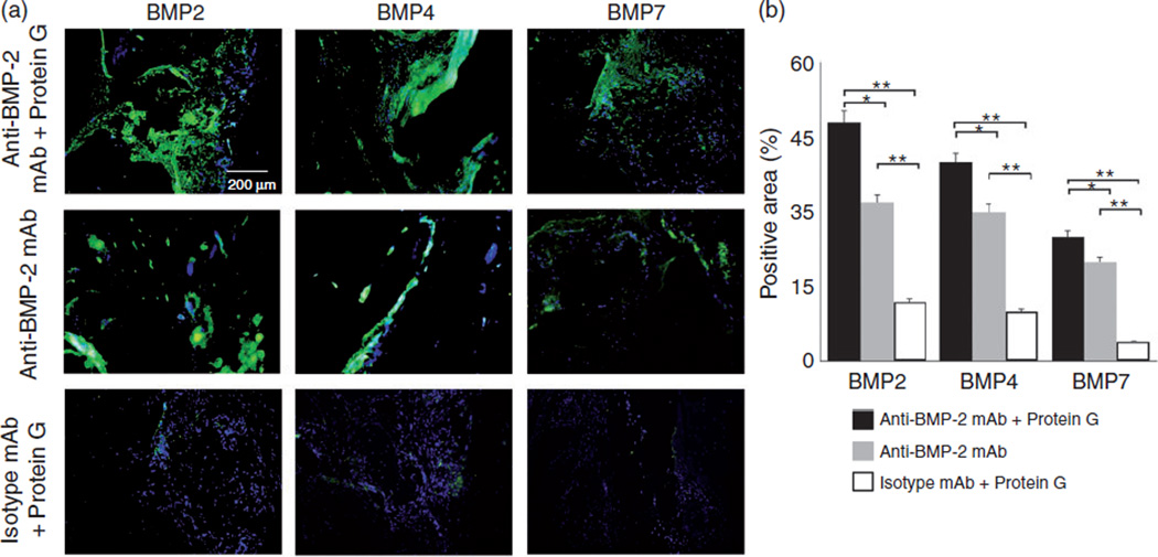
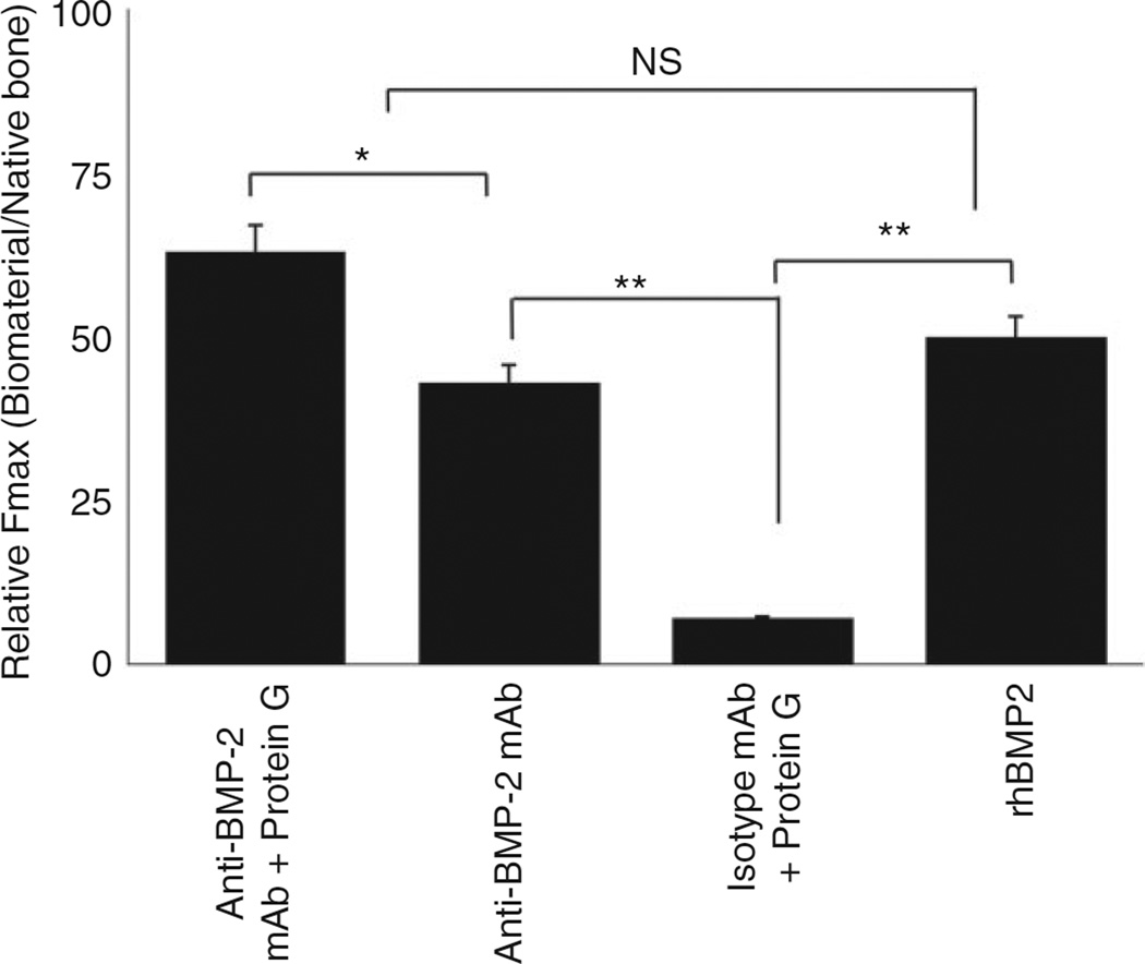
Recently, we have shown that anti-BMP2 monoclonal antibodies (mAbs) can trap endogenous osteogenic BMP ligands, which can in turn mediate osteodifferentiation of progenitor cells. The effectiveness of this strategy requires the availability of the anti-BMP-2 monoclonal antibodies antigen-binding sites for anti-BMP-2 monoclonal antibodies to bind to the scaffold through a domain that will leave its antigen-binding region exposed and available for binding to an osteogenic ligand. We examined whether antibodies bound to a scaffold by passive adsorption versus through Protein G as a linker will exhibit differences in mediating bone formation. In vitro anti-BMP-2 monoclonal antibodies was immobilized on absorbable collagen sponge (ACS) with Protein G as a linker to bind the antibody through its Fc region and implanted into rat calvarial defects. The biomechanical strength of bone regenerated by absorbable collagen sponge/Protein G/anti-BMP-2 monoclonal antibodies immune complex was compared to ACS/anti-BMP-2 monoclonal antibodies or ACS/Protein G/isotype mAb control group. Results demonstrated higher binding of anti-BMP-2 monoclonal antibodies/BMPs to C2C12 cells, when the mAb was initially attached to recombinant Protein G or Protein G-coupled microbeads. After eight weeks, micro-CT and histomorphometric analyses revealed increased bone formation within defects implanted with absorbable collagen sponge/Protein G/anti-BMP-2 monoclonal antibodies compared with defects implanted with absorbable collagen sponge/anti-BMP-2 monoclonal antibodies (p < 0.05). Confocal laser scanning microscopy (CLSM) confirmed increased BMP-2, -4, and -7 detection in sites implanted with absorbable collagen sponge/Protein G/anti-BMP-2 monoclonal antibodies in vivo. Biomechanical analysis revealed the regenerated bone in sites with Protein G/anti-BMP-2 monoclonal antibodies had higher mechanical strength in comparison to anti-BMP-2 monoclonal antibodies. The negative control group, Protein G/isotype mAb, did not promote bone regeneration and exhibited significantly lower mechanical properties (p < 0.05). Altogether, our results demonstrated that application of Protein G as a linker to adsorb anti-BMP-2 monoclonal antibodies onto the scaffold was accompanied by increased in vitro binding of the anti-BMP-2 mAb/BMP immune complex to BMP-receptor positive cell, as well as increased volume and strength of de novo bone formation in vivo.
Keywords: Bone regeneration; Protein G; biomaterials; chimeric anti-BMP2 monoclonal antibodies; tissue engineering.
© The Author(s) 2015.
Conflict of interest statement
None declared.
Figures
References
-
- Chen FM, Zhang J, Zhang M, et al. A review on endogenous regenerative technology in periodontal regenerative medicine. Biomaterials. 2010;31:7892–7927. - PubMed
-
- Monaco E, Bionaz M, Hollister SJ, et al. Strategies for regeneration of the bone using porcine adult adipose-derived mesenchymal stem cells. Theriogenology. 2011;75:1381–1399. - PubMed
-
- Arrington ED, Smith WJ, Chambers HG, et al. Complications of iliac crest bone graft harvesting. Clin Orthop Relat Res. 1996;329:300–309. - PubMed
MeSH terms
Substances
Grants and funding
LinkOut - more resources
Full Text Sources
Other Literature Sources
