. 2015 Dec:89:170-81.
doi: 10.1016/j.freeradbiomed.2015.07.013. Epub 2015 Jul 14.

Modulation of p38 kinase by DUSP4 is important in regulating cardiovascular function under oxidative stress

Affiliations

Modulation of p38 kinase by DUSP4 is important in regulating cardiovascular function under oxidative stress

Alma Barajas-Espinosa et al. Free Radic Biol Med. 2015 Dec.

Abstract

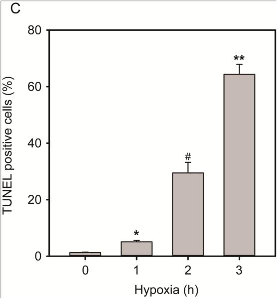
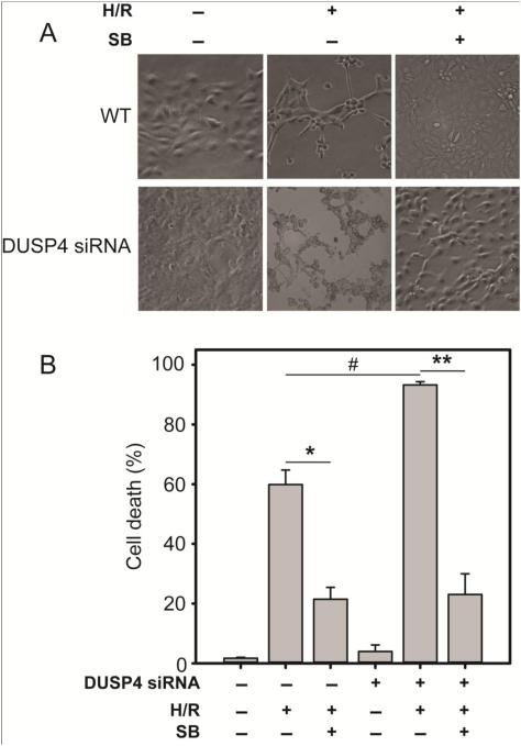
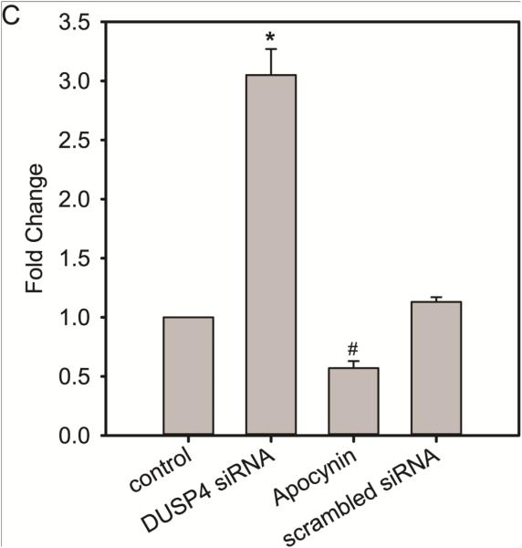
Over-activation of p38 is implicated in many cardiovascular diseases (CVDs), including myocardial infarction, hypertrophy, heart failure, and ischemic heart disease. Numerous therapeutic interventions for CVDs have been directed toward the inhibition of the p38 mitogen-activated protein kinase activation that contributes to the detrimental effect after ischemia/reperfusion (I/R) injuries. However, the efficacy of these treatments is far from ideal, as they lack specificity and are associated with high toxicity. Previously, we demonstrated that N-acetyl cysteine (NAC) pretreatment up-regulates DUSP4 expression in endothelial cells, regulating p38 and ERK1/2 activities, and thus providing a protective effect against oxidative stress. Here, endothelial cells under hypoxia/reoxygenation (H/R) insult and isolated heart I/R injury were used to investigate the role of DUSP4 in the modulation of the p38 pathway. In rat endothelial cells, DUSP4 is time-dependently degraded by H/R (0.25 ± 0.07-fold change of control after 2h H/R). Its degradation is closely associated with hyperphosphorylation of p38 (2.1 ± 0.36-fold change) and cell apoptosis, as indicated by the increase in cells immunopositive for cleaved caspase-3 (12.59 ± 3.38%) or TUNEL labeling (29.46 ± 3.75%). The inhibition of p38 kinase activity with 20 µM SB203580 during H/R prevents H/R-induced apoptosis, assessed via TUNEL (12.99 ± 1.89%). Conversely, DUSP4 gene silencing in endothelial cells augments their sensitivity to H/R-induced apoptosis (45.81 ± 5.23%). This sensitivity is diminished via the inhibition of p38 activity (total apoptotic cells drop to 17.47 ± 1.45%). Interestingly, DUSP4 gene silencing contributes to the increase in superoxide generation from cells. Isolated Langendorff-perfused mouse hearts were subjected to global I/R injury. DUSP4(-/-) hearts had significantly larger infarct size than WT. The increase in I/R-induced infarct in DUSP4(-/-) mice significantly correlates with reduced functional recovery (assessed by RPP%, LVDP%, HR%, and dP/dtmax) as well as lower CF% and a higher initial LVEDP. From immunoblotting analysis, it is evident that p38 is significantly overactivated in DUSP4(-/-) mice after I/R injury. The activation of cleaved caspase-3 is seen in both WT and DUSP4(-/-) I/R hearts. Infusion of a p38 inhibitor prior to ischemia and during the reperfusion improves both WT and DUSP4(-/-) cardiac function. Therefore, the identification of p38 kinase modulation by DUSP4 provides a novel therapeutic target for oxidant-induced diseases, especially myocardial infarction.

Keywords: Cardiovascular diseases; DUSP4; H/R; I/R; MAPK; Oxidative stress; p38.


Figures

Figure 1
Figure 1
Increased cellular oxidative stress after hypoxic exposure (0-3 h) leads to a time-dependent degradation of DUSP4 in RAECs. (A) Upper panel is the immunoblot against DUSP4 and lower panel is the immunoblot against actin as the loading control. 2 and 3 hr hypoxic exposure of cells leads to significant DUSP4 degradation (0.25 ± 0.07 and 0.29 ± 0.07 fold change of control level). * and ** versus control, P < 0.001. Degradation of DUSP4 is correlated to the uncontrolled activation of p38 after H/R insult. (B) Immunoblots of RAECs exposed to either control (no H/R) or 2 h H/R. Actin was used a loading control for DUSP4 and DUSP1, whereas the ratio of p-p38/p38 was used to determine the extent of phosphorylation of p38. (C) Following 2 h H/R, only 0.25 ± 0.07 of DUSP4/actin remains, yet there is no significant change in DUSP1/actin level (0.85 ± 0.17). Phosphorylation of p38 increases more than two fold (2.1 ± 0.36). There is no significant difference in ERK1/2 phosphorylation. No JNK phosphorylation is seen (data not shown). * P < 0.001, and # P < 0.05. Data is expressed as mean ± SEM, n ≥ 3.
Figure 1
Figure 1
Increased cellular oxidative stress after hypoxic exposure (0-3 h) leads to a time-dependent degradation of DUSP4 in RAECs. (A) Upper panel is the immunoblot against DUSP4 and lower panel is the immunoblot against actin as the loading control. 2 and 3 hr hypoxic exposure of cells leads to significant DUSP4 degradation (0.25 ± 0.07 and 0.29 ± 0.07 fold change of control level). * and ** versus control, P < 0.001. Degradation of DUSP4 is correlated to the uncontrolled activation of p38 after H/R insult. (B) Immunoblots of RAECs exposed to either control (no H/R) or 2 h H/R. Actin was used a loading control for DUSP4 and DUSP1, whereas the ratio of p-p38/p38 was used to determine the extent of phosphorylation of p38. (C) Following 2 h H/R, only 0.25 ± 0.07 of DUSP4/actin remains, yet there is no significant change in DUSP1/actin level (0.85 ± 0.17). Phosphorylation of p38 increases more than two fold (2.1 ± 0.36). There is no significant difference in ERK1/2 phosphorylation. No JNK phosphorylation is seen (data not shown). * P < 0.001, and # P < 0.05. Data is expressed as mean ± SEM, n ≥ 3.
Figure 2
Figure 2
Hypoxia/reoxygenation results in endothelial cell death and apoptosis. (A) 20X bright-field images of RAECs exposed to hypoxia (0-3 h), images were captured using the Zeiss Axiovert 135 microscope. Rounded, detached cells were considered dead cells and are expressed as a percentage of total cells in the graph. Cell death also time-dependently increased with all three hypoxic exposure time periods (18.49% ± 0.86% (1 h); 59.85% ± 4.89% (2 h); and 97.12% ± 0.70% (3 h)) compared to normoxic condition. *, #, and ** versus 0 h P < 0.0001. (B) RAEC immunostaining against cleaved caspase-3 and DAPI staining for cell nuclei. Immunopositive cells (red) were considered apoptotic and expressed as a percentage of total cell nuclei (blue). A significantly greater percentage of total cells become positive for apoptotic marker, cleaved caspase-3 at 2 and 3 h post-hypoxia exposure (12.59% ± 3.38% and 28.01% ± 5.62%, respectively) compared to control (0.10% ± 0.03%). * and # versus 0 h P < 0.05. (C) Quantification of TUNEL positive cells revealed a time-dependent increment in apoptotic cells. Exposure to the three H/R time points causes significant increase in TUNEL positive cells (5.09% ± 0.55% (1h); 29.46% ± 3.75% (2h); 64.39% ± 3.52% (3h) compared to control 1.28% ± 0.2%, *, #, and ** versus control P ≤ 0.001). Data is expressed as mean ± SEM, n ≥ 3.
Figure 2
Figure 2
Hypoxia/reoxygenation results in endothelial cell death and apoptosis. (A) 20X bright-field images of RAECs exposed to hypoxia (0-3 h), images were captured using the Zeiss Axiovert 135 microscope. Rounded, detached cells were considered dead cells and are expressed as a percentage of total cells in the graph. Cell death also time-dependently increased with all three hypoxic exposure time periods (18.49% ± 0.86% (1 h); 59.85% ± 4.89% (2 h); and 97.12% ± 0.70% (3 h)) compared to normoxic condition. *, #, and ** versus 0 h P < 0.0001. (B) RAEC immunostaining against cleaved caspase-3 and DAPI staining for cell nuclei. Immunopositive cells (red) were considered apoptotic and expressed as a percentage of total cell nuclei (blue). A significantly greater percentage of total cells become positive for apoptotic marker, cleaved caspase-3 at 2 and 3 h post-hypoxia exposure (12.59% ± 3.38% and 28.01% ± 5.62%, respectively) compared to control (0.10% ± 0.03%). * and # versus 0 h P < 0.05. (C) Quantification of TUNEL positive cells revealed a time-dependent increment in apoptotic cells. Exposure to the three H/R time points causes significant increase in TUNEL positive cells (5.09% ± 0.55% (1h); 29.46% ± 3.75% (2h); 64.39% ± 3.52% (3h) compared to control 1.28% ± 0.2%, *, #, and ** versus control P ≤ 0.001). Data is expressed as mean ± SEM, n ≥ 3.
Figure 3
Figure 3
Endothelial cells with DUSP4 gene silencing are more susceptible to H/R-induced cell death and apoptosis. (A) 20X bright-field images of either control or DUSP4 siRNA-transfected RAECs. (B) H/R increases cell death (H/R 59.85% ± 4.89% compared to control, P < 0.005), pre-treatment with SB203580 prevents death (SB H/R 21.45% ± 3.98%, *P<0.0005). DUSP4 gene silencing significantly enhances H/R-induced death (93.24% ± 1.09%) compared to the H/R treatment group (# P < 0.005). This increased susceptibility is reversed by treatment with SB203580 (23.04% ± 6.93%, **P < 0.005 compared to si H/R). Data is expressed as mean ± SEM, n ≥ 3. (C) DUSP4 gene silencing in RAECs enhances H/R-induced TUNEL positive cells (45.81% ± 5.23% compared to H/R alone 29.46 ± 3.75%, #P < 0.05). However, SB203580 treatment of transfected cells significantly lowers their sensitivity to H/R-induced apoptosis (17.47% ± 1.45%, ** P < 0.005 compared to the si H/R group). Similarly the inhibition of H/R-treated cells with SB203580 also significantly diminishes H/R-induced apoptosis (12.99% ± 1.89%), * P < 0.001. Data is expressed as mean ± SEM, n ≥ 3. There is no significant effect on the scrambled siRNA control group compared to control (data not shown).
Figure 3
Figure 3
Endothelial cells with DUSP4 gene silencing are more susceptible to H/R-induced cell death and apoptosis. (A) 20X bright-field images of either control or DUSP4 siRNA-transfected RAECs. (B) H/R increases cell death (H/R 59.85% ± 4.89% compared to control, P < 0.005), pre-treatment with SB203580 prevents death (SB H/R 21.45% ± 3.98%, *P<0.0005). DUSP4 gene silencing significantly enhances H/R-induced death (93.24% ± 1.09%) compared to the H/R treatment group (# P < 0.005). This increased susceptibility is reversed by treatment with SB203580 (23.04% ± 6.93%, **P < 0.005 compared to si H/R). Data is expressed as mean ± SEM, n ≥ 3. (C) DUSP4 gene silencing in RAECs enhances H/R-induced TUNEL positive cells (45.81% ± 5.23% compared to H/R alone 29.46 ± 3.75%, #P < 0.05). However, SB203580 treatment of transfected cells significantly lowers their sensitivity to H/R-induced apoptosis (17.47% ± 1.45%, ** P < 0.005 compared to the si H/R group). Similarly the inhibition of H/R-treated cells with SB203580 also significantly diminishes H/R-induced apoptosis (12.99% ± 1.89%), * P < 0.001. Data is expressed as mean ± SEM, n ≥ 3. There is no significant effect on the scrambled siRNA control group compared to control (data not shown).
Figure 4
Figure 4
H/R-induced endothelial cell death is modulated by DUSP4 and p38 activity. (A) Immunoblotting of RAECs after H/R. DUSP4 is degraded after 2 h hypoxic treatment. The degradation of DUSP4 is correlated with the over-activation of p38. Phosphorylation of p38 subsequently activates its downstream target, MK2, which can trigger caspase-3 activation and apoptosis. Inhibition of p38 with 20 µM SB203580 prevents H/R-induced apoptosis by blocking the phosphorylation of MK2 at the T334 site. (B) DUSP4 gene silencing leads to more pronounced p38 activation and MK2 phosphorylation at the T334 site after H/R. Similarly the inhibition of p38 kinase activity with 20 µM SB203580 diminishes MK2 activation and can thus prevent H/R-induced apoptosis. * Increased p38 MAPK activity. A negative scrambled siRNA is always performed. No effect from this negative control is seen (data not shown). (C) DUSP4 gene silencing up-regulates superoxide generation from cells (3.05 ± 0.22-fold increase compared to control, * P < 0.001). This increase in superoxide generation is inhibited by the treatment of 100 µM apocynin (0.57 ± 0.06-fold change compared to control, # P < 0.05). There is no significant effect from the negative control of scrambled siRNA. Data is expressed as mean ± SEM, n ≥ 3.
Figure 4
Figure 4
H/R-induced endothelial cell death is modulated by DUSP4 and p38 activity. (A) Immunoblotting of RAECs after H/R. DUSP4 is degraded after 2 h hypoxic treatment. The degradation of DUSP4 is correlated with the over-activation of p38. Phosphorylation of p38 subsequently activates its downstream target, MK2, which can trigger caspase-3 activation and apoptosis. Inhibition of p38 with 20 µM SB203580 prevents H/R-induced apoptosis by blocking the phosphorylation of MK2 at the T334 site. (B) DUSP4 gene silencing leads to more pronounced p38 activation and MK2 phosphorylation at the T334 site after H/R. Similarly the inhibition of p38 kinase activity with 20 µM SB203580 diminishes MK2 activation and can thus prevent H/R-induced apoptosis. * Increased p38 MAPK activity. A negative scrambled siRNA is always performed. No effect from this negative control is seen (data not shown). (C) DUSP4 gene silencing up-regulates superoxide generation from cells (3.05 ± 0.22-fold increase compared to control, * P < 0.001). This increase in superoxide generation is inhibited by the treatment of 100 µM apocynin (0.57 ± 0.06-fold change compared to control, # P < 0.05). There is no significant effect from the negative control of scrambled siRNA. Data is expressed as mean ± SEM, n ≥ 3.
Figure 5
Figure 5
DUSP4−/− mice are more prone to I/R-induced myocardial damage. (A) TTC-stained Langendorff-perfused heart slices from DUSP4−/− versus WT hearts subjected to 30 min global ischemia and 60 min reperfusion demonstrated a significantly greater infarct size (46.75% ± 4.19%) compared to WT (30.31% ± 3.33%). P < 0.05. n = 6 (B-G) Post-ischemic myocardial recovery following 30 min global ischemia and 30 min reperfusion for WT and DUSP4−/− hearts. (B) Rate pressure product (RPP), (C) Left ventricular developed pressure (LVDP), (D) Heart rate (HR), and (E) Coronary flow (CF) are expressed as a percentage of pre-ischemic (PI) value (100 %). (F) dP/dtmax is expressed in mmHg/s. The pre-ischemic (PI) value of dP/dtmax of WT is 4078.4 ± 183.5 mmHg/s, and PI value of dP/dtmax of DUSP4 KO is 5236.8 ± 270.4 mmHg/s. (G) Left ventricular end diastolic pressure (LVEDP) is expressed in mmHg. * P ≤ 0.05, and ** P ≤ 0.01, n > 10.
Figure 5
Figure 5
DUSP4−/− mice are more prone to I/R-induced myocardial damage. (A) TTC-stained Langendorff-perfused heart slices from DUSP4−/− versus WT hearts subjected to 30 min global ischemia and 60 min reperfusion demonstrated a significantly greater infarct size (46.75% ± 4.19%) compared to WT (30.31% ± 3.33%). P < 0.05. n = 6 (B-G) Post-ischemic myocardial recovery following 30 min global ischemia and 30 min reperfusion for WT and DUSP4−/− hearts. (B) Rate pressure product (RPP), (C) Left ventricular developed pressure (LVDP), (D) Heart rate (HR), and (E) Coronary flow (CF) are expressed as a percentage of pre-ischemic (PI) value (100 %). (F) dP/dtmax is expressed in mmHg/s. The pre-ischemic (PI) value of dP/dtmax of WT is 4078.4 ± 183.5 mmHg/s, and PI value of dP/dtmax of DUSP4 KO is 5236.8 ± 270.4 mmHg/s. (G) Left ventricular end diastolic pressure (LVEDP) is expressed in mmHg. * P ≤ 0.05, and ** P ≤ 0.01, n > 10.
Figure 6
Figure 6
DUSP4 gene deletion up-regulates Nox4 expression. (A) Immunoblotting against Nox4 antibody reveals that DUSP4 gene deletion promotes Nox4 protein expression (11.3 ± 3.64-fold change versus control, * P < 0.05) under basal conditions. (B) Quantitative PCR analysis shows that DUSP4 gene deletion increases Nox4 mRNA expression (4.9 ± 1.17-fold change versus control, * P < 0.01) under basal conditions. Data is expressed as mean ± SEM, n ≥ 3.
Figure 7
Figure 7
Molecular alterations in DUSP4−/− hearts after I/R injury. (A) Increased p38 phosphorylation is evident in hearts subjected to I/R. This effect is more pronounced in DUSP4−/− mice. The ratio of p-p38/p38 of WT I/R hearts is set at 1. * versus WT I/R hearts, P < 0.001 n = 5. (B) The increase in p38 activity leads to MK2 phosphorylation at T334 and cleaved caspase-3 activation. Immunoblotting of cleaved caspase-3 is an indicator of apoptosis. WT and DUSP4−/− hearts both show an increase in caspase-3 and cleaved caspase-3 expression. * Increased p38 MAPK activity. (C) Direct inhibition of p38 activity by infusing with 10 µM SB203580 improves cardiac function at the end of 30 min reperfusion. RPP (%), LVDP (%), and CF (%) are dramatically improved for both WT and DUSP4−/− hearts. For (dP/dt)max, only DUSP4−/− hearts show significant recovery after the treatment of p38 inhibitor. There is no significant difference for heart rate and LVEDP at the end of 30 min reperfusion for both WT and DUSP4−/− hearts. * P < 0.05, and ** P < 0.01. n ≥ 7.
Figure 7
Figure 7
Molecular alterations in DUSP4−/− hearts after I/R injury. (A) Increased p38 phosphorylation is evident in hearts subjected to I/R. This effect is more pronounced in DUSP4−/− mice. The ratio of p-p38/p38 of WT I/R hearts is set at 1. * versus WT I/R hearts, P < 0.001 n = 5. (B) The increase in p38 activity leads to MK2 phosphorylation at T334 and cleaved caspase-3 activation. Immunoblotting of cleaved caspase-3 is an indicator of apoptosis. WT and DUSP4−/− hearts both show an increase in caspase-3 and cleaved caspase-3 expression. * Increased p38 MAPK activity. (C) Direct inhibition of p38 activity by infusing with 10 µM SB203580 improves cardiac function at the end of 30 min reperfusion. RPP (%), LVDP (%), and CF (%) are dramatically improved for both WT and DUSP4−/− hearts. For (dP/dt)max, only DUSP4−/− hearts show significant recovery after the treatment of p38 inhibitor. There is no significant difference for heart rate and LVEDP at the end of 30 min reperfusion for both WT and DUSP4−/− hearts. * P < 0.05, and ** P < 0.01. n ≥ 7.

Similar articles

Cited by

References

    1. Global, regional, and national age-sex specific all-cause and cause-specific mortality for 240 causes of death, 1990-2013: a systematic analysis for the Global Burden of Disease Study 2013. Lancet. 2014. - PMC - PubMed
    1. Mendis S, Puska P, Norrving B, World Health Organization. World Heart Federation. World Stroke Organization . Global atlas on cardiovascular disease prevention and control. World Health Organization in collaboration with the World Heart Federation and the World Stroke Organization; Geneva: 2011.
    1. Frank A, Bonney M, Bonney S, Weitzel L, Koeppen M, Eckle T. Myocardial ischemia reperfusion injury: from basic science to clinical bedside. Semin Cardiothorac Vasc Anesth. 2012;16:123–132. - PMC - PubMed
    1. Zweier JL, Talukder MA. The role of oxidants and free radicals in reperfusion injury. Cardiovasc Res. 2006;70:181–190. - PubMed
    1. Aune SE, Yeh ST, Zelinski DP, Angelos MG. Measurement of hydrogen peroxide and oxidant stress in a recirculating whole blood-perfused rat heart model. Resuscitation. 2011;82:222–227. - PubMed

Publication types

MeSH terms

Substances