. 2015 Jul 17:6:7737.
doi: 10.1038/ncomms8737.

Desialylation is a mechanism of Fc-independent platelet clearance and a therapeutic target in immune thrombocytopenia

Affiliations

Desialylation is a mechanism of Fc-independent platelet clearance and a therapeutic target in immune thrombocytopenia

June Li et al. Nat Commun. .

Abstract

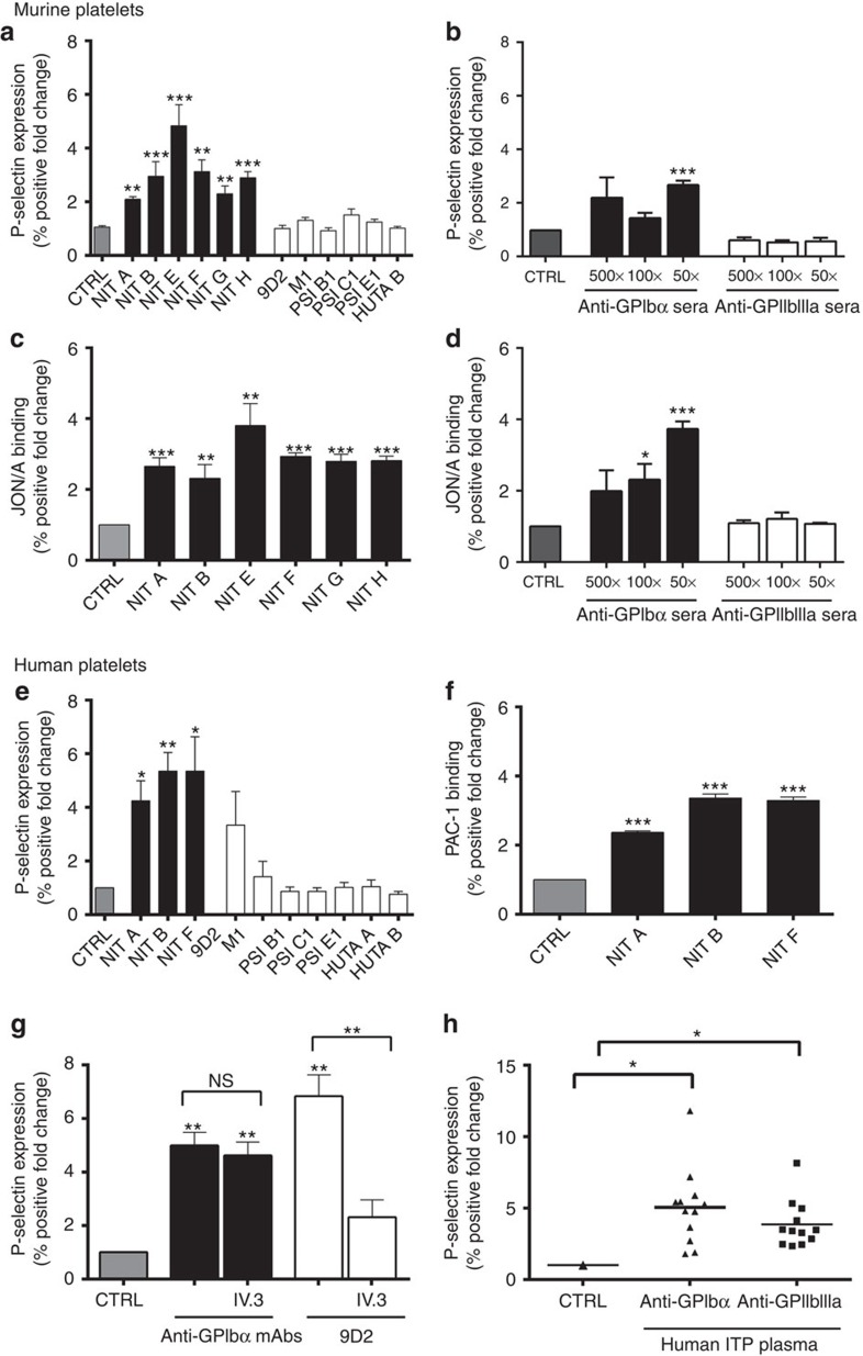
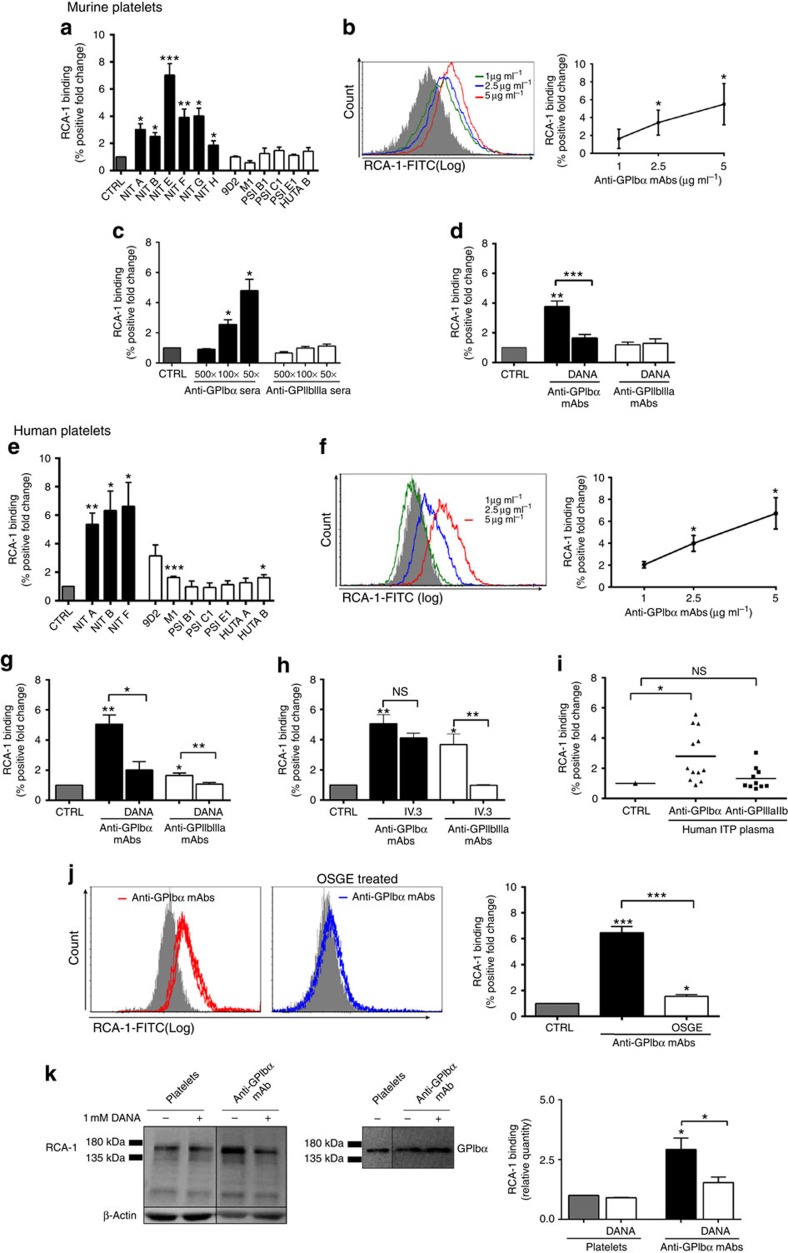
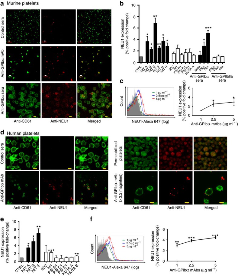
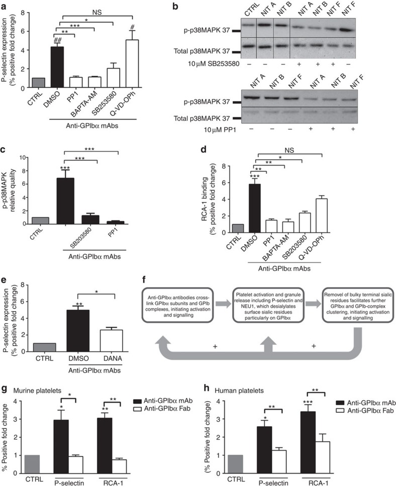
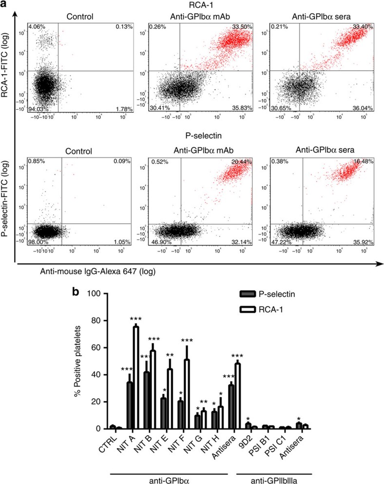
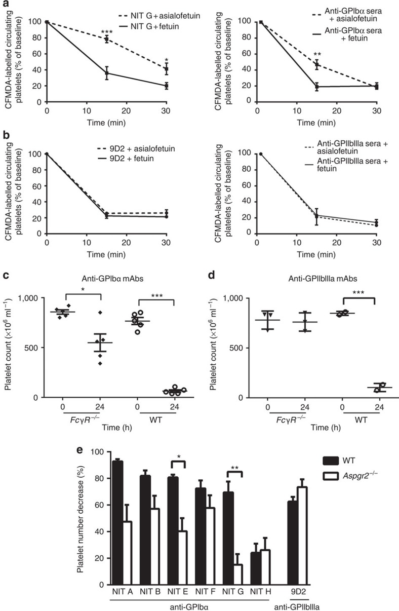
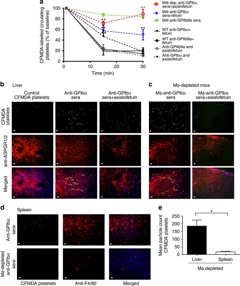
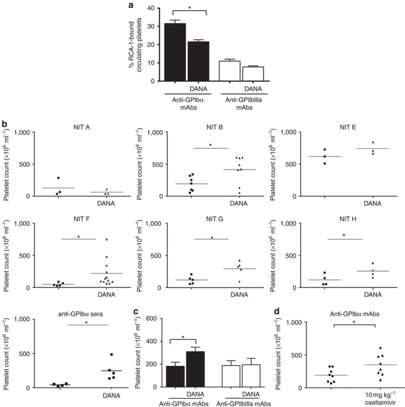
Immune thrombocytopenia (ITP) is a common bleeding disorder caused primarily by autoantibodies against platelet GPIIbIIIa and/or the GPIb complex. Current theory suggests that antibody-mediated platelet destruction occurs in the spleen, via macrophages through Fc-FcγR interactions. However, we and others have demonstrated that anti-GPIbα (but not GPIIbIIIa)-mediated ITP is often refractory to therapies targeting FcγR pathways. Here, we generate mouse anti-mouse monoclonal antibodies (mAbs) that recognize GPIbα and GPIIbIIIa of different species. Utilizing these unique mAbs and human ITP plasma, we find that anti-GPIbα, but not anti-GPIIbIIIa antibodies, induces Fc-independent platelet activation, sialidase neuraminidase-1 translocation and desialylation. This leads to platelet clearance in the liver via hepatocyte Ashwell-Morell receptors, which is fundamentally different from the classical Fc-FcγR-dependent macrophage phagocytosis. Importantly, sialidase inhibitors ameliorate anti-GPIbα-mediated thrombocytopenia in mice. These findings shed light on Fc-independent cytopenias, designating desialylation as a potential diagnostic biomarker and therapeutic target in the treatment of refractory ITP.


Figures

Figure 1
Figure 1. Anti-GPIbα antibodies induce platelet activation.
Surface P-selectin expression on murine (a,b) or human platelets (e,g,h) and JON/A binding on murine (c,d) or PAC-1 binding on human platelets (f) were measured by flow cytometry following incubation with mAbs (a,c,e,f,g) or antisera (b,d). (g) FcγRIIa/III blocker IV.3 was incubated with mAb 9D2 and platelets, and platelet P-selectin was assessed following. Only the healthy donor platelets that were significantly activated in the presence of 9D2 were tested; n=3. (h) P-selectin expression was measured in healthy human platelets following incubation with anti-GPIbα (ITP-1 to ITP-12) or anti-GPIIbIIIa (ITP-a to ITP-l) antibody-positive ITP patient plasma. All flow cytometry data are expressed as fold change from nonspecific murine IgG (murine)- or IVIG (human)-treated control platelets (CTRL). Anti-GPIbα mAbs shown as mean±s.e.m. of individual mAbs. *P<0.05, **P<0.01, ***P<0.001 versus CTRL as analysed by the Student's t-test (af) or one-way analysis of variance followed by Bonferroni post hoc analysis (g,h). NS, not significant.
Figure 2
Figure 2. Antibody-mediated platelet desialylation occurs mainly on the GPIbα subunit.
Galactose exposure was detected with RCA-1 binding and measured by flow cytometry in murine (ad) and human (ej) platelets following incubation with mAbs (a,b,d,e,fh,j,k) or antisera (c) n=10–20. (d,g) Co-incubation with sialidase inhibitor DANA prior to addition of mAbs to murine (d) or human (g) platelets; n=8. (h) FcγRII/III blocker IV.3 was incubated with mAbs (9D2, M1 or HUTA B) and platelets, RCA-1 binding was assessed following. Only the healthy donor platelets that were significantly desialylated in the presence of these mAbs were tested; n=4. (i) RCA-1 binding was measured in healthy human platelets following incubation with anti-GPIbα (ITP-1 to ITP-12) or anti-GPIIbIIIa (ITP-a to ITP-l) antibody-positive ITP patient plasma. (j) anti-GPIbα-mediated RCA-1 binding was assessed following removal of GPIbα with OSGE. (k) Representative western blot of RCA-1 binding (left), and probing with commercial anti-GPIbα antibody (right) to confirm the identity of the RCA-1-positive band following incubation with anti-GPIbα mAb (NIT F). RCA-1 binding on GPIbα was also quantified by protein densitometry in the presence of DANA. All flow cytometry data are expressed as fold change from nonspecific murine IgG (murine)- or IVIG (human)-treated control platelets (CTRL). Anti-GPIbα mAbs shown as mean±s.e.m. of individual mAbs. *P<0.05, **P<0.01, ***P<0.001 versus CTRL as analysed by the Student's t-test (ac,e,f) or one-way analysis of variance followed by Bonferroni post hoc (d,gk). NS, not significant.
Figure 3
Figure 3. Anti-GPIbα antibodies induce surface expression of NEU1.
Representative confocal images of surface expression of NEU1 on murine (a) and human (d) platelets stained with anti-NEU1 and anti-CD61 following incubations with anti-GPIbα mAb (NIT G (murine), NIT B (human)) or anti-GPIbα sera (murine). All other mAbs were also tested with similar results; n=5–8. Total NEU1 was detected in permeabilized human platelets; n=2. White scale bars, 5 μM; yellow scale bars, 2 μM. (b,c,e,f) Flow cytometric analysis of surface NEU1 expression on murine (b,c) and human (e,f) platelets following incubations with anti-GPIbα mAb or sera. Anti-GPIbα mAbs shown as mean±s.e.m. of individual mAbs; n=5–8. All flow cytometry data are expressed as fold change from nonspecific murine IgG (murine)- or IVIG (human)-treated control platelets (CTRL). Anti-GPIbα mAbs shown as mean±s.e.m. of individual mAbs. *P<0.05, **P<0.01, ***P<0.001 versus CTRL as analysed by the Student's t-test.
Figure 4
Figure 4. Anti-GPIbα platelet activation and desialylation is a positive feedback loop.
(a,d) Human platelets were pre-incubated with inhibitors of platelet activation including intracellular Ca2+ flux (BAPTA-AM), phosphorylation of P38MAPK (SB203580) and Src kinase (PP1) prior to addition of anti-GPIbα mAbs. Following which, platelet activation (P-selectin expression) (a) or desialylation (RCA-1 binding) (d) was detected via flow cytometry. # and ## indicate comparison with CTRL for a only; n=8. (b) Representative western blots of whole-platelet lysate following incubations with GPIbα mAbs with or without indicated inhibitors. Membranes were probed for phosphorylated p38MAPK (p-p38MAPK) then stripped and re-probed for total pMAP38K. (c) Densitometry protein quantification of p-P38MAPK detected in b. Data representative of three separate experiments. (e) Sialidase inhibitor DANA was added prior to anti-GPIbα mAb incubation. DANA-mediated inhibition of antibody-induced platelet activation was measured by P-selectin expression and detected via flow cytometry. n=6. (f) Schematic flow chart illustrating platelet activation–desialylation-positive feedback loop. Following initial anti-GPIbα antibody crosslinking of GPIbα subunits, activation signalling occurs leading to surface translocation of NEU1. NEU1 cleavage of terminal sialic residues on a GPIbα subunit facilitates receptor clustering, resulting in amplification of platelet activation. (g,h) NIT A, NIT B and NIT F Fab fragments were generated and their effects on platelet activation and desialylation on murine (g) and human platelets (h) were analysed by flow cytometry. n=5. All flow cytometry data are expressed as fold change from nonspecific murine IgG (murine)- or IVIG (human)-treated control platelets (CTRL), unless otherwise indicated. Anti-GPIbα mAbs shown as mean±s.e.m. of individual mAbs. *,#P<0.05, **,##P<0.01, ***P<0.001 as assessed by one-way analysis of variance followed by Bonferroni post hoc analysis. NS, not significant.
Figure 5
Figure 5. Anti-GPIbα mAbs induce platelet activation and desialylation in vivo.
(a,b) ITP was induced by intraperitoneal injection of anti-GPIbα mAbs (NIT A, NIT B, NIT E, NIT F, NIT G and NIT H) or sera. At 16 h post injection, platelets isolated from the mice were examined for activation (P-selectin) and desialylation (RCA-1) by flow cytometry; n=3 per mAb. (a) Representative dot plots of isolated washed platelets from anti-GPIbα mAb (NIT E)- and sera-injected mice double-stained for anti-mouse IgG (against anti-GPIbα antibodies) (x axis) and RCA-1/P-selectin (y axis).
Figure 6
Figure 6. Fc-independent anti-GPIbα-mediated thrombocytopenia occurs via the AMR.
Mice were co-injected with (a) anti-GPIbα (NIT G) or (b) anti-GPIIbIIIa mAb (9D2)- or polyclonal sera-opsonized CFMDA-labelled platelets with asialofetuin (AMR inhibitor) or fetuin (a nonspecific control). Mice were bled at indicated time points and the percentage of fluorescently labelled platelets remaining in circulation was assessed with flow cytometry; n=6. *P<0.05 versus Fetuin-injected group at same time point as analysed by two-way analysis of variance followed by Bonferroni post hoc analysis. (ce) Same doses of indicated mAb or anti-GPIbα or anti-GPIIbIIIa mAbs (equal ratio mixture of individual antibody clones) were injected into age-matched FcγR−/− (c,d) or Aspgr−/− (e) or WT control mice. Platelet counts were taken immediately prior to antibody injection (time 0) and 24 h later (time 24). (e) Data are represented as a percentage calculated from [(time 0 platelet count−time 24 platelet count)/time 0 platelet count] × 100. *P<0.05, **P<0.01, ***P<0.001 as determined by the Student's t-test.
Figure 7
Figure 7. Anti-GPIbα platelet clearance in macrophage-depleted mice is via the AMR.
(a-e) Macrophages were depleted with clondrate liposomes via intravenous injection prior to study, and CFMDA-labelled antisera-opsonized platelets were injected with asialofeuin or fetuin and circulating platelets were quantified (a) as described in Fig. 6. n=5. ^^P<0.01 versus fetuin-injected group at the same time point. *P<0.05, **P<0.01 versus baseline as determined by two-way analysis of variance followed by Bonferroni post hoc analysis. (bd) Tissue sections of the spleen and liver harvested from normal or macrophage-depleted mice following the above circulation studies and were stained with anti-ASPGR1/2 (liver; red) (b,c) or anti-F4/80 (spleen; red) (d). Nuclei were counterstained with 4′,6-diamidino-2-phenylindole (blue). Arrows indicate co-localization of CFMDA-labelled platelets with ASPGR on hepatocytes. (e) Fluorescent platelet (green) localization was assessed with immunofluorescent microscopy at × 60 magnification and quantified with Image J. White scale bars, 10 μM. *P<0.05 as assessed by the Student's t-test. Data are representative of five randomly selected fluorescent images.
Figure 8
Figure 8. Sialidase inhibition rescues thrombocytopenia in a murine model of ITP.
(ac) DANA or PBS was injected intraperitoneally immediately prior to anti-GPIbα or anti-GPIIbIIIa antibody injection (mAb or sera) to induce thrombocyotopenia. (a) Platelets from antibody-injected mice were analysed for desialylation (RCA-1 binding) via flow cytometry. (b) Platelet numbers were enumerated and compared between DANA-treated or mock (PBS)-treated groups. (c) Cumulative platelet counts from all anti-GPIbα and anti-GPIIbIIIa antibody (9D2, M1, PSI C1 and antisera)-injected mice with or without DANA treatment. For individual platelet counts of anti-GPIIbIIIa-injected mice, please see Supplementary Fig. 7. (d) Oseltamivir phosphate was injected intravenously 2 h after anti-GPIbα mAbs injection (equal ratio mixture of individual mAb clones). Platelet enumeration was as described above. Data are shown as mean±s.e.m. platelet counts of individual mAbs. *P<0.05, ***P<0.001 as determined by the Student's t-test.

References

    1. Rodeghiero F. et al.. Standardization of terminology, definitions and outcome criteria in immune thrombocytopenic purpura of adults and children: report from an international working group. Blood 113, 2386–2393 (2009). - PubMed
    1. McMillan R. The pathogenesis of chronic immune thrombocytopenic purpura. Semin. Hematol. 44, S3–S11 (2007). - PubMed
    1. Cines D. B., Bussel J. B., Liebman H. A. & Luning Prak E. T. The ITP syndrome: pathogenic and clinical diversity. Blood 113, 6511–6521 (2009). - PMC - PubMed
    1. Mathias S. D. et al.. Impact of chronic Immune Thrombocytopenic Purpura (ITP) on health-related quality of life: a conceptual model starting with the patient perspective. Health Qual. Life Outcomes 6, 13 (2008). - PMC - PubMed
    1. Provan D. et al.. International consensus report on the investigation and management of primary immune thrombocytopenia. Blood 115, 168–186 (2010). - PubMed

Publication types

MeSH terms

Substances