. 2015 Jul 14;21(26):8043-51.
doi: 10.3748/wjg.v21.i26.8043.

Contrast-enhanced micro-computed tomography using ExiTron nano6000 for assessment of liver injury

Affiliations

Contrast-enhanced micro-computed tomography using ExiTron nano6000 for assessment of liver injury

Xiang-Wei Hua et al. World J Gastroenterol. .

Abstract

Aim: To explore the potential of contrast-enhanced computed tomography (CECT) using ExiTron nano6000 for assessment of liver lesions in mouse models.

Methods: Three mouse models of liver lesions were used: bile duct ligation (BDL), lipopolysaccharide (LPS)/D-galactosamine (D-GalN), and alcohol. After injection with the contrast agent ExiTron nano6000, the mice were scanned with micro-CT. Liver lesions were evaluated using CECT images, hematoxylin and eosin staining, and serum aminotransferase levels. Macrophage distribution in the injury models was shown by immunohistochemical staining of CD68. The in vitro studies measured the densities of RAW264.7 under different conditions by CECT.

Results: In the in vitro studies, CECT provided specific and strong contrast enhancement of liver in mice. CECT could present heterogeneous images and densities of injured livers induced by BDL, LPS/D-GalN, and alcohol. The liver histology and immunochemistry of CD68 demonstrated that both dilated biliary tracts and necrosis in the injured livers could lead to the heterogeneous distribution of macrophages. The in vitro study showed that the RAW264.7 cell masses had higher densities after LPS activation.

Conclusion: Micro-CT with the contrast agent ExiTron nano6000 is feasible for detecting various liver lesions by emphasizing the heterogeneous textures and densities of CECT images.

Keywords: ExiTron Nano6000; Liver injury; Micro-computed tomography.


Figures

Figure 1
Figure 1
Contrast enhanced computed tomography images of a normal liver. A: 3D image of a liver (arrow); B: Perspective view of a liver (arrow).
Figure 2
Figure 2
Contrast-enhanced computed tomography images of liver lesions induced by bile duct ligation (A), lipopolysaccharide/D-GalN (B) and alcohol (C); density measured in the livers are reported as HU. The black arrows indicate black regions with low densities. Values are represented as the means of triplicate values and presented as the mean ± SD.
Figure 3
Figure 3
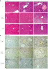
HE stains for liver lesions induced by bile duct ligation, lipopolysaccharide/D-GalN and alcohol (A); Immunostaining of CD68 after liver lesions induced by bile duct ligation, lipopolysaccharide/D-GalN or alcohol (B). The black arrows indicate necrosis, and the white arrows indicate dilated biliary tracts.
Figure 4
Figure 4
ALT levels in mouse models induced by bile duct ligation (A), lipopolysaccharide/D-GalN (B) and alcohol (C), bP < 0.01.
Figure 5
Figure 5
Comparison of the number of CD68+ cells in the injured livers of the three models (A); comparison of the densities of RAW264.7 cell mass co-cultured with nano6000, lipopolysaccharide or both (B). Values are represented as the means of triplicate values and presented as the mean ± SD.

References

    1. Deroose CM, De A, Loening AM, Chow PL, Ray P, Chatziioannou AF, Gambhir SS. Multimodality imaging of tumor xenografts and metastases in mice with combined small-animal PET, small-animal CT, and bioluminescence imaging. J Nucl Med. 2007;48:295–303. - PMC - PubMed
    1. Wurnig MC, Tsushima Y, Weiger M, Jungraithmayr W, Boss A. Assessing lung transplantation ischemia-reperfusion injury by microcomputed tomography and ultrashort echo-time magnetic resonance imaging in a mouse model. Invest Radiol. 2014;49:23–28. - PubMed
    1. Wu X, Yu G, Lindner D, Brady-Kalnay SM, Zhang Q, Lu ZR. Peptide targeted high-resolution molecular imaging of prostate cancer with MRI. Am J Nucl Med Mol Imaging. 2014;4:525–536. - PMC - PubMed
    1. Bastard C, Bosisio MR, Chabert M, Kalopissis AD, Mahrouf-Yorgov M, Gilgenkrantz H, Mueller S, Sandrin L. Transient micro-elastography: A novel non-invasive approach to measure liver stiffness in mice. World J Gastroenterol. 2011;17:968–975. - PMC - PubMed
    1. Martiniova L, Schimel D, Lai EW, Limpuangthip A, Kvetnansky R, Pacak K. In vivo micro-CT imaging of liver lesions in small animal models. Methods. 2010;50:20–25. - PMC - PubMed

Publication types

MeSH terms