

doi: 10.1155/2015/482342.
Epub 2015 Jun 21.
A Rare Case of a Primary Squamous Cell Carcinoma of the Stomach Presenting as a Submucosal Mass
Affiliations
- PMID: 26185704
- PMCID: PMC4491394
- DOI: 10.1155/2015/482342
Item in Clipboard
A Rare Case of a Primary Squamous Cell Carcinoma of the Stomach Presenting as a Submucosal Mass
Case Rep Surg.
2015.
Abstract
We report a case of a 70-year-old man, with a status after aortic valve replacement, who presented with melena and hypotension. On physical examination, he was hypotensive, but he responded to resuscitation. Esophagogastroduodenoscopy revealed a submucosal mass in the gastric fundus. Imaging of the chest, abdomen, and pelvis showed no evidence of local or distant metastasis. He underwent a partial diaphragmatic resection, gastrectomy, lymphadenectomy, and Roux-en-Y esophagojejunostomy. Pathology showed a gastric squamous cell carcinoma (SCC) invading the diaphragm, with negative margins of resection, and one positive perigastric lymph node. He received chemoradiation, but the patient expired 27 months after surgery.
Figures
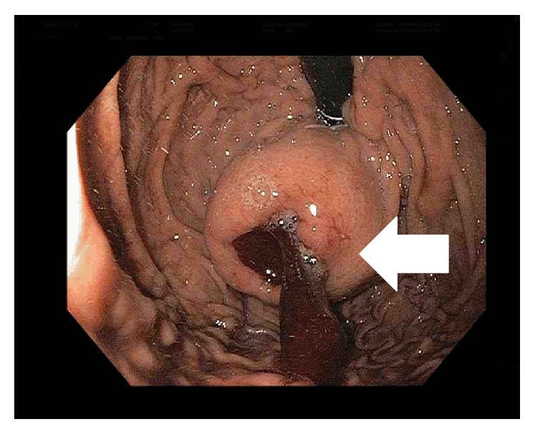
Esophagogastroduodenoscopy (EGD) showing an ulcerated submucosal mass in the fundus of the stomach (white arrow).
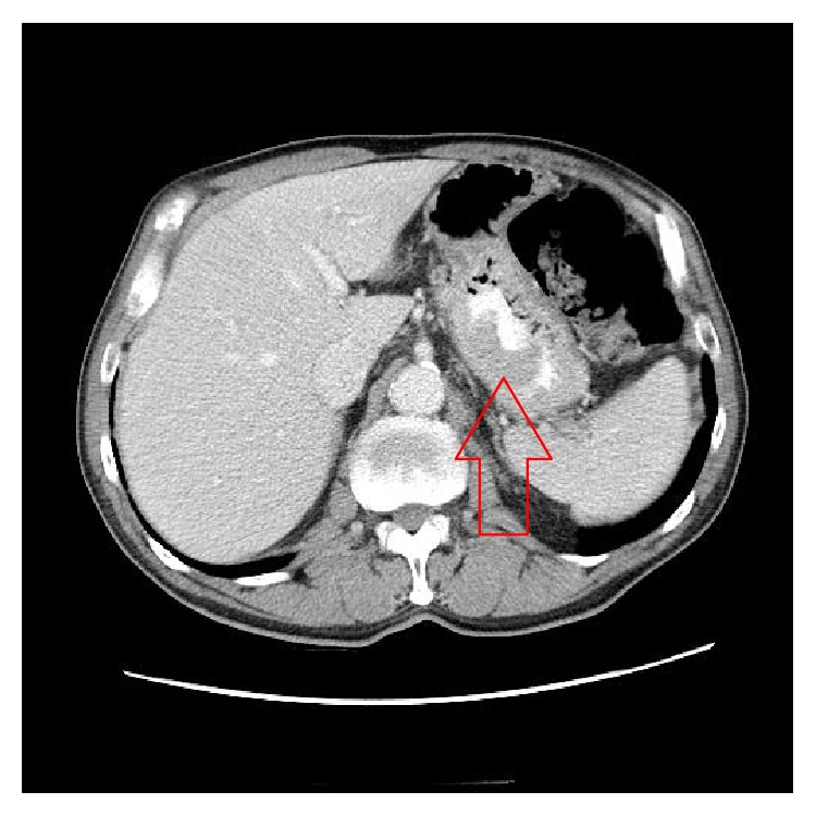
Computerized axial tomography scan with IV and PO contrast showing a mass in the gastric fundus (red arrow).
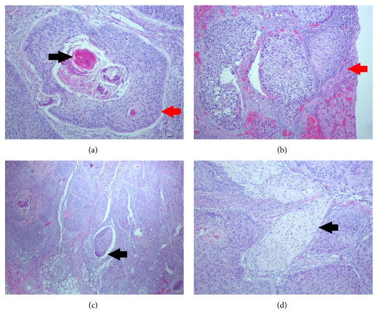
(a) Histopathological examination showing a moderately differentiated squamous cell carcinoma with keratinization (×100). Keratin pearl (black arrow). Mosaic cell arrangement with sharp border (red arrow). (b) Tumor invasion of the diaphragm (×100) (red arrow). (c) Lymphovascular invasion is present (×40) (black arrow). (d) Tumor with nerve invasion (×40) (black arrow).
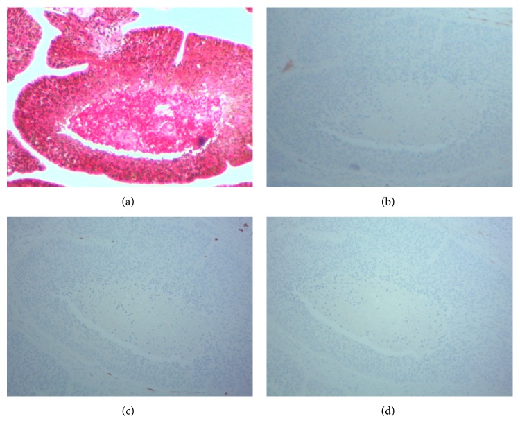
Tumor cells showing strong coexpression of p63 (nuclear stain, brown) and cytokeratin 5/6 (membrane stain, red) (a) and negative expression of p16 (b), CD117 (c), and CK7 (d).
References
-
- Muto M., Hasebe T., Muro K., et al. Primary squamous cell carcinoma of the stomach: a case report with a review of Japanese and Western literature. Hepato-Gastroenterology. 1999;46(29):3015–3018. - PubMed
-
- Bonnheim D. C., Sarac O. K., Fett W. Primary squamous cell carcinoma of the stomach. The American Journal of Gastroenterology. 1985;80(2):91–94. - PubMed
-
- Schmidt C., Schmid A., Lüttges J. E., Kremer B., Henne-Bruns D. Primary squamous cell carcinoma of the stomach. Report of a case and review of literature. Hepato-Gastroenterology. 2001;48(40):1033–1036. - PubMed
LinkOut - more resources
Full Text Sources
Other Literature Sources
Research Materials
