. 2015;2(3):141-164.
doi: 10.3390/jcdd2030141.

Vascular Calcification and Stone Disease: A New Look towards the Mechanism

Affiliations

Vascular Calcification and Stone Disease: A New Look towards the Mechanism

Allen J Yiu et al. J Cardiovasc Dev Dis. 2015.

Abstract

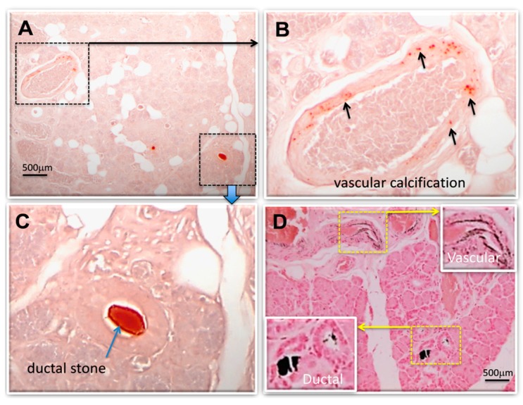
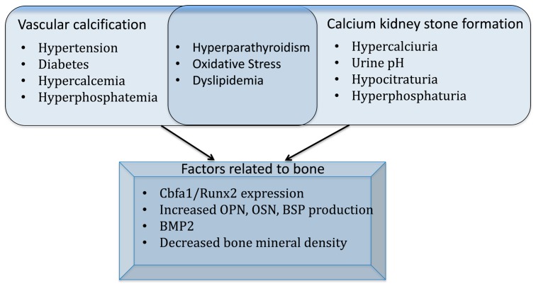
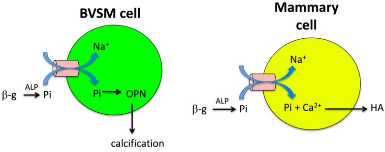
Calcium phosphate (CaP) crystals are formed in pathological calcification as well as during stone formation. Although there are several theories as to how these crystals can develop through the combined interactions of biochemical and biophysical factors, the exact mechanism of such mineralization is largely unknown. Based on the published scientific literature, we found that common factors can link the initial stages of stone formation and calcification in anatomically distal tissues and organs. For example, changes to the spatiotemporal conditions of the fluid flow in tubular structures may provide initial condition(s) for CaP crystal generation needed for stone formation. Additionally, recent evidence has provided a meaningful association between the active participation of proteins and transcription factors found in the bone forming (ossification) mechanism that are also involved in the early stages of kidney stone formation and arterial calcification. Our review will focus on three topics of discussion (physiological influences-calcium and phosphate concentration-and similarities to ossification, or bone formation) that may elucidate some commonality in the mechanisms of stone formation and calcification, and pave the way towards opening new avenues for further research.

Keywords: calcification; calcium phosphate; calculi; ectopic; kidney; ossification; physiologic.


Conflict of interest statement

The authors declare no conflict of interest.

Figures

Figure 1
Figure 1
Vascular and ductal calcifications were found in salivary gland from stone patient. Human submandibular gland (SMG) sections from stone patient were used to detect calcified material. Calcified blood vessel and intra-ductal stones were stained with the: (A) alizarin red method [48] for examining the calcified deposits; enlarged in (B) showing vascular calcification; and (C) showing intra-ductal stone (pictures are representative of n = 6); no such staining was found in control SMG (n = 4). Von Kossa [49] stained SMG (D) confirms the presence of calcification, both vascular and stone, in same SMG tissue. Specimens are formalin fixed paraffin embedded (FFPE) de-identified tissue section (biopsy sample, not individually identifiable to any person) from tissue bank through an exempt Institutional Review Board (IRB) protocol.
Figure 2
Figure 2
Identified instigators of vascular calcification and stone formation, highlighting common factors, and several commonalities related to bone mineralization/de-mineralization. Risk factors for vascular calcification include both the “traditional” (such as hypertension and diabetes) and the “non-traditional” (i.e., abnormal mineral metabolism such as hypercalcemia and hyperphosphatemia) [123]. A majority of calcium kidney stones are predominantly calcium oxalate, with CaP admixed in small amounts [4]. Thus, factors associated with the predominant form of calcium stone formation include: hypercalciuria; urine pH, high (pH > 6.3) for CaP stone formation, and for CaP deposits that may be the initial nidus for calcium oxalate stones, and low for calcium oxalate stone formation; and hypocitraturia, which increases the chance for calcium to bind with oxalate or phosphate [4]. Hyperphosphaturia may increase the risk of calcium kidney stone formation by urinary calcium excretion and urinary saturation [56]. Common risk factors for both vascular calcification and calcium kidney stone formation include hyperparathyroidism [4,123], oxidative stress [99,123], and dyslipidemia [37,123]. Interestingly, studies have found bone related proteins and other bone related factors in vascular calcification, calcium deposits and calcium kidney stone formation as discussed in the review.
Figure 3
Figure 3
In bovine vascular smooth muscle (BVSM) cell and mammary cells, studies have suggested that Na-Pi co-transporters can induce calcification and hydroxyapatite (HA) crystal production, respectively. In BVSM and mammary cells, β-glycerophosphate (β-g) via alkaline phosphatase (ALP) hydrolyzes pyrophosphate (Ppi) into Pi, where Pi is transported into the cell via Na-Pi co-transporters [136,144]. In BVSM cells, the increase in Pi induces osteopontin (OPN) expression and calcification [144]. In mammary cells, increased in intracellular Pi combines with Ca2+ to product HA crystals; however, further research is needed to determine the mechanism in which HA crystals are transported into the extracellular matrix [136].

References

    1. Metheny N.M. Fluid and Electrolyte Balance: Nursing Considerations. 5th ed. Jones & Bartlett Learning; St. Louis, MO, USA: 2010.
    1. Clapham D.E. Calcium signaling. Cell. 2007;131:1047–1058. doi: 10.1016/j.cell.2007.11.028. - DOI - PubMed
    1. Hopkins C.R., Walker A.M. Calcium as a second messenger in the stimulation of luteinizing hormone secretion. Mol. Cell. Endocrinol. 1978;12:189–208. doi: 10.1016/0303-7207(78)90114-4. - DOI - PubMed
    1. Worcester E.M., Coe F.L. Nephrolithiasis. Prim. Care. 2008;35:369–391. doi: 10.1016/j.pop.2008.01.005. - DOI - PMC - PubMed
    1. Moe S.M., Chen N.X. Mechanisms of vascular calcification in chronic kidney disease. J. Am. Soc. Nephrol. 2008;1:213–216. doi: 10.1681/ASN.2007080854. - DOI - PubMed

LinkOut - more resources