. 2015:2015:293570.
doi: 10.1155/2015/293570. Epub 2015 Jun 22.

Regenerating Salivary Glands in the Microenvironment of Induced Pluripotent Stem Cells

Affiliations

Regenerating Salivary Glands in the Microenvironment of Induced Pluripotent Stem Cells

Hitomi Ono et al. Biomed Res Int. 2015.

Abstract

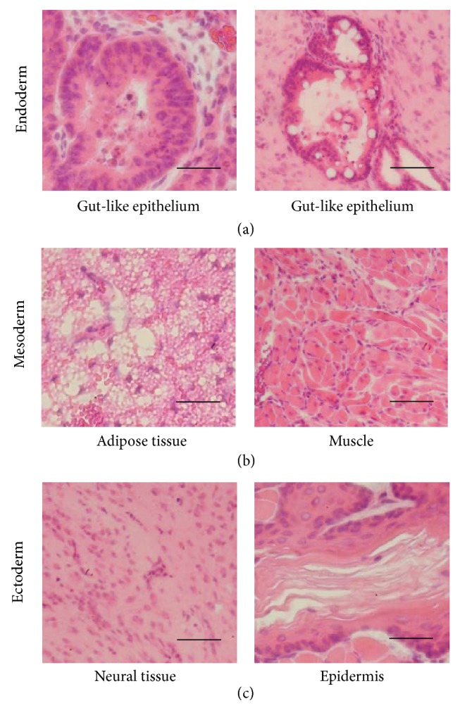
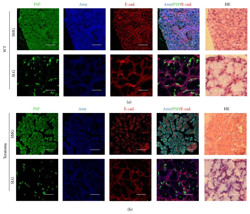
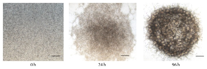
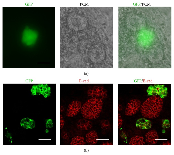
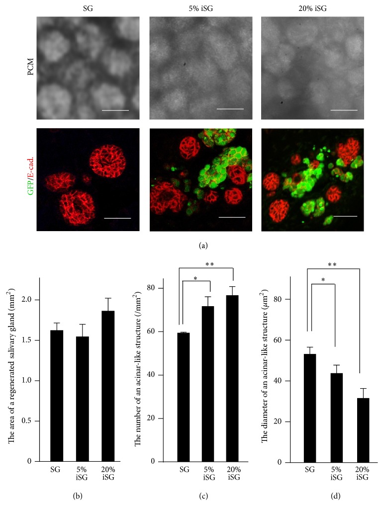
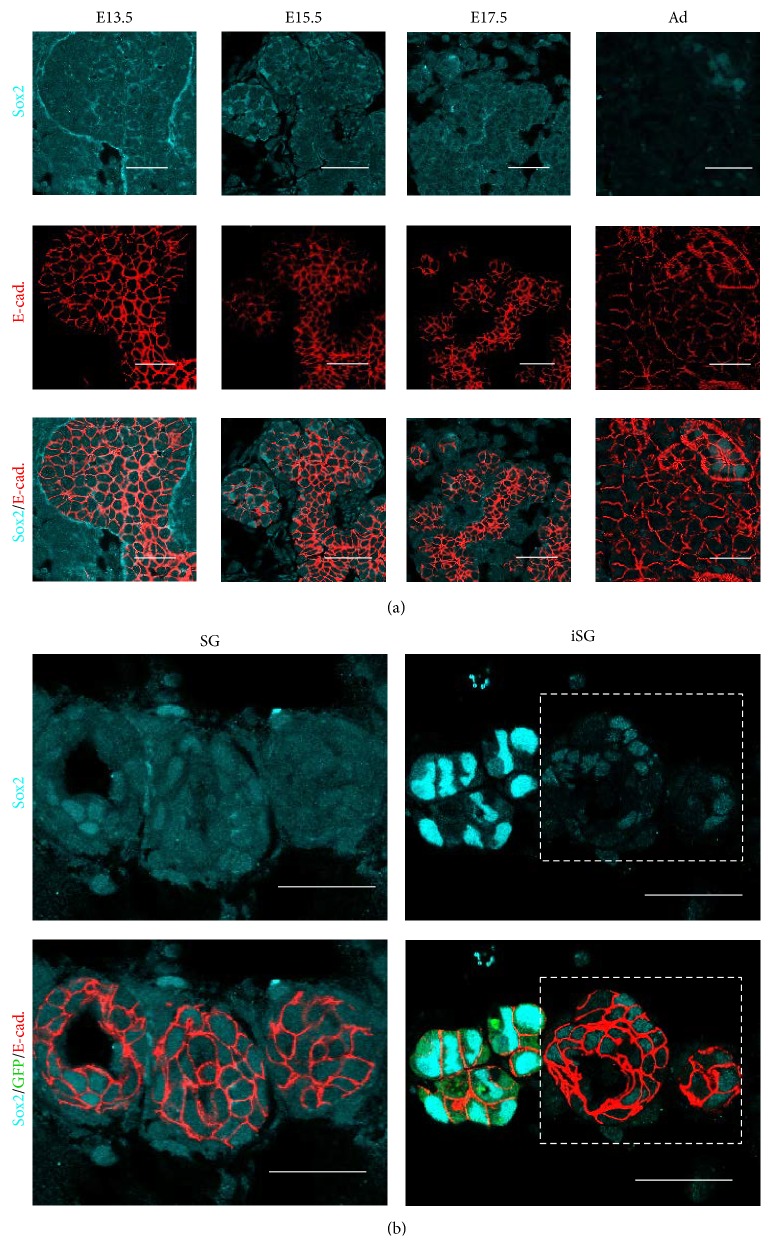
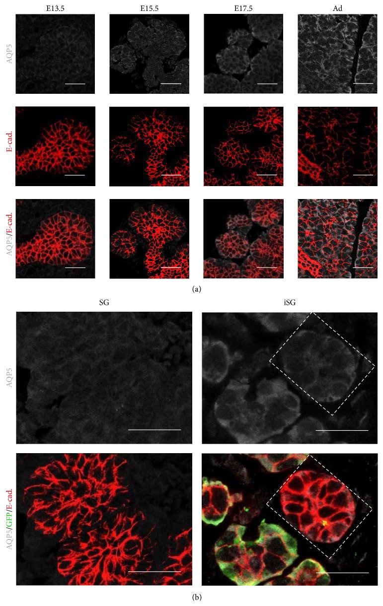
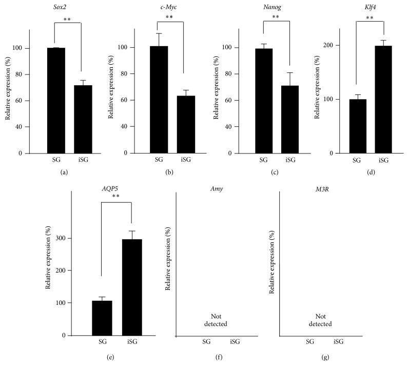
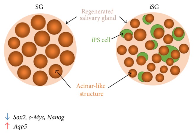
This report describes our initial attempt to regenerate salivary glands using induced pluripotent stem (iPS) cells in vivo and in vitro. Glandular tissues that were similar to the adult submandibular glands (SMGs) and sublingual glands could be partially produced by the transplantation of iPS cells into mouse salivary glands. However, the tumorigenicity of iPS cells has not been resolved yet. It is well known that stem cells affect their microenvironment, known as a stem cell niche. We focused on the niche and the interaction between iPS cells and salivary gland cells in our study on salivary gland regeneration. Coculture of embryonic SMG cells and iPS cells have better-developed epithelial structures and fewer undifferentiated specific markers than monoculture of embryonic SMG cells in vitro. These results suggest that iPS cells have a potential ability to accelerate differentiation for salivary gland development and regeneration.


Figures

Figure 1
Figure 1
Histological analysis of teratoma formation after transplantation of GFP-iPS cells. Images of sections of teratomas formed after transplantation of GFP-iPS cells into salivary glands of SCID mice. A teratoma formed from transplantation of 5.0 × 10⁵ GFP-iPS cells. H&E stained sections of a teratoma showing derivatives of all three germ layers, including gut-like epithelium tissues ((a) endoderm), adipose tissues and muscles ((b) mesoderm), and neural tissues and keratin-containing epidermal tissues ((c) ectoderm). Scale bars, 50 µm.
Figure 2
Figure 2
Histological analysis of salivary gland-like tissue in teratomas formed after transplantation of GFP-iPS cells. Immunofluorescence and H&E staining of sections showed submandibular glands (SMGs) and sublingual glands (SLGs) of adult mouse. PSP (green), Amy (blue), and E-cadherin (red) (a). Scale bars, 25 µm. Immunofluorescence and H&E staining of sections of GFP-iPS-grafted teratoma revealed salivary gland-like tissue (b). Scale bars, 25 µm.
Figure 3
Figure 3
Aggregation of SMG cells in SG. Phase-contrast image of regenerated SMG in organ culture for 0, 24, and 96 h. The regenerated SMG contained many acinar-like structures. Scale bar, 0.5 mm.
Figure 4
Figure 4
Aggregation of SMG cells in iSG. SMG cells and GFP-iPS cells were cocultured with DMEM/F12 for 96 h. Localization of GFP-iPS cells in iSG. iSG consists of 20% iPS cells and 80% SMG cells. Phase-contrast image and immunological stained image GFP-iPS (green) and pericentriolar material (PCM) (a). Scale bar, 25 µm. Immunofluorescence of paraffin-embedded tissue of regenerated salivary glands. GFP-iPS (green) and E-cadherin (red) (b). Scale bar, 25 µm.
Figure 5
Figure 5
Morphological analysis of regenerated SMGs (SG, 5% iSG, and 20% iSG). PCM and immunostained images indicated a reduction in size and an increase in the number of acinar-like structures in regenerated salivary glands (a). Size of whole regenerated salivary glands (n = 3) (b). Number of acinar-like structures in regenerated salivary glands (n = 3) (c). Size of acinar-like structures in regenerated salivary glands (n = 24) (d). p < 0.05; ∗∗ p < 0.01 versus control.
Figure 6
Figure 6
Distribution of Sox2 in developing SMG and in regenerated SMGs (SG and iSG). Sox2 is expressed in the cytoplasm of both epithelial cells and mesenchymal cells in E13.5, E15.5, and E17.5 developing SMGs and is gradually decreased in later embryonic stages. Sox2 is expressed in the nucleus of epithelial cells in both postnatal and adult SMGs. Sox2 (cyan) and E-cadherin (red) (a). Scale bar, 25 µm. Sox2 expression in the cytoplasm of both epithelial cells and mesenchymal cells in iSG (20% iSG) is less than that in SG. The nucleus of GFP-iPS cells highly expressed Sox2 as a positive control. Sox2 (cyan), GFP (green), and E-cadherin (red) (b). Scale bar, 25 µm.
Figure 7
Figure 7
Distribution of AQP5 in developing SMG and regenerated SMGs (SG and iSG). AQP5 is gradually increased in the epithelial cells of SMGs in E13.5, E15.5, E17.5, and adult. AQP5 is expressed in the cell membrane of epithelial cells in iSG (20% iSG) more than that in SG. AQP5 (white) and E-cadherin (red) (a). Scale bar, 25 µm. Distribution on AQP5 in regenerated SMG (b). AQP5 (white), GFP (green), and E-cadherin (red). Scale bar, 25 µm.
Figure 8
Figure 8
Gene expression of salivary gland cells in regenerated SMGs between SG and iSG. Gene expression of Sox2 (a), c-Myc (b), and Nanog (c) was decreased in iSG, and gene expression of Klf4 (d) was increased in iSG. Aqp5 (e) gene expression was increased in iSG, but Amy (f) and M3r (g) gene expression were not detected by qPCR, similar to early embryonic SMG cells. ∗∗ p < 0.01 versus control.
Figure 9
Figure 9
Effect of iPS cells on aggregation of SMG cells. iPS cells (green balls) reduced the size of the epithelial tissue, but acinar-like structure (orange balls) increased their number. iPS cells cannot mix completely with SMG cells and, instead, surround the epithelium of SMG. Stem cell markers, such as Sox2, c-Myc, and Nanog, are decreased, but Aqp5 is increased in iSG.

References

    1. Humphrey S. P., Williamson R. T. A review of saliva: normal composition, flow, and function. Journal of Prosthetic Dentistry. 2001;85(2):162–169. doi: 10.1067/mpr.2001.113778. - DOI - PubMed
    1. Jensen S. B., Pedersen A. M. L., Vissink A., et al. A systematic review of salivary gland hypofunction and xerostomia induced by cancer therapies: management strategies and economic impact. Supportive Care in Cancer. 2010;18(8):1061–1079. doi: 10.1007/s00520-010-0837-6. - DOI - PubMed
    1. Lim J.-Y., Ra J. C., Shin I. S., et al. Systemic transplantation of human adipose tissue-derived mesenchymal stem cells for the regeneration of irradiation-induced salivary gland damage. PLoS ONE. 2013;8(8) doi: 10.1371/journal.pone.0071167.e71167 - DOI - PMC - PubMed
    1. Takahashi K., Tanabe K., Ohnuki M., et al. Induction of pluripotent stem cells from adult human fibroblasts by defined factors. Cell. 2007;131(5):861–872. doi: 10.1016/j.cell.2007.11.019. - DOI - PubMed
    1. Kamao H., Mandai M., Okamoto S., et al. Characterization of human induced pluripotent stem cell-derived retinal pigment epithelium cell sheets aiming for clinical application. Stem Cell Reports. 2014;2(2):205–218. doi: 10.1016/j.stemcr.2013.12.007. - DOI - PMC - PubMed

Publication types

MeSH terms

LinkOut - more resources