

Review
doi: 10.1016/j.cell.2015.06.067.
Clarifying Tissue Clearing
Affiliations
- PMID: 26186186
- PMCID: PMC4537058
- DOI: 10.1016/j.cell.2015.06.067
Item in Clipboard
Review
Clarifying Tissue Clearing
Cell.
.
Abstract
Biological specimens are intrinsically three dimensional; however, because of the obscuring effects of light scatter, imaging deep into a tissue volume is problematic. Although efforts to eliminate the scatter by "clearing" the tissue have been ongoing for over a century, there have been a large number of recent innovations. This Review introduces the physical basis for light scatter in tissue, describes the mechanisms underlying various clearing techniques, and discusses several of the major advances in light microscopy for imaging cleared tissue.
Copyright © 2015 Elsevier Inc. All rights reserved.
Conflict of interest statement
The authors have no conflicts of interest to declare.
Figures
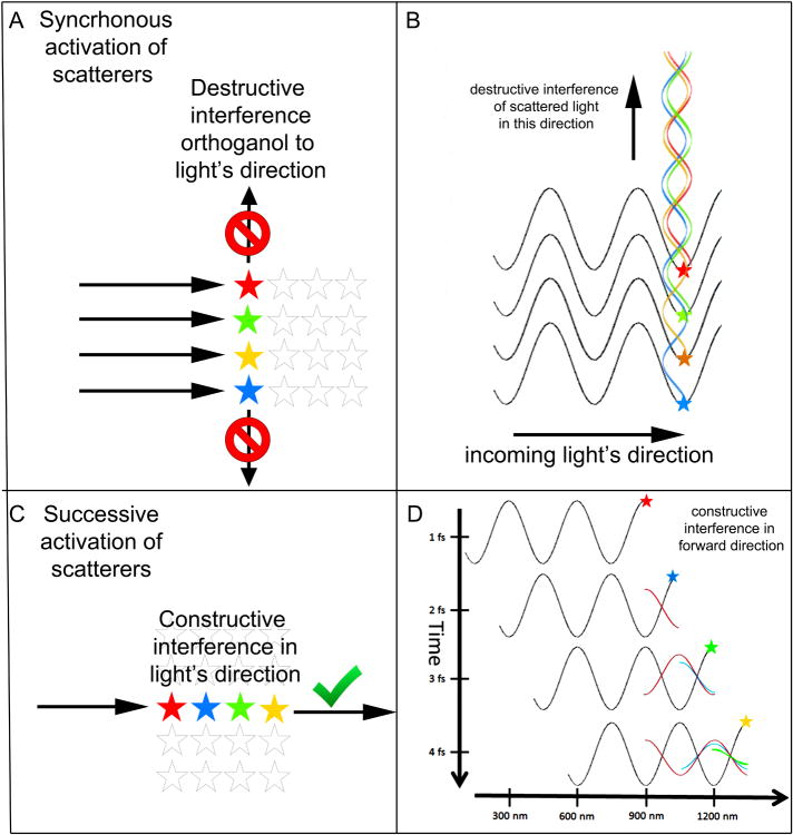
(A) In a material with a uniform density of scattering molecules (shown here as stars)
such as glass, water, or air, light is not scattered orthogonal to the direction of the
incoming light wave (arrows). In this situation all the scattering molecules that are
excited simultaneously (colored stars) are aligned orthogonal to the direction of the
plane wave. (B) A wave view to explain what is happening in panel A. Each of these
simultaneously excited molecules generate wave energy in all directions but by virtue of
their different positions there is always a molecule that is exactly ½ a
wavelength out of phase with every other molecule in the vertical direction causing
destructive interference. (C) The scatters that are aligned in the direction of the
incoming light wave (arrow) are activated sequentially (colored stars). Their excitations
sum constructively in the forward direction causing no attenuation of the light moving
through the medium. (D) A wave view to explain what is happening in panel C. Shown is the
same light wave a four successive time points 1 fs apart. At the first time point (top)
the wave imparts vibrational energy to a scattering molecule (red star). 1 fs later the
molecule emits the absorbed energy (red wave, 1/2 phase delay from incoming light). At
this time another molecule (blue star) is first excited by the incoming light wave. At 2
fs the molecule denoted by the blue star emits its scattered light (blue wave) that is in
phase with the scattered light from the red wave and a third molecule (green star) is
excited by the incoming light wave. At 3 fs the light from all three scatters are
vibrating in phase (superimposition of the red, blue and green waves) and another molecule
(yellow star) absorbs energy from the incoming light wave. In this way all forward moving
scattered light remains in phase causing no attenuation in light energy in this
direction.
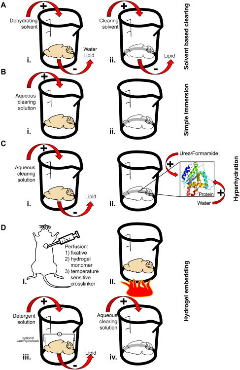
(A) Left, Solvent based clearing is a two-step process. First, the tissue is dehydrated
and lipid is removed. Second, the tissue is moved to a high refractive index solvent where
additional lipid solvation and clearing occurs. Right, Molecules commonly used for solvent
based clearing along with the refractive indices (RI) of the pure chemical. (B) Left, For
simple immersion, the tissue to be cleared is placed in an aqueous clearing solution for
days to months. During this time the solution is exchanged repeatedly. Right, Molecules
commonly used for simple immersion along with the refractive indices (RI) at the commonly
used concentration. (C) Hyperhydration involves submerging the sample in an aqueous
solution and allowing it to passively clear. During this clearing step, urea or formamide
in the clearing solution can enter tightly folded regions of high refractive index
proteins creating an osmotic gradient that pulls in water as well. This partially
denatures the protein, hydrates it and decreases its overall refractive index. Some
hyperhydration methods contain detergent which is used to disrupt membranes and remove
lipid from the sample. (D) Left, Hydrogel embedding is most often performed on an entire
animal by perfusing with a fixative, a temperature sensitive crosslinker, and the hydrogel
monomer. Alternatively, these chemicals can be passively diffused into an isolated tissue
sample. Once fixed, the tissue of interest is warmed to induce hydrogel crosslinking. The
sample is then placed in a detergent solution to remove lipid material passively or via an
electrophoretic charge. Finally the lipid-free sample is placed in a high-refractive index
matching solution for clearing. Histodenz is one high refractive index molecule that can
be a component of this clearing solution. Glycerol, TDE or Diatrizoic acid also can play
this role.
References
-
- Alnuami AA, Zeedi B, Qadri SM, Ashraf SS. Oxyradical-induced GFP damage and loss of fluorescence. International journal of biological macromolecules. 2008;43:182–186. - PubMed
-
- Booth MJ. Adaptive optics in microscopy. Philosophical transactions Series A, Mathematical, physical, and engineering sciences. 2007;365:2829–2843. - PubMed
Publication types
MeSH terms
Grants and funding
LinkOut - more resources
Full Text Sources
Other Literature Sources
