Cyclophosphamide-Induced Morphological Changes in Dental Root Development of ICR Mice
- PMID: 26186337
- PMCID: PMC4506128
- DOI: 10.1371/journal.pone.0133256
Cyclophosphamide-Induced Morphological Changes in Dental Root Development of ICR Mice
Abstract
Background: Survivors of childhood cancer are at risk of late dental development. Cyclophosphamide is one of the most commonly used chemotherapeutic agents against cancer in children. The aim of this study was to investigate the effects of cyclophosphamide on root formation in the molars of growing mice and to assess the morphological changes in these roots using three-dimensional structural images.
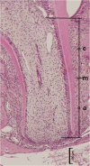
Methods: We treated 16 12-day-old ICR mice with cyclophosphamide (100 mg/kg, i.p.) and 16 control mice with saline. At 16, 20, 24, and 27 days of age, the mandibular left first molars were scanned using soft micro-computed tomography. After scanning, the structural indices were calculated using a three-dimensional image analysis system, and the images were subjected to three-dimensional reconstruction. The length and apical foramen area of all distal roots were assessed. Histological changes in the apical region were then assessed via hematoxylin and eosin staining.
Results: The mandibular molars of all experimental mice showed evidence of cytotoxic injury, which appeared in the form of anomalous root shapes. Although all roots developed further after cyclophosphamide injection, the three-dimensional structural images showed that the roots in the experimental group tended to develop more slowly and were shorter than those in the control group. At 27 days of age, the mean root length was shorter in the experimental group than in the control group. Conversely, the apical foramen of the roots in the experimental group tended to close faster than that of roots in the control group. In addition, hematoxylin and eosin staining of the distal roots in the experimental group showed increased dentin thickness in the apical region.
Conclusion: Our results suggest that cyclophosphamide can result in short root lengths and early apical foramen closure, eventually leading to V-shaped or thin roots.
Conflict of interest statement
Figures


References
-
- Maeda M. Late effects of childhood cancer: life-threatening issues. J Nippon Med Sch. 2008;75: 320–324. - PubMed
-
- Mertens AC, Yong J, Dietz AC, Kreiter E, Yasui Y, Bleyer A, et al. Conditional survival in pediatric malignancies: analysis of data from the Childhood Cancer Survivor Study and the Surveillance, Epidemiology, and End Results Program. Cancer. 2015;121: 1108–1117. 10.1002/cncr.29170 - DOI - PMC - PubMed
-
- Sonis AL, Tarbell N, Valachovic RW, Gelber R, Schwenn M, Sallan S. Dentofacial development in long-term survivors of acute lymphoblastic leukemia. A comparison of three treatment modalities. Cancer. 1990;66: 2645–2652. - PubMed
Publication types
MeSH terms
Substances
LinkOut - more resources
Full Text Sources
Other Literature Sources

