. 2015 Sep;75(1):38-50.
doi: 10.1016/j.cyto.2015.05.023. Epub 2015 Jul 14.

Commentary: IL-4 and IL-13 receptors and signaling

Affiliations

Commentary: IL-4 and IL-13 receptors and signaling

Sarah M McCormick et al. Cytokine. 2015 Sep.

Abstract

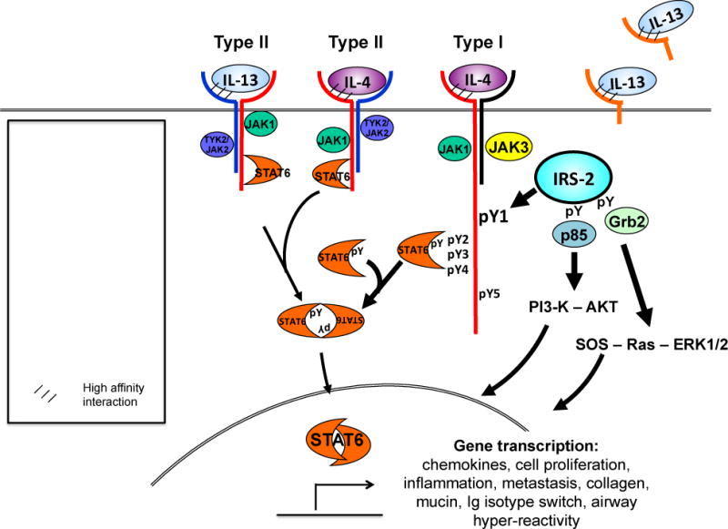
Interleukin (IL)-4 and IL-13 were discovered approximately 30years ago and were immediately linked to allergy and atopic diseases. Since then, new roles for IL-4 and IL-13 and their receptors in normal gestation, fetal development and neurological function and in the pathogenesis of cancer and fibrosis have been appreciated. Studying IL-4/-13 and their receptors has revealed important clues about cytokine biology and led to the development of numerous experimental therapeutics. Here we aim to highlight new discoveries and consolidate concepts in the field of IL-4 and IL-13 structure, receptor regulation, signaling and experimental therapeutics.

Keywords: Interleukin-13; Interleukin-4; Receptor; Regulation; Singling.


Figures

Figure 1
Figure 1. IL-4 and IL-13 receptor structure
IL-4 signals through two possible receptor complexes composed of a heterodimer of the IL-4Rα (140 kDa) and γc chain (60 kDa); type I receptor or the IL-4Rα and IL-13Rα1 (65–70 kDa) chain; type II receptor. IL-4 binds IL-4Rα with high affinity, triggering dimerization with the secondary signaling chain. IL-13 binds IL-13Rα1 which complexes with the IL-4Rα, forming the type II receptor. Signaling through the type I receptor leads to activation JAKs and downstream signaling adaptor molecules STAT6 and IRS-2, whereas signaling through the type II receptor predominantly activates STAT6. IL-4 signaling also activates PI3-K and AKT. IL-13 also binds cell-surface and soluble forms of the IL-13Rα2, this so-called inhibitory subunit binds IL-13 with greater affinity than IL-13Rα1 and acts as a “cytokine sink”.
Figure 2
Figure 2. Regulation of signaling receptors
IL-4Rα expression is regulated in the steady state by Hepatocyte growth factor-regulated tyrosine substrate (Hrs) (A). Hrs binding targets the IL-4Rα to the late endosome for degradation. Downregulation of IL-4Rα occurs through STUB1 binding the cytoplasmic domain of the receptor through Hsp70 (B). Subsequent ubiquitination and degradation the IL-4Rα reduces cell surface expression and cellular responsiveness to IL-4. The IL-13Rα2 acts as a negative regulator of IL-4- but not IL-13-induced signaling through the type II IL-4 receptor (C). The IL-13Rα2 acts as a negative regulator of IL-4- but not IL-13-induced signaling through the type II IL-4 receptor although the precise mechanisms of this inhibition are not understood. IL-13Rα2 expression is induced by TNFα in fibroblasts and leads to AP-1 driven pro-fibrotic reprogramming (D). YKL40 binds binds to the IL-13Rα2 chain leading to serine 473 phosphorylation in addition to triggering ERK signaling (E). AKT phosphorylation following binding of YKL40 to IL-13Rα2 activates a myriad of signaling pathways including FoxO1/3 and the Wnt/b-catenin pathway. Hepatocyte growth factor-regulated tyrosine substrate, Hrs: STIP1 homology and U-Box containing protein 1, STUB1; heat shock protein 70, Hsp70; forkhead box protein O, FoxO; transforming growth factor-β, TGF-β; activator protein-1, AP-1.
Figure 3
Figure 3. Regulation of signaling in response to IL-4 and IL-13
Engagement of the type I IL-4 receptor by IL-4 brings about tyrosine phosphorylation of the associated JAKs, key tyrosine residues (pY1-5) in the cytoplasmic domain in IL-4Ra, STAT6 and IRS-2. STAT6 is dephosphorylated by SHP1, a protein tyrosine phosphatase (A). De-phosphorylation of STAT6 is critical to limit autoimmune and Th2 allergic inflammation, mast cell, B cell and epithelial cell activation. IRS-2 binds the regulatory p85 subunit of PI3-K, activating the enzyme to generate PIP3. In turn, PIP3 activates PDK1 and mTORC2. The proteins comprising the two mTOR complexes 1 and 2 are shown (B). TORC2 kinase phosphorylates AKT on serine 473 and to SGK1 in CD4+ T cells, leading to Th2 polarization. AKT inactivation of the Tsc1/2 complex prevents hydrolysis of GTP bound to Rheb. GTP-bound Rheb activates mTORC1, triggering a negative feedback loop via multiple pathways on IL-4-activated IRS-2/PI3-K/AKT signaling (C). The composition of the mTORC1 complex is shown. The mTORC1 complex also regulates macrophage polarization, as Raptor-deficient macrophages have enhanced M1 and M2 responses. The mTORC1 and mTORC2 complexes also regulate by unknown mechanisms, the polarization of T cells [adapted from (170)]. An additional negative regulatory pathway triggered by IL-4 is the induction of the SOCS proteins. SOCS1 binds IRS-2 and targets it for ubiquitin-mediated degradation (D). Phosphotyrosine, pY; phosphatidylinositol-4,5-bisphosphate 3-kinase, PI3-K; phosphatidylinositol (3,4,5)-trisphosphate, PIP3; 3-phosphoinositide dependent protein kinase-1, PDK1; mTOR complex 2, mTORC2; serum and glucocorticoid-inducible kinase 1, SGK1; Ras homolog enriched in brain, RHEB; tuberous sclerosis complex 1, TSC1; regulatory-associated protein of mTOR, Raptor; mammalian lethal with Sec13 protein 8, mLST8; proline-rich Akt substrate of 40 kDa, PRAS40; DEP-domain-containing mTOR-interacting protein, DEPTOR; rapamycin-insensitive companion of TOR, RICTOR; mammalian stress-activated protein kinase-interacting protein 1, mSIN1; protein observed with RICTOR, PROTOR; suppressor of cytokine signaling 1, SOCS1.

References

    1. Gadani SP, Cronk JC, Norris GT, Kipnis J. Interleukin-4: A Cytokine to Remember. Journal of immunology (Baltimore, Md. : 1950) 2012;189:4213–4219. - PMC - PubMed
    1. McKenzie Andrew NJ, Spits H, Eberl G. Innate Lymphoid Cells in Inflammation and Immunity. Immunity. 2014;41:366–374. - PubMed
    1. Hams E, Fallon PG. Innate type 2 cells and asthma. Current Opinion in Pharmacology. 2012;12:503–509. - PubMed
    1. Junttila IS, Creusot RJ, Moraga I, Bates DL, Wong MT, Alonso MN, Suhoski MM, Lupardus P, Meier-Schellersheim M, Engleman EG, Utz PJ, Fathman CG, Paul WE, Garcia KC. Redirecting cell-type specific cytokine responses with engineered interleukin-4 superkines. Nat Chem Biol. 2012;8:990–998. - PMC - PubMed
    1. Heller NM, Dasgupta P, Dorsey NJ, Chapoval SP, Keegan AD. The Type I and Type II Receptor Complexes for IL-4 and IL-13 Differentially Regulate Allergic Lung Inflammation, Allergic Diseases. Pereira C, editor. Highlights in the Clinic, Mechanisms and Treatment. 2012 http://www.intechopen.com/books/allergic-diseases-highlights-in-the-clin....

MeSH terms