. 2015 Sep 4;290(36):21845-56.
doi: 10.1074/jbc.M115.676221. Epub 2015 Jul 17.

Zeta Inhibitory Peptide Disrupts Electrostatic Interactions That Maintain Atypical Protein Kinase C in Its Active Conformation on the Scaffold p62

Affiliations

Zeta Inhibitory Peptide Disrupts Electrostatic Interactions That Maintain Atypical Protein Kinase C in Its Active Conformation on the Scaffold p62

Li-Chun Lisa Tsai et al. J Biol Chem. .

Abstract

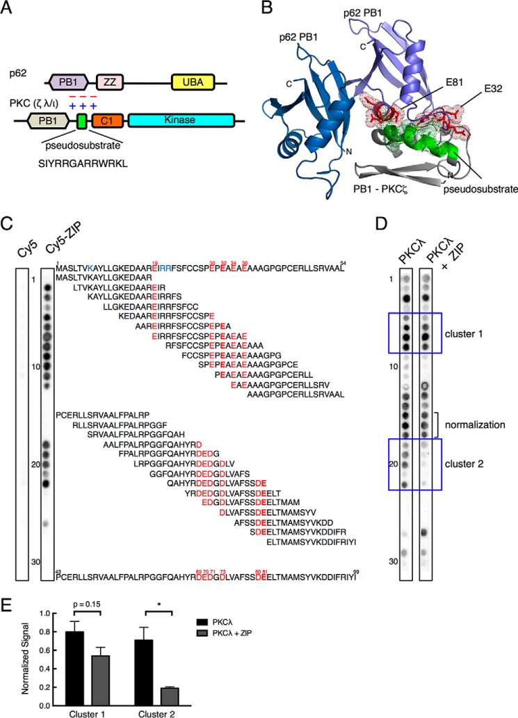
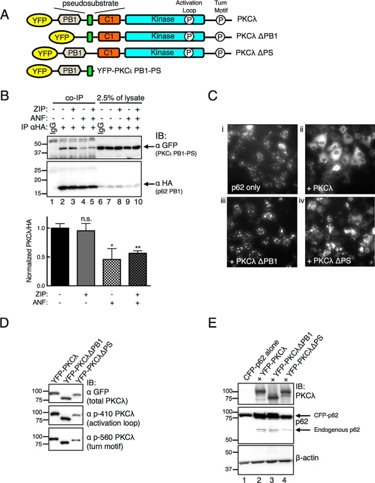
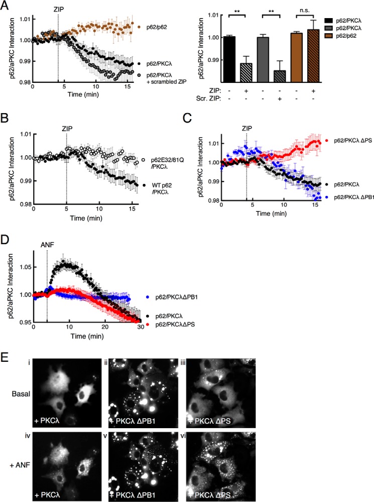
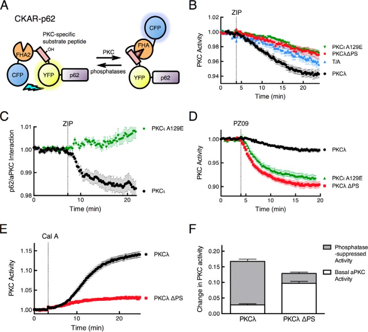
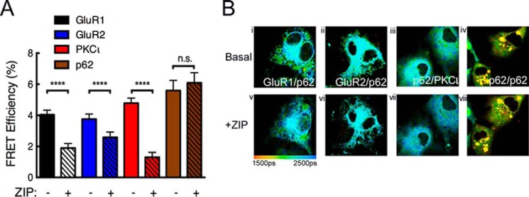
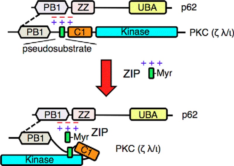
Atypical protein kinase C (aPKC) enzymes signal on protein scaffolds, yet how they are maintained in an active conformation on scaffolds is unclear. A myristoylated peptide based on the autoinhibitory pseudosubstrate fragment of the atypical PKCζ, zeta inhibitory peptide (ZIP), has been extensively used to inhibit aPKC activity; however, we have previously shown that ZIP does not inhibit the catalytic activity of aPKC isozymes in cells (Wu-Zhang, A. X., Schramm, C. L., Nabavi, S., Malinow, R., and Newton, A. C. (2012) J. Biol. Chem. 287, 12879-12885). Here we sought to identify a bona fide target of ZIP and, in so doing, unveiled a novel mechanism by which aPKCs are maintained in an active conformation on a protein scaffold. Specifically, we used protein-protein interaction network analysis, structural modeling, and protein-protein docking to predict that ZIP binds an acidic surface on the Phox and Bem1 (PB1) domain of p62, an interaction validated by peptide array analysis. Using a genetically encoded reporter for PKC activity fused to the p62 scaffold, we show that ZIP inhibits the activity of wild-type aPKC, but not a construct lacking the pseudosubstrate. These data support a model in which the pseudosubstrate of aPKCs is tethered to the acidic surface on p62, locking aPKC in an open, signaling-competent conformation. ZIP competes for binding to the acidic surface, resulting in displacement of the pseudosubstrate of aPKC and re-engagement in the substrate-binding cavity. This study not only identifies a cellular target for ZIP, but also unveils a novel mechanism by which scaffolded aPKC is maintained in an active conformation.

Keywords: atypical protein kinase C (aPKC); enzyme mechanism; p62 (sequestosome 1(SQSTM1)); protein-protein interaction; scaffold protein; serine/threonine protein kinase.


Figures

FIGURE 1.
FIGURE 1.
The basic pseudosubstrate of aPKC interacts with an acidic surface on the PB1 domain of the scaffold p62. A, schematic representation showing the domain structure of atypical PKC members (PKCλ/ι and PKCζ) and p62, each of which contains a PB1 domain (lavender and tan, respectively). aPKCs also have a pseudosubstrate segment (green rectangle; sequence indicated below) immediately followed by a C1 domain (orange) and a kinase domain (cyan). p62 also has a ZZ domain (pink) and ubiquitin-associated (UBA) domain (yellow). B, structural modeling showing predicted binding pose of PKCζ pseudosubstrate to a PB1 domain dimer of p62 (rat; Protein Data Bank ID 2KTR). A fragment of PKCζ sequence (residues 79–146) containing the pseudosubstrate segment (green helix) flanked by a partial C1 domain and a partial PB1 (gray) was docked onto the structure of a PB1 dimer of p62 (blue and purple). The positively charged Arg and Lys residues (shown in dot representation) within the pseudosubstrate are predicted to form an electrostatic interaction interface with the negatively charged Glu and Asp residues of the p62 PB1 (shown in red stick and dot representation); these residues include Glu81 and Glu32 on two separate segments of the p62 PB1 domain. C, peptide overlay of the p62 PB1 domain with Cy5-ZIP or Cy5 and PKCλ. Overlapping 18-mer peptides covering the PB1 domain of p62 (residues 1–105) and staggered by three amino acids were overlaid with 5 μm Cy5 or Cy5-ZIP, and binding was detected by a FluorChem Q imaging system. The sequence of residues 1–54 is shown above and that for residues 43–99 below the array. Glu residues present in the spots to which ZIP bound are indicated in red; Glu32 and Glu81 are indicated in bold. Lys residues known to bind the PB1 domain of p62 are colored in blue. Spots are numbered on the left. D, peptide overlay of the p62 PB1 domain with PKCλ (27 nm) and Cy5 or PKCλ and Cy5-ZIP (5 μm). PKCλ binding to the array was detected with an anti-PKCλ antibody. Spots in clusters 1 and 2 (blue boxes) were quantified in E. The graph presents the mean ± S.E. of three separate strips. The data were analyzed by non-paired Student's t test: *, p < 0.05.
FIGURE 2.
FIGURE 2.
Atypical PKC physically interacts with scaffold p62. A, schematic of YFP-tagged PKCλ constructs used in this study: PKCλ with PB1 domain deleted (PKCλ ΔPB1), PKCλ with pseudosubstrate deleted (PKCλ ΔPS), and N-terminal portion of PKCι containing PB1 domain through pseudosubstrate (PKCι PB1-PS). Indicated are the phosphorylation sites (P) at the activation loop and turn motif sites; occupancy of these sites serves as a measure for proper folding of the enzyme (see Refs. and 6). B, COS-7 cells co-expressing YFP-tagged PKCι PB1-PS and HA-tagged p62 PB1 were treated with 5 μm ZIP for 20 min and/or 10 μm ANF for 1 h before lysing. Immunoprecipitation (IP) was performed with a HA monoclonal antibody or IgG control. Immunoprecipitates (entire sample) and 2.5% of total lysates were analyzed by Western blots (IB) probing for a GFP antibody to detect PKCι and a HA antibody to detect the PB1 domain of p62. The PKCι PB1-PS detected in the immunoprecipitates was normalized to the HA signal in the immunopellet and quantified in the graph below. Data represent the mean ± S.E. of 6 independent experiments and were analyzed by one-way analysis of variance with Holm-Šídák multiple comparison test: n.s., no significance; *, p < 0.05; **, p < 0.01. C, fluorescence micrographs of COS-7 cells showing the localization of CFP-tagged p62 expressed alone (panel i) or with PKCλ (panel ii), PKCλ ΔPB1 (panel iii) or PKCλ ΔPS (panel iv). Images were adjusted for an optimal dynamic range using ImageJ. D, Western blot analysis of YFP-tagged constructs showing immunoreactivity with antibodies to the activation loop (Thr410) and turn motif sites (Thr560) as well as total GFP to detect the YFP-tagged PKCλ. p-410 PKCλ, phospho-PKCλ (Thr410); p-560 PKCλ, phospho-PKCλ (Thr560). E, Western blot of COS-7 cells expressing the constructs in panel C and probed with antibodies to PKCλ, p62, and β-actin.
FIGURE 3.
FIGURE 3.
ZIP disruption of p62/aPKC complex requires an intact pseudosubstrate segment. A, the ability of ZIP to disrupt the p62/PKCλ complex was assessed by monitoring real-time FRET between fluorescent pair CFP and YFP using live cell imaging. The FRET/CFP ratio of COS-7 cells co-expressing CFP-p62 and YFP-PKCλ (black circles, n = 26) or YFP-p62 as control (brown circles, n = 13) was monitored before and after treatment with 5 μm ZIP or scrambled ZIP (Scr. ZIP) (gray circles, n = 24). Data were plotted as a p62-aPKC interaction and represent the normalized FRET/CFP ratio mean ± S.E. of the indicated number of cells from at least 3 independent experiments. The graph shows the ratio values immediately prior to ZIP treatment (basal) and 10 min past treatment (+ ZIP) analyzed by paired Student's t test: n.s., no significance; *, p < 0.05; **, p < 0.01. B, FRET/CFP ratio changes were monitored as in panel A for COS-7 cells co-expressing YFP-PKCλ and either CFP-tagged wild-type p62 (black circles, n = 26) or a construct in which Glu32 and Glu81 were mutated to Gln (p62E32/81Q; open circles, n = 24); cells were treated with 5 μm ZIP. C, FRET/CFP ratios were monitored for COS-7 cells expressing CFP-p62 and YFP-PKCλ ΔPS (red circles, n = 31), PKCλ ΔPB1 (blue circles, n = 12), or full-length YFP-PKCλ (black circles, n = 26). D, FRET/CFP ratios were measured for COS-7 cells expressing CFP-p62 and YFP-PKCλ (black circles, n = 15), YFP-PKCλ ΔPS (red circles, n = 16), or CFP-PKCλ ΔPB1 (blue circles, n = 15) before and after treatment with 10 μm ANF. E, fluorescence micrographs of COS-7 cells showing the localization of CFP-tagged p62 co-expressed with PKCλ (panels i and iv), PKCλ ΔPB1 (panels ii and v), or PKCλ ΔPS (panels iii and vi) before or after 1 h ANF treatment.
FIGURE 4.
FIGURE 4.
ZIP releases the pseudosubstrate of PKCλ from p62 to allow intramolecular autoinhibition of the kinase. A, schematic showing fusion of the PKC activity reporter CKAR to the N terminus of p62. Phosphorylation causes an intramolecular clamp of the phosphorylated segment (pink) with a phospho-peptide-binding FHA2 module (orange) that results in a decrease in FRET between the flanking CFP (blue) and YFP (yellow) (see Ref. for more details). B, COS-7 cells co-expressing CKAR-p62 and mCherry-tagged wild-type PKCλ (black circles, n = 44), PKCλ ΔPS (red squares, n = 19), or PKCι A129E (green triangles, n = 41) were pretreated with 1 μm Gö6983, an inhibitor of conventional and novel PKC isozymes, and then monitored for CFP/FRET ratio changes following 5 μm ZIP treatment. The control CKAR-p62 T/A (blue triangles, n = 23), where its phospho-acceptor Thr was mutated to Ala, was also examined under the same experimental conditions. Data were plotted as PKC activity and represent the normalized FRET/CFP ratio mean ± S.E. of the indicated number of cells from at least 3 independent experiments. C, the FRET/CFP ratio of COS-7 cells co-expressing CFP-p62 and YFP-PKCι (black circles, n = 31) or YFP-PKCι A129E (green diamonds, n = 28) was monitored before and after 5 μm ZIP treatment. Data were plotted as a p62-aPKC interaction and represent the normalized FRET/CFP ratio mean ± S.E. of the indicated number of cells from at least 3 independent experiments. D, COS-7 cells co-expressing CKAR-p62 and mCherry-tagged wild-type PKCλ (black circles, n = 28), PKCλ ΔPS (red squares, n = 30), or PKCι A129E (green triangles, n = 18) were monitored for CFP/FRET ratio changes before and after treatment with 5 μm PZ09, an atypical PKC-selective inhibitor (48). E, COS-7 cells co-expressing CKAR-p62 and either mCherry-tagged wild-type PKCλ (black circles, n = 33) or PKCλ ΔPS (red squares, n = 38) were monitored for CFP/FRET ratio changes when treated with 50 nm calyculin A, a Ser/Thr protein phosphatase inhibitor (56). Data were plotted as PKC activity and represent the normalized FRET/CFP ratio mean ± S.E. of the indicated number of cells from at least 3 independent experiments. F, quantification of basal and phosphatase-suppressed activities of PKCλ and PKCλ ΔPS was calculated as a fraction of FRET ratio change. The data were extrapolated from the plateau portion of the curves in panels D and E, 20 min after the addition of PZ09 or calyculin A.
FIGURE 5.
FIGURE 5.
ZIP targets the association of GluR1 or GluR2 with the p62 scaffold. A, FRET efficiency was measured from COS-7 cells co-expressing mCherry-p62 and GFP-tagged GluR1 (white bars, n = 114), GluR2 (blue bars, n = 167), PKCι (red bars, n = 94), or p62 (brown bars, n = 53), both before and after 5 μm ZIP treatment. Solid bars represent before ZIP treatment, and bars with black slashes indicate FRET efficiency of post-ZIP treatment. The data were analyzed by paired Student's t test: n.s., no significance; ****, p < 0.0001. B, representative fluorescence lifetime images of COS-7 cells expressing the indicated constructs before and after ZIP treatment. Pseudocolor scale indicates GFP lifetime at each pixel.
FIGURE 6.
FIGURE 6.
The acidic surface of p62 tethers the pseudosubstrate segment of aPKCs to maintain its active conformation. The schematic shows the canonical interaction of the PB1 domain of aPKCs with that of p62 (dotted line) and the previously undescribed interaction between the basic pseudosubstrate and acidic surface on p62. This interaction tethers the pseudosubstrate out of the substrate-binding cavity of aPKCs, allowing constitutive activity on the scaffold. The positively charged peptide ZIP competes with the pseudosubstrate for binding to the acidic surface of p62, displacing the pseudosubstrate and allowing it to re-engage in the substrate-binding site of aPKC to affect intramolecular autoinhibition of the kinase. UBA, ubiquitin-associated domain; Myr, myristoylated peptide.

Similar articles

Cited by

References

    1. Wu-Zhang A. X., Schramm C. L., Nabavi S., Malinow R., Newton A. C. (2012) Cellular pharmacology of protein kinase Mζ (PKMζ) contrasts with its in vitro profile: implications for PKMζ as a mediator of memory. J. Biol. Chem. 287, 12879–12885 - PMC - PubMed
    1. Langeberg L. K., Scott J. D. (2015) Signalling scaffolds and local organization of cellular behaviour. Nat. Rev. Mol. Cell Biol. 16, 232–244 - PMC - PubMed
    1. Rosse C., Linch M., Kermorgant S., Cameron A. J., Boeckeler K., Parker P. J. (2010) PKC and the control of localized signal dynamics. Nat. Rev. Mol. Cell Biol. 11, 103–112 - PubMed
    1. Antal C. E., Newton A. C. (2013) Spatiotemporal dynamics of phosphorylation in lipid second messenger signaling. Mol. Cell. Proteomics 12, 3498–3508 - PMC - PubMed
    1. Newton A. C. (2010) Protein kinase C: poised to signal. Am. J. Physiol. Endocrinol. Metab. 298, E395–E402 - PMC - PubMed

Publication types

MeSH terms

Associated data