Review
. 2015 Aug 28;290(35):21244-51.
doi: 10.1074/jbc.R115.675165. Epub 2015 Jul 17.

Regulating the Regulators: Recent Revelations in the Control of E3 Ubiquitin Ligases

Affiliations
Review

Regulating the Regulators: Recent Revelations in the Control of E3 Ubiquitin Ligases

Vinayak Vittal et al. J Biol Chem. .

Abstract

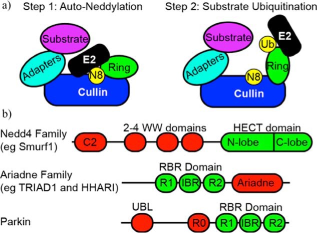
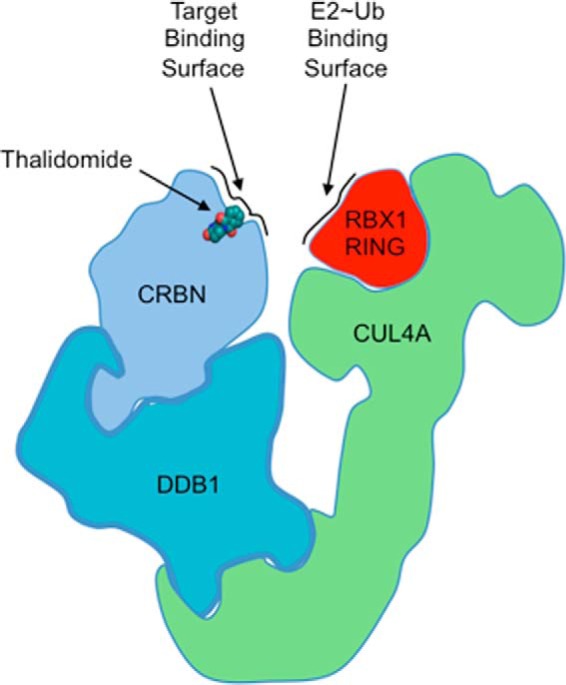
Since its discovery as a post-translational signal for protein degradation, our understanding of ubiquitin (Ub) has vastly evolved. Today, we recognize that the role of Ub signaling is expansive and encompasses diverse processes including cell division, the DNA damage response, cellular immune signaling, and even organismal development. With such a wide range of functions comes a wide range of regulatory mechanisms that control the activity of the ubiquitylation machinery. Ub attachment to substrates occurs through the sequential action of three classes of enzymes, E1s, E2s, and E3s. In humans, there are 2 E1s, ∼ 35 E2s, and hundreds of E3s that work to attach Ub to thousands of cellular substrates. Regulation of ubiquitylation can occur at each stage of the stepwise Ub transfer process, and substrates can also impact their own modification. Recent studies have revealed elegant mechanisms that have evolved to control the activity of the enzymes involved. In this minireview, we highlight recent discoveries that define some of the various mechanisms by which the activities of E3-Ub ligases are regulated.

Keywords: E3 ubiquitin ligase; HECT; RING; cullin; parkin; ubiquitin; ubiquitin ligase; ubiquitin regulation; ubiquitylation (ubiquitination).


Figures

FIGURE 1.
FIGURE 1.
Domain architecture of regulated E3 ligases. a, left, schematic of a cullin ligase bound to an E2∼Nedd8 conjugate. The RING domain of the cullin is positioned to allow specific neddylation. Right, upon neddylation, the RING adopts a new conformation that allows for interaction with an incoming E2∼Ub conjugate that can facilitate substrate ubiquitylation. b, domain architecture of the Nedd4, Ariadne family, and Parkin E3 ligases. IBR, in between ring domain.
FIGURE 2.
FIGURE 2.
Ubiquitin regulation by supramolecular assembly. a, EM structure (Protein Data Bank (PDB) 3J92) of the 60S ribosome subunit bound to peptidyl-tRNA (orange), NEMF (blue), and Listerin (red). The C-terminal region of Listerin, and specifically the RING domain, is positioned just outside the exit tunnel (orange) to facilitate ubiquitinylation of the nascent chain. The RWD domain of Listerin binds to one interaction surface of the 60S ribosome. NEMF directly interacts with the peptidyl-tRNA and the N terminus of Listerin to help anchor the RQC and prevent reassembly with the 40S subunit. b, structure of the PRC1-nucleosome complex (PDB 4R8P). The members of the PRC1 complex, UbcH5c (green), RING1a (blue), and Bmi1 (gray), sit atop the nucleosome, positioning the active site of the E2 adjacent to Lys-119 of histone H2A (orange). Other histone components, histone H2B 1.1, (yellow), histone H3.2 (dark pink), and histone H4 (maroon), play roles in positioning the complex for proper ubiquitylation. c, the inactive RING domain of Bmi1 makes direct contacts with histones H3.2 and H4 that also stabilize the interaction between PRC1 and the nucleosome. Side chain residues of Lys-62 and Arg-64 hydrogen-bond with Glu-74 in histone H4 and Asp-77 of H3.2, respectively. d, β-sheet residues in UbcH5c that are distant from the active site play a role in positioning the E2 on the NCP. Side chain residues of His-32 and Lys-66 make hydrogen-bonding contacts with backbone phosphate groups from the nucleosome DNA.
FIGURE 3.
FIGURE 3.
Architectural schematic of the putative CUL4, RBX1, DDB1, CRBN complex (CRL4CRBN) bound to thalidomide (PDB 4C1I for DDB1-CRBNThalidomide complex). Thalidomide occupies a position in the putative CRBN-substrate interface and can positively or negatively modulate target substrate interactions. CRBN sits adjacent to RBX1, bringing E2∼Ub conjugates and substrate together.
FIGURE 4.
FIGURE 4.
An allosteric switch: the structure of the RNF146-UbcH5a complex. a, left, selected member of the solution ensemble of the RNF146 RING domain showing the E2 binding-incompetent state (PDB 2D8T). Right, crystal structure of the RNF146 RING-WWE domains bound to iso-ADPR (PDB 4QPL). Iso-ADPR binds between the RING and WWE domains. Binding of iso-ADPR results in the repositioning of Trp-66 and the extension of the helical region of the RING domain. b, UbcH5a binds to the RING domain of RNF146 when iso-ADPR binds between the RING and WWE domains. Displacement of Trp-66 allows for the interaction between RING and E2.

References

    1. Berndsen C. E., Wolberger C. (2014) New insights into ubiquitin E3 ligase mechanism. Nat. Struct. Mol. Biol. 21, 301–307 - PubMed
    1. Mattiroli F., Sixma T. K. (2014) Lysine-targeting specificity in ubiquitin and ubiquitin-like modification pathways. Nat. Struct. Mol. Biol. 21, 308–316 - PubMed
    1. Metzger M. B., Pruneda J. N., Klevit R. E., Weissman A. M. (2014) RING-type E3 ligases: master manipulators of E2 ubiquitin-conjugating enzymes and ubiquitination. Biochim. Biophys. Acta 1843, 47–60 - PMC - PubMed
    1. Pruneda J. N., Littlefield P. J., Soss S. E., Nordquist K. A., Chazin W. J., Brzovic P. S., Klevit R. E. (2012) Structure of an E3:E2∼Ub complex reveals an allosteric mechanism shared among RING/U-box ligases. Mol. Cell 47, 933–942 - PMC - PubMed
    1. Plechanovová A., Jaffray E. G., Tatham M. H., Naismith J. H., Hay R. T. (2012) Structure of a RING E3 ligase and ubiquitin-loaded E2 primed for catalysis. Nature 489, 115–120 - PMC - PubMed

Publication types

LinkOut - more resources