. 2015 Jul 18:13:52.
doi: 10.1186/s12915-015-0162-0.

Macrolides rapidly inhibit red blood cell invasion by the human malaria parasite, Plasmodium falciparum

Affiliations

Macrolides rapidly inhibit red blood cell invasion by the human malaria parasite, Plasmodium falciparum

Danny W Wilson et al. BMC Biol. .

Abstract

Background: Malaria invasion of red blood cells involves multiple parasite-specific targets that are easily accessible to inhibitory compounds, making it an attractive target for antimalarial development. However, no current antimalarial agents act against host cell invasion.

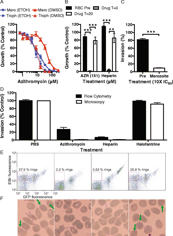
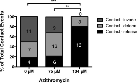
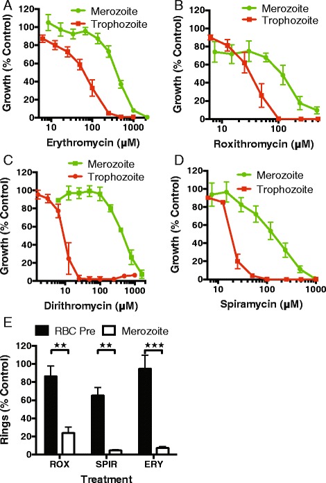
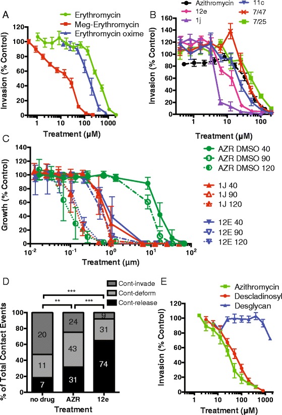
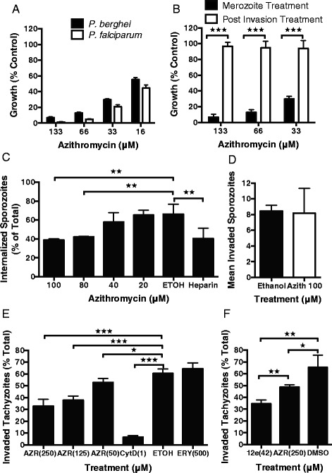
Results: Here, we demonstrate that the clinically used macrolide antibiotic azithromycin, which is known to kill human malaria asexual blood-stage parasites by blocking protein synthesis in their apicoplast, is also a rapid inhibitor of red blood cell invasion in human (Plasmodium falciparum) and rodent (P. berghei) malarias. Multiple lines of evidence demonstrate that the action of azithromycin in inhibiting parasite invasion of red blood cells is independent of its inhibition of protein synthesis in the parasite apicoplast, opening up a new strategy to develop a single drug with multiple parasite targets. We identified derivatives of azithromycin and erythromycin that are better invasion inhibitors than parent compounds, offering promise for development of this novel antimalarial strategy.

Conclusions: Safe and effective macrolide antibiotics with dual modalities could be developed to combat malaria and reduce the parasite's options for resistance.


Figures

Fig. 1
Fig. 1
Drug treatment strategies used in this study. The lifecycle stage of drug treatment is represented in the first box for each panel and the stage of parasitaemia measurements are highlighted by red boxes with yellow background. (a) Merozoite; successful invasion of drug-treated purified merozoites was measured at ring-stage immediately after addition of erythrocytes and merozoite invasion (<1 hour rings) or after washing out the drug (denoted by green dashed line) and growing parasites through to late trophozoite stage (40 hours post-invasion). (b) In cycle; early ring-stage parasites (<4 hours post-invasion) were drug-treated and the resulting growth inhibition was assessed at late trophozoite stage (40 hours post-invasion). (c) 1 cycle; early ring-stage parasites (<4 hours post-invasion) were drug-treated and the resulting growth inhibition was assessed 90 hours later, after 1 cycle of reinvasion, at late trophozoite stage. These assays are a longer duration, but nonetheless equivalent in terms of including 1 cycle of reinvasion and development, to 1 cycle assays reported in other studies [–44]. (d) 2 cycle (delayed death); early ring-stage parasites (<4 hours post-invasion) were drug-treated and the drug-treated parasites were grown for 80 hours prior to washing out the drug in fresh media (denoted by green dashed line). Growth inhibition was assessed approximately 40 hours later after a second cycle of reinvasion (120 hours post-invasion). (e) Live filming; mature schizont-stage parasites were incubated with azithromycin and the success of merozoite invasion was recorded and quantified by live filming (green arrow, successful invasion; blue arrow, deformation of the erythrocyte but no invasion; red arrow, attachment and release or attachment with failure to deform erythrocyte and no release) (see Additional file 1: Video S1 and Additional file 2: Video S2). White box-drug treatment; yellow box-analysis of parasitaemia; green dashed line-drug washout
Fig. 2
Fig. 2
Structure of macrolide antibiotics. (a) Structure of the 15-membered macrolide, azithromycin, and its modified analogues (names used in the text underlined, in brackets). (b) Structure of the 14-membered macrolide, erythromycin A, and its modified analogues. (c) Structure of the 16-membered macrolide, spiramycin. (d) Structure of the non-macrolide antibiotic, clindamycin
Fig. 3
Fig. 3
Azithromycin inhibits P. falciparum merozoite invasion. (a) The potency of azithromycin in ethanol or DMSO as vehicle was compared for invasion inhibition (unbroken line, 10 minute merozoite treatment, parasitaemia measured 40 hours later) and 1 cycle growth inhibition assays (broken line, treatment rings to trophozoites’ next cycle). The invasion inhibitory IC50 of azithromycin prepared in ethanol (blue, IC50 10 μM) was similar to that for growth inhibition assays (IC50 7 μM; P = 0.0743, Log IC50 same between data sets, extra sum of squares F-test). The invasion inhibitory activity of azithromycin in DMSO (red, IC50 38 μM) was 5-fold higher than 1 cycle growth assays (IC50 7 μM; P <0.0001, Log IC50 different between data sets). (b) Inhibition profiles for pretreated erythrocytes (RBC Pre), merozoite treatment (T = 0; drug added at time zero) and rings treated for <1 hour (T = 20; drug added 20 minutes post-invasion) were identical between azithromycin (in DMSO) and the invasion inhibitor heparin (IC80 concentration). (c) Increasing the concentration of azithromycin (in ethanol) to 10 × IC80 (380 μM) did not result in substantial inhibition of invasion into pretreated cells compared to treatment of merozoites. (d) Flow cytometry and microscopy assessments confirmed that azithromycin (IC80 in ethanol) and heparin, but not the trophozoite-targeting antimalarial halofantrine (2 × IC80 ring-stage treatment (46 nM) [13]), inhibit merozoite invasion and establishment of ring stages in erythrocytes. Representative (e) flow cytometry plots (GFP high and EtBr low ring-stage parasites represented by square gate) and (f) microscopy thin smears (rings highlighted by green arrows) show absence of ring-stage parasites for azithromycin and heparin compared to non-invasion inhibitory controls (labeling as per Fig. 3d). Experiments represent the mean and SEM of three or more experiments. Significance was tested using an unpaired t-test (*P = 0.01–0.05, **P ≤0.01, ***P ≤0.001)
Fig. 4
Fig. 4
Azithromycin inhibits the early steps of invasion. Video microscopy of merozoite invasion of erythrocytes was performed in the presence of 75 and 134 μM azithromycin (in ethanol) compared to a no drug control (0 μM). Five schizont ruptures were observed for each treatment. Of the merozoites that contacted erythrocytes, some were observed to deform erythrocytes and then successfully invade their host cells (contact–invade), while others did not progress beyond initial attachment (contact–detach) or progressed to deformation but did not invade (contact–deform). From several rupturing schizonts, the number of merozoites exhibiting each of these steps was counted for each drug treatment and the percentages are shown along with the number of events in the column boxes. A Chi-squared test was performed to indicate significant differences at the following levels (**P ≤0.01, ***P ≤0.001)
Fig. 5
Fig. 5
Related macrolides inhibit merozoite invasion. The 14-membered macrolides (a) erythromycin A (IC50 mero 420 μM), (b) roxithromycin (83 μM), (c) dirithromycin (521 μM) and (d) the 16-membered macrolide spiramycin (123 μM) had variable levels of invasion inhibitory activity (green) and a higher IC50 than that achieved for 1 cycle assays (red). (e) The invasion inhibitory activity of erythromycin A, roxithromycin and spiramycin at an IC80 concentration was confirmed by flow cytometry assessment of ring stages with minimal inhibition evident for pretreated erythrocytes. All experiments represent the mean and SEM of three or more experiments. Significance of differences was compared using an unpaired t-test (**P ≤0.01, ***P ≤0.001)
Fig. 6
Fig. 6
The mechanism of invasion inhibition is unlikely to target the apicoplast ribosome. (a) Clindamycin targets the same subunit of the apicoplast ribosome but was found to have a much higher IC80 for apparent merozoite invasion inhibition (2,972 μM). There was evidence of non-specific inhibition of invasion as pretreatment of erythrocytes with clindamycin gave significant inhibition, which was not seen for azithromycin (prepared in ethanol; mean and SEM of four or more experiments; significance of differences tested with an unpaired t-test; **P ≤0.01) (38 μM). (b) The D10-AZRr line showed up to a 57-fold higher tolerance of azithromycin in 2 cycle (delayed death) apicoplast-targeting drug inhibition assays compared to D10 parental line. In contrast, the IC50 for purified merozoite invasion inhibitory activity differed by less than 2.5-fold between the D10-AZRr line and the D10-PfPHG line for (c) azithromycin (IC50: PfPHG 10 μM; D10-AZRr 25 μM), (d) erythromycin A (IC50: PfPHG 420 μM; D10-AZRr 732 μM) and (e) clindamycin (IC50: PfPHG 743 μM; D10-AZRr 557 μM). Data represent the mean of two or more experiments in at least duplicate. D10-AZRr, D10 azithromycin-resistant
Fig. 7
Fig. 7
Macrolide modification lowers invasion inhibitory IC50, but not apicoplast-targeting ‘delayed death’ inhibition. (a) Addition of an L-megosamine sugar [41] to form Meg-erythromycin (6-O-megosaminyl erythromycin A, IC50 13 μM) or an oxime group (150 μM) lowered the invasion inhibitory IC50 activity compared to the parent drug erythromycin A (IC50 420 μM). (b) Screening of an azithromycin analogue panel identified three compounds (12e, 15 μM; 1j, 7 μM; 11c, 28 μM) with up to 5-fold lower invasion inhibitory IC50 compared to the parent azithromycin (in DMSO, 38 μM). (c) Treatment of parasites during in cycle (40 hours, rings to schizonts), 1 cycle (90 hours, 1 cycle of replication) and 2 cycle (120 hours, 2 cycles of replication) assays with azithromycin (in DMSO) and analogues (12e, 1j), indicated that the IC50 of 1 cycle (40 hour and 90 hour, high drug concentration) inhibition was greatly reduced for the analogues compared to azithromycin. In contrast, the IC50 of the delayed death phenotype was almost identical for azithromycin and its analogues. d) Video microscopy of merozoite invasion was performed in the presence of a no drug control (0 μM), 122 μM of analogue 12e (2 × IC80) and azithromycin (AZR; both in DMSO). Merozoites that contacted the erythrocyte and i) invaded (cont–invade), ii) deformed but did not invade (cont–deform), or iii) released without deforming or invading (cont–detach) were tallied and analyzed as per Fig. 4. (e) Removal of the cladinosyl sugar (azithromycin descladinosyl, 50 μM) from azithromycin increased the invasion inhibitory IC50 compared to azithromycin (10 μM). The additional removal of the desosaminyl sugar (azithromycin desglycan, IC50 >1,600 μM) resulted in loss of invasion inhibitory activity compared to azithromycin. Data represent the mean and SEM of three or more experiments (**P ≤0.01, ***P ≤0.001)
Fig. 8
Fig. 8
Azithromycin inhibits P. berghei merozoite and Toxoplasma gondii tachyzoite, but not sporozoite, invasion of host cells. (a) Azithromycin inhibited purified P. berghei merozoite invasion at similar concentrations compared to P. falciparum. (b) Treatment of P. berghei-infected erythrocytes immediately after invasion with azithromycin for 30 minutes did not result in a loss of late-stage parasites detected by flow cytometry, confirming that azithromycin inhibited invasion and not parasite growth. (c) Azithromycin was found to have a small but significant effect on the number of P. berghei sporozoites that had entered or traversed host cells. However, using a more specific assay that measures successfully invaded hepatocytes containing a developing parasite (d), there was no significant inhibition in sporozoites that had invaded and formed a parasitophorous vacuole. (e) Azithromycin treatment of T. gondii tachyzoites at concentrations of 250, 125 and 50 μM resulted in a dose-dependent inhibition of host cell invasion. The invasion inhibitory control cytochalasin D (1 μM) was a considerably more potent inhibitor of tachyzoite invasion, while erythromycin A, as was found for P. falciparum, showed no evidence of invasion inhibitory activity at concentrations up to 500 μM. (f) Azithromycin analogue 12e (42 μM) was significantly more inhibitory to Toxoplasma tachyzoite invasion than azithromycin (250 μM, both solubilized in DMSO) even when tested at a 6-fold lower concentration. Data represent the mean and SEM of three or more experiments, significance of differences was tested using an unpaired t-test (*P = 0.01 to 0.05, **P ≤0.01, ***P ≤0.001)

Comment in

References

    1. World Health Organization (WHO) World malaria report 2014. Geneva: WHO; 2014.
    1. Ferone R, Burchall JJ, Hitchings GH. Plasmodium berghei dihydrofolate reductase. Isolation, properties, and inhibition by antifolates. Mol Pharmacol. 1969;5:49–59. - PubMed
    1. Zarchin S, Krugliak M, Hinsburg H. Digestion of the host erythrocyte by malaria parasites is the primary target for quinoline containing antimalarials. Biochem Pharmacol. 1986;35:2435–42. doi: 10.1016/0006-2952(86)90473-9. - DOI - PubMed
    1. Loria P, Miller S, Foley M, Tilley L. Inhibition of the peroxidative degradation of haem as the basis of action of chloroquine and other quinoline antimalarials. Biochem J. 1999;339:363–70. doi: 10.1042/0264-6021:3390363. - DOI - PMC - PubMed
    1. Krishna S, Uhlemann AC, Haynes RK. Artemisinins: mechanisms of action and potential for resistance. Drug Resist Updat. 2004;7:233–44. doi: 10.1016/j.drup.2004.07.001. - DOI - PubMed

Publication types