. 2015 Jul 18:14:280.
doi: 10.1186/s12936-015-0801-5.

Plasmodium falciparum coronin organizes arrays of parallel actin filaments potentially guiding directional motility in invasive malaria parasites

Affiliations

Plasmodium falciparum coronin organizes arrays of parallel actin filaments potentially guiding directional motility in invasive malaria parasites

Maya A Olshina et al. Malar J. .

Abstract

Background: Gliding motility in Plasmodium parasites, the aetiological agents of malaria disease, is mediated by an actomyosin motor anchored in the outer pellicle of the motile cell. Effective motility is dependent on a parasite myosin motor and turnover of dynamic parasite actin filaments. To date, however, the basis for directional motility is not known. Whilst myosin is very likely orientated as a result of its anchorage within the parasite, how actin filaments are orientated to facilitate directional force generation remains unexplained. In addition, recent evidence has questioned the linkage between actin filaments and secreted surface antigens leaving the way by which motor force is transmitted to the extracellular milieu unknown. Malaria parasites possess a markedly reduced repertoire of actin regulators, among which few are predicted to interact with filamentous (F)-actin directly. One of these, PF3D7_1251200, shows strong homology to the coronin family of actin-filament binding proteins, herein referred to as PfCoronin.

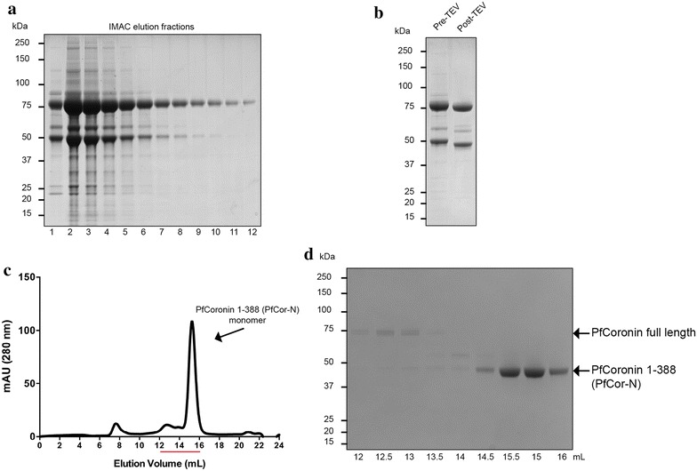
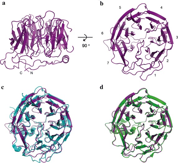
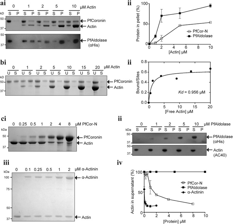
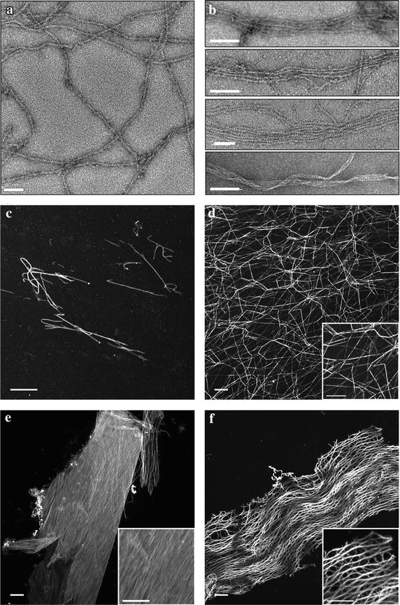
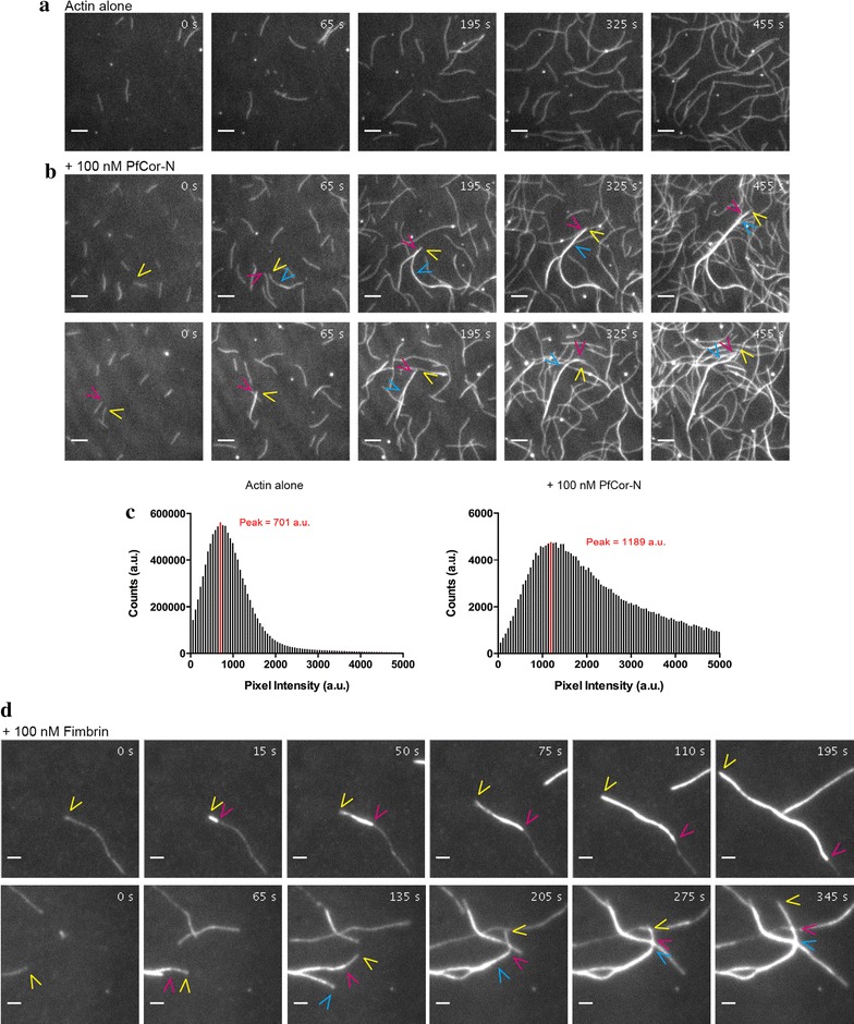
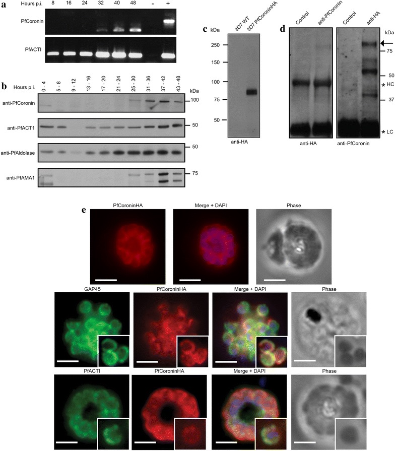
Methods: Here the N terminal beta propeller domain of PfCoronin (PfCor-N) was expressed to assess its ability to bind and bundle pre-formed actin filaments by sedimentation assay, total internal reflection fluorescence (TIRF) microscopy and confocal imaging as well as to explore its ability to bind phospholipids. In parallel a tagged PfCoronin line in Plasmodium falciparum was generated to determine the cellular localization of the protein during asexual parasite development and blood-stage merozoite invasion.

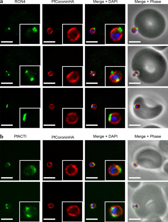
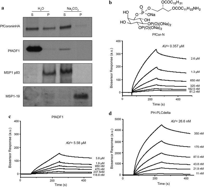
Results: A combination of biochemical approaches demonstrated that the N-terminal beta-propeller domain of PfCoronin is capable of binding F-actin and facilitating formation of parallel filament bundles. In parasites, PfCoronin is expressed late in the asexual lifecycle and localizes to the pellicle region of invasive merozoites before and during erythrocyte entry. PfCoronin also associates strongly with membranes within the cell, likely mediated by interactions with phosphatidylinositol-4,5-bisphosphate (PI(4,5)P2) at the plasma membrane.

Conclusions: These data suggest PfCoronin may fulfil a key role as the critical determinant of actin filament organization in the Plasmodium cell. This raises the possibility that macro-molecular organization of actin mediates directional motility in gliding parasites.


Figures

Figure 1
Figure 1
Purification of PfCoronin. a IMAC elution fractions 1–12. b PfCoronin pre- and post-removal of the N-terminal 6×His tag with TEV protease. c Size exclusion chromatography elution profile of PfCoronin. d Size exclusion chromatography elution fractions between 12 and 16 mL as indicated by the red line in (c) with PfCor-N eluting as monomer.
Figure 2
Figure 2
Homology model of PfCoronin. a Side view of homology model for PfCoronin generated using the I-TASSER server. b Top view of model. Potential blades within the propeller numbered 1–7 as per MmCoro1A and TgCoronin. c Alignment of PfCoronin homology model (purple) with MmCoro1A structure (2AQ5.pdb, [29]) (blue). d Alignment of PfCoronin homology model (purple) with TgCoronin structure (4OZU.pdb, [32]) (green).
Figure 3
Figure 3
PfCor-N binds to and bundles F-actin. ai High speed sedimentation of PfCor-N (2 µM) or PfAldolase (2 µM) with 0–10 μM pre-assembled F-actin. Supernatant (S) and pellet (P) fractions shown for representative gel of PfCor-N (Coomassie stained) or PfAldolase (Western, probed with anti-His). ii Densitometry showing % protein co-pelleting with F-actin, values = mean ± SEM (n = 3). bi Supernatant depletion assay showing unspun (U) and spun (S) samples PfCor-N (2 μM) incubated with pre-assembled F-actin (0–20 μM) ii Densitometry of bound PfCor-N based on amount of free actin in solution post-centrifugation, providing a dissociation constant (Kd) = 0.956 μM. c Low-speed sedimentation assay of 2 uM F-actin assembled in presence of 0–10 μM PfCor-N (i), PfAldolase (ii) or α-Actinin with densitometry (iv) shown as % actin remaining in supernatant post-centrifugation, values = mean ± SEM (n = 3).
Figure 4
Figure 4
PfCor-N organizes F-actin into bundles and higher order sheets and networks. TEM of F-actin (2 μM) alone (a) or in presence of 0.2 μM PfCor-N (b, top three panels) and 1 μM PfCor-N (b, lower panel). Scale bars 50 nm (a), 100 nm (b). cf Confocal micrographs of Phalloidin-488 labelled F-actin (1.5 μM) (c) alone, or in the presence of d 0.5 μM α-actinin, (ef) 0.5 μM PfCor-N. Scale bars 10 μm.
Figure 5
Figure 5
PfCor-N produces parallel actin bundles observed by TIRF microscopy. 2 μM Mg-ATP-actin with 1 μM Oregon Green Mg-ATP-actin (a) alone (b) + 0.1 μM PfCor-N. Arrowheads indicate growing ends of filaments. Scale bars 5 μm. c Pixel intensity quantification of a and b across 45 frames represented as measure of filament bundling. Actin alone pixel intensity mode = 701 arbitrary units (au) (red line). 100 nM PfCor-N pixel intensity mode = 1,189 au (red line). d Actin assembled in presence of 100 nM Fimbrin. Top panel antiparallel bundle, bottom panel parallel bundle. Arrowheads indicate growing ends of the filaments. Scale bar 15 μm.
Figure 6
Figure 6
PfCoronin is expressed in schizonts/merozoites and is located at the periphery of mature merozoites. a RT-PCR of P. falciparum cDNA for PfCoronin and PfACTI genes across asexual life-cycle post invasion (pi). gDNA positive control (with introns). Negative control—no reverse transcriptase. b Western blots across asexual life-cycle post invasion (pi), probed with anti-PfCoronin, anti-PfACT1 (anti-Act239-253 (rabbit), [10]), anti-PfAldolase [40] and anti-PfAMA1 [60]. c Western blot WT 3D7 P. falciparum versus PfCoroninHA, probed with anti-HA. d Reciprocal western blots of immunoprecipitations from 3D7 PfCoroninHA schizont lysates with anti-PfCoronin bait, probed with anti-HA (left panel) or anti-HA bait, probed with anti-PfCoronin (right panel). Control lanes = WT 3D7 P. falciparum. Arrow indicates PfCoroninHA. Asterisks heavy and light chain cross-reactivity. e IFA of schizonts probed with anti-HA (Coronin, red), DAPI (nucleus, blue), anti-GAP45 (IMC, green) (middle panel) [40] or anti-PfACTI (IMC/cytosol green) (bottom panel). Scale bar 2 μm.
Figure 7
Figure 7
PfCoroninHA remains peripherally located in merozoites throughout erythrocyte invasion. a Invading merozoite IFA early (top panel), mid (middle panel) and late (bottom panel) in invasion. Labeling is anti-RON4 (tight junction, green), anti-HA (Coronin, red) and DAPI (nucleus, blue). Scale bar 2 μm. b Invading merozoite IFA early (top panel) and midway (bottom panel) through invasion. Labeling is anti-PfActin (IMC/cytosol, green), anti-HA (Coronin, red) and DAPI (nucleus, blue). Scale bar 2 μm.
Figure 8
Figure 8
Membrane association of PfCoroninHA is mediated by PI(4,5)P2. a Western blot of PfCoroninHA. Lanes 1 and 2 supernatant (S) and pellet (P) post hypotonic lysis. Lanes 3 and 4 S and P post Na2CO3 treatment. Top panel probed with anti-HA (Coronin). Second panel probed with anti-PfADF1 [63]. Third panel probed with anti-MSP1p83 (non-membrane associated protein). Fourth panel probed with anti-MSP1-19 (membrane associated GPI anchor). bd SPR sensograms of (b) PfCor-N (c) PfADF1 and (d) PH-PLCdelta binding to immobilized PI(4,5)P2, shown above panel b. Concentrations of each analyte displayed by curves. Determined Kds = inset in graph.

References

    1. Cowman AF, Berry D, Baum J. The cellular and molecular basis for malaria parasite invasion of the human red blood cell. J Cell Biol. 2012;198:961–971. doi: 10.1083/jcb.201206112. - DOI - PMC - PubMed
    1. Prudencio M, Rodriguez A, Mota MM. The silent path to thousands of merozoites: the Plasmodium liver stage. Nat Rev Microbiol. 2006;4:849–856. doi: 10.1038/nrmicro1529. - DOI - PubMed
    1. Baum J, Gilberger T-W, Frischknecht F, Meissner M. Host-cell invasion by malaria parasites: insights from Plasmodium and Toxoplasma. Trends Parasitol. 2008;24:557–563. doi: 10.1016/j.pt.2008.08.006. - DOI - PubMed
    1. Morrissette NS, Sibley LD. Cytoskeleton of apicomplexan parasites. Microbiol Mol Biol Rev. 2002;66:21–38. doi: 10.1128/MMBR.66.1.21-38.2002. - DOI - PMC - PubMed
    1. Frénal K, Polonais V, Marq J-B, Stratmann R, Limenitakis J, Soldati-Favre D. Functional dissection of the apicomplexan glideosome molecular architecture. Cell Host Microbe. 2010;8:343–357. doi: 10.1016/j.chom.2010.09.002. - DOI - PubMed

Publication types

MeSH terms