. 2015 Oct;73(7):ftv046.
doi: 10.1093/femspd/ftv046. Epub 2015 Jul 17.

Contribution of the platelet activating factor signaling pathway to cerebral microcirculatory dysfunction during experimental sepsis by ExoU producing Pseudomonas aeruginosa

Affiliations

Contribution of the platelet activating factor signaling pathway to cerebral microcirculatory dysfunction during experimental sepsis by ExoU producing Pseudomonas aeruginosa

Maria Cristina Plotkowski et al. Pathog Dis. 2015 Oct.

Abstract

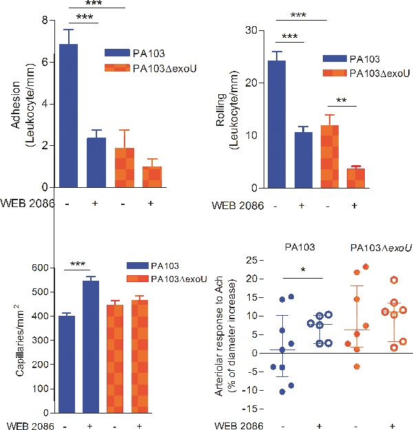
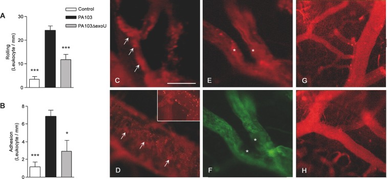
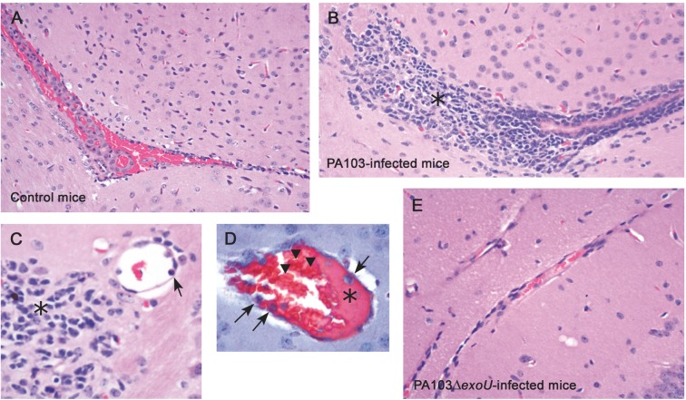
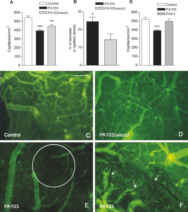
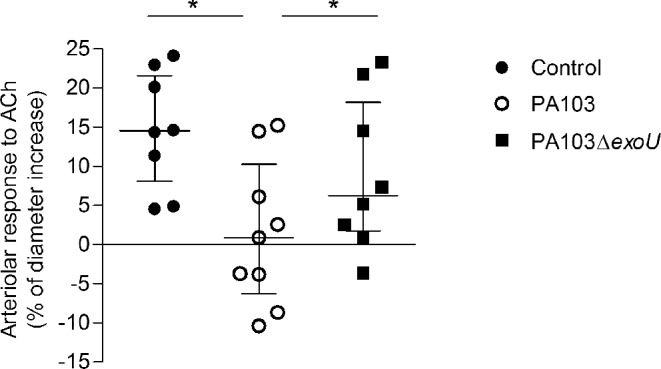
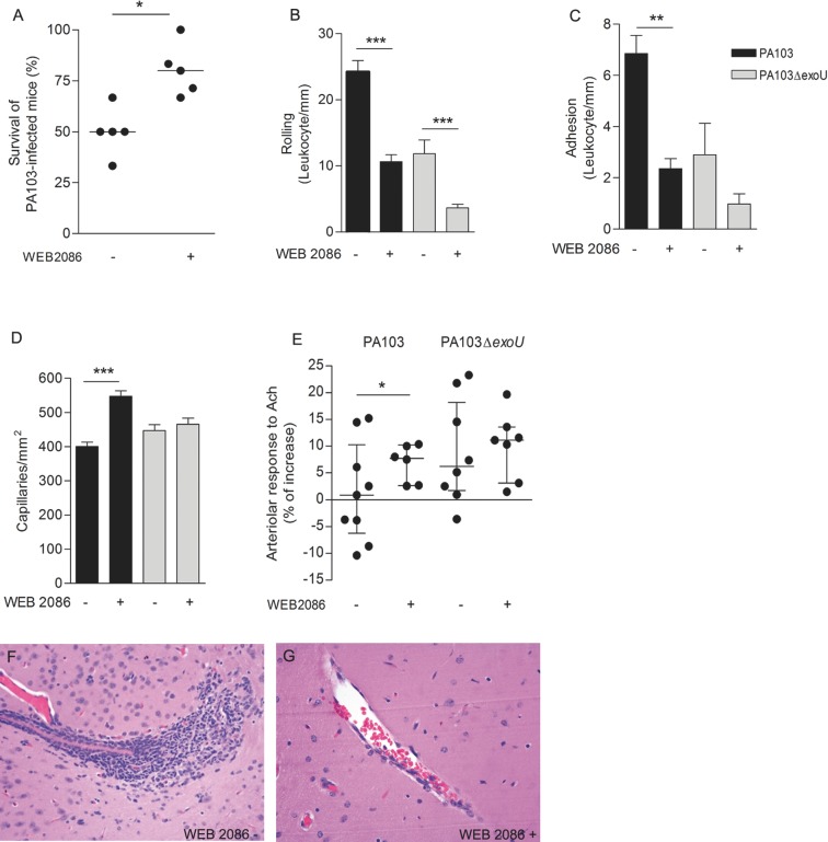
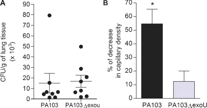
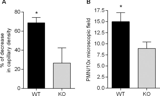
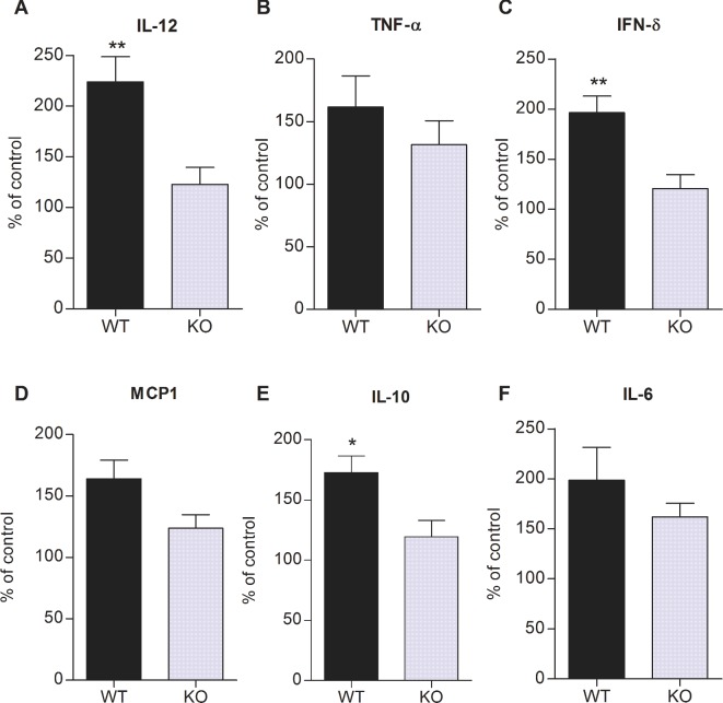
Intravital microscopy was used to assess the involvement of ExoU, a Pseudomonas aeruginosa cytotoxin with phospholipase A2 activity, in dysfunction of cerebral microcirculation during experimental pneumosepsis. Cortical vessels from mice intratracheally infected with low density of the ExoU-producing PA103 P. aeruginosa strain exhibited increased leukocyte rolling and adhesion to venule endothelium, decreased capillar density and impaired arteriolar response to vasoactive acetylcholine. These phenomena were mediated by the platelet activating factor receptor (PAFR) pathway because they were reversed in mice treated with a PAFR antagonist prior to infection. Brains from PA103-infected animals exhibited a perivascular inflammatory infiltration that was not detected in animals infected with an exoU deficient mutant or in mice treated with the PAFR antagonist and infected with the wild type bacteria. No effect on brain capillary density was detected in mice infected with the PAO1 P. aeruginosa strain, which do not produce ExoU. Finally, after PA103 infection, mice with a targeted deletion of the PAFR gene exhibited higher brain capillary density and lower leukocyte adhesion to venule endothelium, as well as lower increase of systemic inflammatory cytokines, when compared to wild-type mice. Altogether, our results establish a role for PAFR in mediating ExoU-induced cerebral microvascular failure in a murine model of sepsis.

Keywords: PAF–PAFR signaling pathway; Pseudomonas aeruginosa type III secretion toxin; cerebral intravital microscopy; cerebral microvascular failure; experimental Pseudomonas aeruginosa sepsis.


Figures

Graphical Abstract Figure.
During Pseudomonas aeruginosa experimental sepsis, cerebral microcirculatory dysfunction induced by the toxin ExoU depends on increased PAF relase and activation of the PAF receptor signaling pathway.
Figure 1.
Figure 1.
ExoU effect on blood cell–endothelium interaction. Leukocyte rolling on (A) and adhesion to (B) the endothelium was significantly higher in PA103-infected mice than in PA103ΔexoU-infected or in control animals. Data are means (and SEM) of the results obtained with six animals from each group, and are representative of the results obtained in two different experiments. *P < 0.05 and ***P < 0.001 when the values obtained from PA103ΔexoU-infected and control mice were compared with those obtained from animals infected with the ExoU-producing PA103 strain. In panels CF microvessels from PA103-infected mice are shown. Note the presence of rhodamin-labeled cell aggregates of different sizes in the blood vessel lumina (arrows in panels C and D) or adherent to the endothelium (asterisk in panel E), forming a structure that narrowed the blood vessel lumen. In this figure, panel F exhibits the same vessels as shown in panel E, labeled with the FITC-dextran complex. Note the absence of perfusion in the area labeled with the asterisks. The inset included in panel D shows leukocyte rolling/adhesion to brain venule endothelium from PA103-infected mice. In panels G and H, microvessels from PA103ΔexoU-infected and control non-infected mice are shown, respectively. Note the absence of cell aggregates in the microvessel lumina. Final magnification, × 200; scale bar: 100 μm.
Figure 2.
Figure 2.
ExoU effect on brain histopathology. Light micrographs of brain sections from control mice (A) as well as from animals infected with the ExoU-producing PA103 (BD) or non-producing PA103ΔexoU P. aeruginosa (E), stained with hematoxylin-eosin (A–C and E) or Masson´s trichrome stain (D). Note in panels B and C, the huge perivascular inflammatory infiltration (asterisks) and inflammatory cells adherent to microvessel endothelium (arrow in panel C). Panel D shows the presence of a proteinaceous material (asterisk) and of inflammatory cells (arrows) adherent to blood vessel endothelium, as well as of aggregated erythrocytes (arrowhead). Note in panels A and E, the absence of perivascular inflammatory infiltration is shown. Original magnifications: A, B and E: × 200; C: ×400; D: ×1000.
Figure 3.
Figure 3.
Effect of infection on brain capillary density. (A) The mean number of perfused capillaries in cerebral tissue of control mice was significantly higher than in brains of animals from the other two groups (**P < 0.01 and ***P < 0.001). Panel B showsthe percentage of decrease in capillary density detected in PA103- and PA103ΔexoU-infected mice, considering the mean number of capillaries of control mice (548.0) as 100%. The decrease in mice infected with the WT bacteria was significantly higher (*P < 0.05), as determined by the Mann Whitney test. Data are mean (and SEM) of the results obtained with six animals from each group, and are representative of the results obtained in two different experiments. In panels CF, micrographs of microvessels from control (C), PA103ΔexoU- (D) and PA103-infected mice (E and F) labeled with the fluorescein isothiocyanate-dextran complex are shown. Note in panel E, the presence of an area of capillary rarefaction (inside the circle) and in panel F, stopped-flow capillaries (arrows). Panel G shows that the mean number of perfused capillaries in PA103-infected animals was significantly lower (***P < 0.001) than in mice infected with PAO1 or in control mice. Data are mean (and SEM) of the results obtained in two different assays, carried out with 10 animals from each group. Final magnification, ×100; scale bar: 100 μm.
Figure 4.
Figure 4.
Effect of ExoU on the arteriolar response to acetylcholine. The percentages of increase of arteriole diameters in control animals and in mice infected with the bacterial mutant were significantly higher than in PA103-infected mice (*P < 0.05), as determined by the Wilcoxon rank test. Data are median with interquartile range of the results obtained in two different experiments.
Figure 5.
Figure 5.
Effect of WEB 2086 treatment prior to infection. Mice treatment with the PAFR inhibitor prior to PA103 infection increased significantly the animal survival (A) and the number of perfused capillaries (D), and reduced significantly the leukocyte rolling on (B) and adhesion (C) to the capillary endothelium. The PAFR inhibitor also increased significantly the arteriolar responsiveness to topically applied acetylcholine (E). Panel A shows that the median of the percentage of survivors in PA103-infected animals previously treated with WEB was significantly increased, as determined in six different assays, each one carried out with six to eight animals, and analyzed with the Mann Whitney test. Data in panels B–D are means (and SEM) of the results obtained in two different experiments, carried out with six to eight animals from each group, whereas panel E shows the median and the interquartile range of the results obtained with six to nine animals. *P < 0.05; **P < 0.01; ***P < 0.001 when the results obtained from WEB-treated and WEB-untreated mice were compared with each other. (F and G) correspond to light micrographs of brain sections of PA103-infected mice. No perivascular infiltration of inflammatory cells, detected in brains from untreated mice (F), was detected in sections from WEB-treated animals (G). Original magnifications: ×200.
Figure 6.
Figure 6.
Bacterial load in mice lungs and decrease in pial capillary density 6 h after infection. Panel A shows that the bacterial concentrations in lungs of mice infected with the ExoU-producing PA103 strain or with the PA103ΔexoU mutant did not differ from each other. Data represent means and SEM of the results obtained in eight mice infected with each bacteria, assessed in two different assays. Panel B shows the decrease in cerebral capillary density in infected mice, considering the mean number of capillaries/mm2 of control mice as 100%. The decrease in mice infected with PA103 was significantly higher (P < 0.05, as determined by the Mann Whitney test). Data are mean (and SEM) of the results obtained in five mice infected with each bacteria.
Figure 7.
Figure 7.
Involvement of PAFR signaling in ExoU-induced microvascular disturbance. Panel A shows the percentage of decrease in capillary density detected in PA103-infected WT and KO mice, considering the mean number s of capillaries of control non-infected WT and KO mice, respectively, as 100%. The decrease in WT animals was significantly higher (*P < 0.05). Panel B shows that leukocyte adhesion to venule endothelium was significantly higher (*P < 0.05) in WT than in KO mice. Data in A and B are means (and SEM) of the results obtained with at least five animals (range 5–12) from each group and significance of the differences was determined with the Mann Whitney test.
Figure 8.
Figure 8.
Role of PAFR signaling in ExoU-induced citokine production. Data represent the percentages of increase in serum cytokine concentrations in WT and KO mice after PA103 infection, considering the mean cytokine concentrations in control non-infected WT and KO mice, respectively, as 100%. Data are means (and SEM) of the results obtained with seven to eight animals from each group and significance of the differences was determined with the Mann Whitney test. *P < 0.05 and **P < 0.01.

References

    1. Araújo CV, Estato V, Tibiriçá E, et al. PPAR gamma activation protects the brain against microvascular dysfunction in sepsis. Microvasc Res. 2012;84:218–21. - PubMed
    1. Castor MG, Rezende BM, Resende CB, et al. Platelet-activating factor receptor plays a role in the pathogenesis of graft-versus-host disease by regulating leukocyte recruitment, tissue injury and lethality. J Leukocyte Biol. 2012;91:629–39. - PubMed
    1. Correa-Costa M, Andrade-Oliveira V, Braga TT, et al. Activation of platelet-activating factor receptor exacerbates renal inflammation and promotes fibrosis. Lab Invest. 2014;94:455–66. - PubMed
    1. Croner RS, Hoerer E, Kulu Y, et al. Hepatic platelet and leukocyte adherence during endotoxemia. Crit Care. 2006;10:R15. - PMC - PubMed
    1. De Backer D, Creteur J, Preiser JC, et al. Microvascular blood flow is altered in patients with sepsis. Am J Resp Crit Care. 2002;166:98–104. - PubMed

Publication types

Substances