. 2015 Sep 30;43(17):8325-39.
doi: 10.1093/nar/gkv737. Epub 2015 Jul 17.

FANCD2 and REV1 cooperate in the protection of nascent DNA strands in response to replication stress

Affiliations

FANCD2 and REV1 cooperate in the protection of nascent DNA strands in response to replication stress

Yeran Yang et al. Nucleic Acids Res. .

Abstract

REV1 is a eukaryotic member of the Y-family of DNA polymerases involved in translesion DNA synthesis and genome mutagenesis. Recently, REV1 is also found to function in homologous recombination. However, it remains unclear how REV1 is recruited to the sites where homologous recombination is processed. Here, we report that loss of mammalian REV1 results in a specific defect in replication-associated gene conversion. We found that REV1 is targeted to laser-induced DNA damage stripes in a manner dependent on its ubiquitin-binding motifs, on RAD18, and on monoubiquitinated FANCD2 (FANCD2-mUb) that associates with REV1. Expression of a FANCD2-Ub chimeric protein in RAD18-depleted cells enhances REV1 assembly at laser-damaged sites, suggesting that FANCD2-mUb functions downstream of RAD18 to recruit REV1 to DNA breaks. Consistent with this suggestion we found that REV1 and FANCD2 are epistatic with respect to sensitivity to the double-strand break-inducer camptothecin. REV1 enrichment at DNA damage stripes also partially depends on BRCA1 and BRCA2, components of the FANCD2/BRCA supercomplex. Intriguingly, analogous to FANCD2-mUb and BRCA1/BRCA2, REV1 plays an unexpected role in protecting nascent replication tracts from degradation by stabilizing RAD51 filaments. Collectively these data suggest that REV1 plays multiple roles at stalled replication forks in response to replication stress.


Figures

Figure 1.
Figure 1.
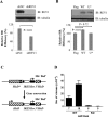
REV1 is involved in gene conversion in mammalian cells. (A) REV1-depleted 293T cells were co-transfected with DR-GFP reporter and I-SceI-IRES-DsRedNLS plasmid. Two days later, the frequency of HR-mediated repair events was calculated by analyzing GFP-positive cells out of the DsRed-positive cells in flow cytometry analysis. The extent of repair is shown relative to the repair observed in shRNA-SHC002 (shNC) knockdown cells. Data from three independent experiments were used to generate the histogram. Error bars indicate standard error. P values derived from a student's t-test are shown at the top. The shREV1-mediated knockdown efficiency was validated by western blot using antibodies against REV1. β-Tubulin: loading control. (B) REV1-depleted 293T cells were co-transfected with DR-GFP reporter, I-SceI-IRES-DsRedNLS plasmid and either Flag or Flag-REV1 (WT) or Flag-REV1 UBMs* (U*). The frequency of HR-mediated repair event was calculated as in (A). The extent of repair is shown relative to the repair observed in cells transfected with control Flag vector. The expression of WT or U* REV1 was validated by western blot using antibodies against REV1. (C) Reporter construct to determine reversion of a defective HisD gene (HisD*) to a functional HisD gene using HisD donor sequences (3HisD) in cells. Cells carrying a stably integrated reporter construct are sensitive to histidinol (His) and resistant to blasticidin (Bsd+). Bsr, blasticidin selection gene. (D) Frequencies of spontaneous His+Bsd+ clones in two independent Rev1 WT (7 and 18) and KO MEFs (6 and 9). Frequencies are corrected for the clone forming ability of the cells. All experiments were carried out in triplicate. Error bars indicate the standard error.
Figure 2.
Figure 2.
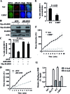
RAD18 and UBMs in REV1 are required for the optimal accumulation of GFP-REV1 at HR processing sites. (A) HCT116 cells transfected with WT and UBM* GFP-REV1 were microirradiated with a pulsed nitrogen laser. Thirty minutes later, cells were fixed and stained with anti-γH2AX antibody. Nuclei were stained with DAPI. (B) and (C) RAD18 stable knockdown and control cells were transfected with GFP-REV1 and Myc-RAD18. (B) The levels of RAD18 and Myc-RAD18 were analyzed by western blot. β-actin, loading control. (C) The cells were microirradiated and the proportion of cells with REV1 accumulation was quantified. Error bars indicate standard error. P values derived from a student's t-test are shown at the top. (D) Kinetic analysis of GFP-REV1 intensity at laser-irradiated sites in RAD18 knockdown and control cells. Error bars represent standard error based on 10 independent measurements. (E) Percentage of HCT116 cells expressing WT and UBM* GFP-REV1 in which the protein was localized at microirradiated sites. P value derived from a student's t-test is shown at the top. (F) Kinetic analysis of WT and UBM* GFP-REV1 intensity at laser-irradiated sites. (G) UBMs are required for the optimal recruitment of REV1 to I-SceI-induced DSBs in CHIP assay. The y-axis represents the relative enrichment of the indicated proteins compared to the IgG control (after normalization to total input). Mutation of UBMs in REV1 significantly decreases REV1 recruitment to I-SceI-induced DSBs as calculated using a Student's t-test (P < 0.01). All the data are from three independent experiments (±SEM, n = 3).
Figure 3.
Figure 3.
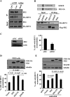
FANCD2 physically interacts with REV1 and modulates REV1 recruitment to laser-induced sites of damage. (A) Co-IP showing FANCD2 interacts with REV1. 293T cells transfected with a HA-REV1 expression vector were treated with CPT (5 μM) for 2 h and crosslinked with DTBP. The lysates were immunoprecipitated with an anti-FANCD2 antibody. Immunoprecipitates (IP) and inputs were analyzed via western blot using antibodies against HA or FANCD2 (FD2), respectively. Con, untreated cell; Mock, the lysate was immunoprecipitated with an anti-Flag antibody.(B) Top: Schematic diagrams of the FANCD2-K561R mutant (K561R) and FANCD2-Ub (FD2-Ub) constructs. Bottom: Co-IP experiment showing enhanced interaction between FANCD2-Ub and REV1. 293T cells were transfected with plasmids expressing HA-REV1 and SFB-FANCD2-K561R or SFB-FANCD2-Ub and analyzed by co-IP in the presence of EtBr. Immunoprecipitates were analyzed by western blot with antibodies against HA or Flag. (C) FANCD2 depletion impairs the recruitment of REV1 to laser-activated sites. U2OS cells were transfected with siFANCD2 (siFD2) or siNC. Three days later, the cells were harvested and the levels of FANCD2 were detected by western blot. β-Actin, loading control. The cells were further transfected with GFP-REV1 and microirradiated. The proportion of cells with REV1 accumulation was quantified. (D) PD20 cells were transfected with GFP-REV1 and FANCD2 WT (FD2), K561R or a FD2-Ub chimera. Levels of FANCD2 in cell lysates were detected by western blot with antibodies against Flag. β-tubulin, loading control. Cells were microirradiated. The proportion of cells containing accumulated REV1 was quantified. (E) Complementation with a FANCD2-Ub chimera can rescue the aberrant REV1 recruitment in the RAD18-depleted cells. RAD18 stable knockdown or control cells were transfected with GFP-REV1 and FD2 WT, K561R or a FD2-Ub chimera. Levels of FANCD2 and RAD18 in cell lysates were detected by western blot with antibodies against either Flag or RAD18. β-Tubulin, loading control. Cells were microirradiated. The proportion of cells with REV1 accumulation was quantified. Error bars indicate standard error. P values derived from a student's t-test are shown at the top.
Figure 4.
Figure 4.
REV1 is involved in cellular response to CPT exposure. (A) WT and Rev1 KO MEFs were treated with CPT for 2 h and further incubated in fresh medium for 7–10 days. The number of cell clones was determined. Surviving fraction was expressed as a percentage of mock-treated cells. Error bars: SD, n = 3. (B) WT and Rev1 KO MEFs were treated with HU for 2 h. Cell survival was analyzed as in (A). (C) WT and Rev1 KO MEFs were transfected with siFancd2 (siFD2) or siNC. The levels of REV1 and FANCD2 were analyzed by western blot. β-Tubulin, loading control. (D) WT and Rev1 KO MEFs transfected with siFancd2 (siFD2) or siNC, were treated with CPT for 2 h and analyzed as in (A). Values are means of three independent experiments. Error bars indicate standard error. Depletion of FANCD2 in Rev1 KO MEFs did not enhance cellular sensitivity to CPT as calculated using a Student's t- test (P > 0.05).
Figure 5.
Figure 5.
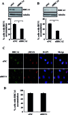
BRCA1 and BRCA2 modulate the assembly of REV1 at laser-induced sites of damage. (A) U2OS cells were transfected with siBRCA1 or siNC. Three days later, the level of BRCA1 was analyzed by western blot. Microirradiation was performed as in Figure 3C. A Student's t-test was used to calculate the P value (P < 0.05). (B) U2OS cells were transfected with siBRCA2 or siNC. The level of BRCA2 was analyzed by western blot. Microirradiation was performed as in Figure 3C. β-Tubulin, loading control. (C) U2OS cells were transfected with siREV1 or siNC. Three days later, the cells were microirradiated. Thirty minutes later, cells were fixed and stained with antibodies against BRCA1 and γH2AX. Nuclei were stained with DAPI. (D) The proportion of cells with BRCA1 accumulation was quantified. Error bars indicate standard error.
Figure 6.
Figure 6.
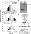
REV1 protects nascent replication tracts at stalled replication forks after exposure to CPT. (A) Scheme of experimental design for fork stability assay in Rev1 WT and KO MEFs. Length of nascent replication tracts (labeled with IdU and CIdU) was measured by DNA spreading after 5 h of replication stalling with CPT. Representative, individual DNA fibers for each experimental condition are shown. (B) Nascent tract length distributions were measured in WT (Top panel) and KO (Bottom panel) with 5 μM CPT or not. Median lengths of nascent replication tracts are given in parentheses. P-value is derived from the Mann-Whitney test. The nascent tract lengths were comparable in mock-treated WT and KO cells.
Figure 7.
Figure 7.
REV1 protects nascent replication tracts intact by stabilizing RAD51 filaments. (A) Complementation of REV1 or RAD51 in Rev1 KO MEFs can rescue the replication fork instability upon CPT. Rev1 KO MEFs transfected with GFP-REV1, or GFP-RAD51 or GFP were labeled with IdU and CIdU and then treated with CPT. GFP-positive cells were sorted with MoFlo XDP High-Speed Cell Sorter (Beckman Coulter) for the DNA spreading assay. Nascent tract length distributions in these cells were measured as in Figure 6. (B) Expression of GFP or GFP-REV1 in the sorted Rev1 KO MEFs was detected through western blot by immunoblotting with anti-GFP antibodies. ß-tubulin was used as loading control. (C) Model of REV1 targeting to DNA breaks to prevent chromosome instability.

Similar articles

Cited by

References

    1. Guo C., Kosarek-Stancel J.N., Tang T.S., Friedberg E.C. Y-family DNA polymerases in mammalian cells. Cell Mol. Life Sci. 2009;66:2363–2381. - PMC - PubMed
    1. Waters L.S., Minesinger B.K., Wiltrout M.E., D'Souza S., Woodruff R.V., Walker G.C. Eukaryotic translesion polymerases and their roles and regulation in DNA damage tolerance. Microbiol. Mol. Biol. Rev. 2009;73:134–154. - PMC - PubMed
    1. Sale J.E., Lehmann A.R., Woodgate R. Y-family DNA polymerases and their role in tolerance of cellular DNA damage. Nat. Rev. Mol. Cell Biol. 2012;13:141–152. - PMC - PubMed
    1. Tissier A., Kannouche P., Reck M.P., Lehmann A.R., Fuchs R.P., Cordonnier A. Co-localization in replication foci and interaction of human Y-family members, DNA polymerase pol eta and REVl protein. DNA Repair. 2004;3:1503–1514. - PubMed
    1. Guo C., Tang T.S., Bienko M., Parker J.L., Bielen A.B., Sonoda E., Takeda S., Ulrich H.D., Dikic I., Friedberg E.C. Ubiquitin-binding motifs in REV1 protein are required for its role in the tolerance of DNA damage. Mol. Cell Biol. 2006;26:8892–8900. - PMC - PubMed

Publication types

MeSH terms