. 2016 Jul;221(6):3027-65.
doi: 10.1007/s00429-015-1086-8. Epub 2015 Jul 19.

Radial and tangential migration of telencephalic somatostatin neurons originated from the mouse diagonal area

Affiliations

Radial and tangential migration of telencephalic somatostatin neurons originated from the mouse diagonal area

Luis Puelles et al. Brain Struct Funct. 2016 Jul.

Abstract

The telencephalic subpallium is the source of various GABAergic interneuron cohorts that invade the pallium via tangential migration. Based on genoarchitectonic studies, the subpallium has been subdivided into four major domains: striatum, pallidum, diagonal area and preoptic area (Puelles et al. 2013; Allen Developing Mouse Brain Atlas), and a larger set of molecularly distinct progenitor areas (Flames et al. 2007). Fate mapping, genetic lineage-tracing studies, and other approaches have suggested that each subpallial subdivision produces specific sorts of inhibitory interneurons, distinguished by differential peptidic content, which are distributed tangentially to pallial and subpallial target territories (e.g., olfactory bulb, isocortex, hippocampus, pallial and subpallial amygdala, striatum, pallidum, septum). In this report, we map descriptively the early differentiation and apparent migratory dispersion of mouse subpallial somatostatin-expressing (Sst) cells from E10.5 onward, comparing their topography with the expression patterns of the genes Dlx5, Gbx2, Lhx7-8, Nkx2.1, Nkx5.1 (Hmx3), and Shh, which variously label parts of the subpallium. Whereas some experimental results suggest that Sst cells are pallidal, our data reveal that many, if not most, telencephalic Sst cells derive from de diagonal area (Dg). Sst-positive cells initially only present at the embryonic Dg selectively populate radially the medial part of the bed nucleus striae terminalis (from paraseptal to amygdaloid regions) and part of the central amygdala; they also invade tangentially the striatum, while eschewing the globus pallidum and the preoptic area, and integrate within most cortical and nuclear pallial areas between E10.5 and E16.5.

Keywords: Cortex; Entopeduncular area; Forebrain interneurons; Medial ganglionic eminence; Pallidum; Preoptic area; Secondary prosencephalon; Striatum; Subpallium.


Figures
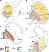
Fig. 1
Fig. 1
- Schemas illustrating the relative topography of subpallial subdivisions. a Schema of a left-side view of the embryonic brain, indicating in colors the telencephalic region. The pallium (orange) is separated from the subpallium by a black line. The subpallium appears divided into four domains: striatum (yellow St), pallidum (pink Pal), diagonal area (green Dg), and preoptic area (blue POA). The preopto-hypothalamic area (dark green POH), a part of POA, abbuts the telencephalic/preoptic border with respect to the terminal and peduncular parts of the hypothalamus (THy, PHy); The POH is continuous laterally across the hemispheric stalk with the subpallial and pallial amygdala (all three enclosed by the dash line). The longitudinal alar/basal boundary of the forebrain is depicted as a dotted line; the forebrain floor plate is marked by a thick black line; p2–p3 refer to diencephalic prosomeres. The coronal (Cor), horizontal (Hor), and transverse (Transv) planes of section used in this study are indicated; note the Hor and Transv planes are oriented relative to the prosomeric length axis and floor plate, while the Cor plane is arbitrary, and corresponds to the coronal section schema in (d). b Two-dimensional schema looking down on a flattened view of the right hemisphere, in which the four subpallial domains are mapped relative to the septal region, the hypothalamus, the pallium, and the medial, lateral and caudal ganglionic eminences (red contour; MGE, LGE, CGE; note the mixed composition of the MGE). The color code, areal names, and dash line used in (a) apply here to the ‘central’ or principal region of the subpallium in (b). The septo-amygdaloid axis can be imagined, with an obliquity that characterizes particularly the evaginated subpallial domains (St, Pal; less so Dg, or POA). The septal end of the subpallium (Se) is strictly septal, and contains in principle the topological dorsal end of all four subpallial domains (note the commissural septal midline lies at the telencephalic roof plate). Intercalated between the septal (Se) and the central subpallial sectors (C) there appears the paraseptal subpallial sector (PSe), which connects them (passing under the interventricular foramen), whereas the subpallial amygdala found within the CGE represents the amygdaloid subpallial sector (Amygd). The four main subpallial domains thus stretch from the septum into the amygdala. The Dg domain, of particular interest in the present study, lies precisely at the hemispheric stalk. The level of the schematic coronal section shown in (d) is indicated. c This schema is basically a reproduction of (b), used for tentative mapping of the Se, MGE, and POA progenitor domains distinguished by Flames et al. (2007) relative to the ganglionic eminences (blue contour line); the color code indicated for these areas is slightly modified (for visibility) from that used by these authors; note the LGE progenitor areas are not represented (not needed in the present context). d Conventional schema of a coronal cross section through the telencephalon, in which the central subpallial domains are intersected side by side—see section plane in (a) and (b). Comparison of the dashed ventricular contour of the sectioned Dg (pMGE5) area with the dash line in (b) illustrates our idea that, in three dimensions, this domain is not a localized neuroepithelial patch, but an obliquely elongated band. POA and Dg converge rostrodorsally at the crossing of the anterior commissure (septocommissural preoptic area)
Fig. 2
Fig. 2
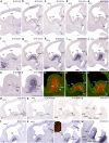
Lateromedial series of sagittal (adjacent) cryostat sections through the MGE at E10.5, illustrating the topography of the earliest Sst cells relative to other markers, Dlx5 and Shh: ae, k Sst; fj, l Dlx5; mo pseudo-color overlap of Sst and Dlx5 images for the indicated levels; ps Sst (different specimens); tw Shh; inset to v pseudo-color overlap of r and v. Note the Sst cells clearly occupy a restricted domain within the Dlx5-positive MGE mantle, which is intercalated between the pallidal and preoptic domains labeled with Shh signal
Fig. 3
Fig. 3
Lateromedial series of sagittal (adjacent) cryostat sections through the MGE at E10.5, illustrating the topography of the earliest Sst cells relative to other markers, Dlx5 and Gbx2: ad Dlx5; eh Sst; il Gbx2; d′, h′, l′, m′–p′ various pseudocolor overlap comparisons; the markers and levels are indicated. Note lack of overlap between Sst cells and the pallidal expression of Gbx2 (which does overlap with Dlx5; see n)
Fig. 4
Fig. 4
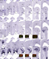
Rostrocaudal series of topologically transversal cryostat sections through the MGE (see plane in Fig. 1a) at E10.5, illustrating in correlative adjacent sections the topography of the earliest Sst cells relative to other markers, Gbx2 and Shh: af Gbx2; gl Sst; inset l′ detail of section caudal to (l); mr Shh; insets i′, j′, p′ pseudocolor overlaps of the indicated markers and levels. Note lack of overlap of Sst cells with the Gbx2- and Shh-positive pallidal mantle elements. Pallidal Gbx2 cells occupy in general more rostral levels than Sst cells, and tend to respect the pallidal marginal stratum. Note also the incipient subpial migration of Sst cells across the marginal pallidal stratum into the striatum (jl). As regards Shh expression, the POA1 ventricular zone appears strongly labeled, and clearly produces a migrating cell stream in the mantle that invades selectively the pallidum (n, o). The adjacent Dg ventricular zone shows less intense and patchy Shh signal as well, all the way to the septal end of this band, which diminishes gradientally toward the amygdaloid pole of the MGE, but does not seem to contribute to the preoptic pallidopetal cell stream in the mantle (mo)
Fig. 5
Fig. 5
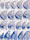
Rostrocaudal series of topologically transversal cryostat sections through the MGE (see plane in Fig. 1a) at E11.5, illustrating in correlative adjacent sections the topography of the Sst cells relative to other markers, Gbx2, Shh, and Nkx2.1: ah Gbx2; ip Sst; qx Shh; y, aa, ac, ae, af Sst; z Nkx2.1; ab, ad Shh; insets l′, m′, o′) pseudocolor overlap of the indicated markers and levels. The red straight lines entered into panels qx indicate the midplane. At this stage, the Gbx2-positive pallidal population extends farther caudalwards, but still essentially does not overlap with the Sst cells at the Dg mantle; isolated Sst cells that apparently do overlap with Pal are marked with arrows in o, p. Note progression of subpial migratory invasion of the striatum by Sst cells (np, y, aa, ac, ae, af). Shh labeling of POA1, Dg, and Pal agrees with the description in Fig. 4; the pallidal Shh-positive mantle respects the marginal stratum occupied by migrating Sst cells (abae)
Fig. 6
Fig. 6
Rostrocaudal series of topologically transversal cryostat sections through the MGE (see plane in Fig. 1a) at E10.5, illustrating in correlative adjacent sections the topography of the Sst cells relative to other markers, Nkx5.1 and Shh: ah Nkx5.1; ip Sst; qx Shh; insets m′–o′, u′–w′ pseudocolor overlap of the indicated markers and levels. There are abundant Nkx5.1 cells in the preoptic and diagonal-septal neighborhoods (ad), as well as in the POA1 mantle layer (eh), without significant overlap with Sst cells. In p, a particularly favourable section plane demonstrates the continuity of Sst cells originated selectively at the Dg domain with the incipient migratory phenomenon at the marginal stratum, without apparent implication of the pallidal domain. Note some Sst cells are adjacent to the POA1 mantle, without intermixing (insets m′–o′). In contrast preoptic Nkx5.1 mantle cells are continuous (and partly mixed) with the pallidopetal Shh-positive migrating cells in the mantle (insets u′–w′). The ventricular zone of Dg clearly expresses patchily Shh (in a septo-amygdaloid decreasing gradient; sx)
Fig. 7
Fig. 7
Examples of sagittal and horizontal sections through E12.5 embryonic brains, showing the migratory dispersion of Sst cells at this stage. ac Lateral to medial set of selected sagittal sections showing the progress of the invasion of the olfactory tuberculum (OT) and striatum (St), as well as the incipient subpial tangential invasion of the pallium (mainly olfactory and insular cortex primordia); the medialmost section c shows a restricted topography of Sst cells within a central area of the Dg, eschewing the larger pallidal mantle, except at its marginal stratum seen in (b). dk Ventrodorsal series of horizontal sections, illustrating the septo-amygdaloid dimension of the studied distribution of Sst cells; the striatal, pallidal, and diagonal domains are delimited tentatively one from another by oblique white or black dash lines. The marginal stratum of the whole olfactory tuberculum is occupied by the dense subpial subpallial migratory stream, where Sst cells stemming from the Dg domain are seen to arrive (SSpM; Dg; dg). Rostrally, labeled cells extend into frontal cortex (FCx); caudally large subpial and subventricular streams of Sst cells invade non-homogeneously the pallial amygdala, beyond the DgA region of the subpallial amygdala (large arrows in eh). Note as well the existence of Sst cells migrating subventricularly across the pallidum into the striatum (small arrows; f, g). The piriform cortex primordium (largely prospective layer III) appears strongly labeled (PirCx; hj)
Fig. 8
Fig. 8
Examples of oblique transversal sections through an E13.5 embryonic brain, showing the migratory dispersion of Sst cells at this stage. The plane of section is indicated by red lines in the inset to a (the section in a corresponds to the right hemisphere, while the other sections illustrate the opposite side). a The obliquity of this section (see inset) aligns the strongly labeled Dg source of Sst cells (Dg) with the path of their migration deep and superficial to the eschewed globus pallidus (curved arrows GP), finally converging at the SSpM (at the olfactory tuberculum), and advancing into the olfactory cortex (PirCx), as well as into subpial and subventricular streams targetting cortical pallium (SvSpM). bj The oblique septo-amygdaloid section plane obtained in these images (compare inset at a) is aligned with the diagonal domain, and in general with the three evaginated subpallial domains (delineated as a whole by a white contour line, with dashed internal limits); the series starts caudally close to the lateral ventricle (b; see the CGE, LGE, MGE bulges) and progresses into the olfactory tuberculum at i, j. The maximal density of labeled cells coincides with the Dg ventricular zone (b, c) and associated periventricular stratum, which forms the supracapsular arch of the medial bed nucleus striae terminalis complex (BSTMsc) over the internal capsule (ic; d, e); beyond this level, the amygdaloid end of the Dg arch displays a very dense aggregate of Sst cells, identified first as the amygdaloid BSTM nucleus (BSTMa) and then as the CA (the primordium of the central amygdaloid nucleus, lateral part) (eg). The latter is continuous superficially—close to the SSpM and the OT—with the diagonal magnocellular nucleus (DgMC; h, i; this was classically misidentified as ‘preoptic magnocellular nucleus’). The other end of the Dg arch constitutes the paraseptal region of the BSTM, which limits with the septum (Se) and the preoptic area (POA) (BSTMps; ce; compare Fig. 1 b). Ventral to the globus pallidus there are dispersed Sst cells within the substantia innominata and the horizontal part of the diagonal band formation (SI; HDB; ej). The pallial amygdala shows substantial invasion by Sst cells of its amygdalo-hippocampal and basolateral/basomedial areas (AHi; cg); in contrast, the medial amygdala (MA) and the posteromedial corticoid area (PMCo) largely remain devoid of these cells (di)
Fig. 9
Fig. 9
Examples of sagittal sections through an E13.5 embryonic brain, showing more advanced migratory dispersion of Sst cells (ac, g, h), and the relationship of the Dg radial domain with the domain of expression of Nkx2.1 (df, j, k); i, l pseudocolor overlap of both markers at the indicated levels. Note that at this stage, the Dg ventricular zone and at least part of its periventricular BSTM formation fall outside the domain of expression of Nkx2.1. Many Sst cells have invaded the isocortical plate at superficial and deep strata
Fig. 10
Fig. 10
Examples of transversal (an) and horizontal (os) sections through E14.5 embryonic brains, showing the migratory dispersion and increasing differentiative stabilization of Sst cells at this stage. an The rostral sections clearly display the superficial subpallial migratory stream advancing from the olfactory tuberculum, just deep to the lateral olfactory tract into the deep stratum of the olfactory cortex and then largely subpially into the insula and isocortical plate (SSpM; lot; CL/I; CP; af); some cells enter medially the septum (Se). The striatal primordium still shows more cells superficially than next to the internal capsule (scarce Sst cells at subventricular levels). The pallidum is practically devoid of labeled cells at the globus pallidus, but shows some small elements within its periventricular (supracapsular) BSTL nucleus (GP; BSTL; fi). As regards the Dg domain, the periventricular BSTM arch is cut obliquely in this plane of section; we see first the paraseptal component, which approaches the crossing of the anterior commissure and the preoptic area (BSTMps; so-called ‘anterior’ BST; fh); the caudal end, composed by the amygdaloid lateral CA and DgMC nuclei, appears close by, internally to the position of the lateral olfactory tract (CA, DgMC; g, h); both elements are soon united by the intermediate supracapsular portion of the BSTM complex (BSTMsc; hj), at shortly thereafter we also see the labeled amygdaloid BST nucleus (BSTMa; jl); the radial continuity of the Dg ventricular zone with its pial surface is populated throughout by Sst cells dispersed in the substantia innominata, converging at the brain surface upon the diagonal band nucleei (vz; SI; DB; fl). Caudolaterally to the CA there appears the pallial amygdala, including the strongly labeled lateral nucleus and the poorly labeled basolateral nucleus (L, BL; il) and the strongly labeled amygdalo-hippocampal area (AHi; m, n). The medial amygdala (MA) only contains scattered labeled cells. os These horizontal sections offer a complementary view of the same anatomic details distinguished in transversal sections
Fig. 11
Fig. 11
Digitally superposed images of adjacent sagittal sections at 6 section levels through an E15.5 embryonic brain, showing the distribution of Sst cells relative to the NKX2.1-immunoreactive domain (inset a′ shows a section lying lateral to a, reacted only for Sst ISH). (a, a′) Lateral sections pass through the pallial amygdala, laterally to the globus pallidus; the central lateral amygdalar nucleus is densely labeled (CA; prospective CeL); note sparse Sst cells within the neighboring basolateral amygdalar nucleus (BL) and more abundant cells at the lateral and basomedial amygdalar nuclear primordia (L, BM); migrating Sst cells have reached the entorhinal cortex, but not yet the hippocampus (ERh; Hi; in b, d). Labeled cells also aggregate more rostrally within the bed nucleus of the external capsule (BEC). bd Section b shows, superposed in green, the image of Shh expression within the medial posteroventral amygdaloid nucleus, a site with scarce Sst cells. These more medial sections also intersect the striatum (dispersed labeled cells in a ventrodorsal gradient); next to it appears the globus pallidus (GP) and the supracapsular lateral BST nucleus (BSTLsc), both of which are NKX2.1 immunoreactive and poor in Sst cells. The pallidal radial domain ending superficially at the olfactory tuberculum (with many labeled cells), and limiting at the ventricular zone with the Sst-positive Dg domain, is tentatively enclosed by black lines in c, d. These lines suggest that the NKX2.1-immunoreactive GP is divided topographically into an external (pallidal) portion and an internal (diagonal) counterpart (EGP, IGP; bd). The diagonal complex (Dg) includes the densely labeled periventricular BSTMsc formation, the IGP, the NKX2.1-immunoreactive substantia innominata (with relatively few intermixed Sst cells) and the superficial horizontal nucleus of the diagonal band (HDB; this has as many labeled cells as the olfactory tuberculum and also contains some NKX2.1-immunoreactive cells). e, f At these more medial section levels, we reach the medial end of the supracapsular BSTM arch (BSTMsc), moving into its paraseptal portion (BSTMps), next to the anterior commissure (ac) and the preoptic area (POA)
Fig. 12
Fig. 12
Selected horizontal sections through an E16.5 embryonic brain, showing the telencephalic distribution of Sst cells at this stage. The subpallial complex—St, Pal, Dg—is contoured by a continuous white line, and white dash lines separate these three domains. a, b The olfactory tuberculum (with many Sst cells) and the basalmost part of the amygdala are cut tangentially (the latter with labeling of the anterior amygdala, AA, and the posterolateral and posteromedial cortical nuclei, PLCo, PMCo). The diagonal band is partly seen (the asterisk marks an artefactual distortion). c, d Levels of section through the crossing of the anterior commissure (ac): intense labeling of the piriform cortex (prospective layer III) continues rostrally into the posterior part of the anterior olfactory area; few cells reach the olfactory bulb, and septal labeling is limited to medial portions (Se). The ventral striatal mantle is well populated by Sst cells (VSt), contrasting with the sparser population within the ventral pallidum (VPal). The diagonal domain is represented by the diagonal band (DB), the substantia innominata (SI), and the paraseptal BSTM nucleus (BSTMps). The subpallial amygdala displays the strongly labeled DgMC and CA derivatives, rostromedially to the relatively unlabeled BL nucleus, whereas the amygdalopiriform area (APi), BM, and AHi amygdalar areas are strongly labeled. e, f The temporal fibers of the anterior commissure traverse the positive IPAC formation (interstitial nucleus of the posterior limb of the anterior commissure) at the back of the striatal mantle, which is limited laterally by a dense population at the bed nucleus of the external capsule, a ventral pallium derivative, jointly with the piirform cortex (BEC; PirCx); caudally, BEC seems continuous with the larger lateral amygdalar nucleus (L), which is also densely labeled. At the interface between L and CA there appears a round or elongated comet-shaped domain devoid of Sst cells that corresponds to the migrating primordium of the nucleus of the lateral olfactory tract (NLOTm). At the rostromedial end of the supracapsular BSTM arch there appears the Dg paraseptal BSTM derivative (BSTMps), which now shows several subdivisions. g, h At these dorsal levels the l amygdalar nucleus diminishes in size, still showing continuity with the BEC, deep to the piriform cortex (PirCx) and next to the striatum (St). A linear claustral aggregate can be distinguished in h (CL). The amygdaloid and paraseptal ends of the BSTM arch meet over the internal capsule and the IGP, forming the supracapsular region (BSTMsc)

References

    1. Anderson SA, Marín O, Horn C, Jennings K, Rubenstein JL. Distinct cortical migrations from the medial and lateral ganglionic eminences. Development. 2001;128:353–363. - PubMed
    1. Bardet SM, Cobos I, Puelles E, Martinez-de-la-Torre M, Puelles L. Chicken lateral septal organ and other circumventricular organs form in a striatal subdomain abutting the molecular striatopallidal border. J Comp Neurol. 2006;499:745–767. doi: 10.1002/cne.21121. - DOI - PubMed
    1. Bardet SM, Ferran JL, Sanchez-Arrones L, Puelles L. Ontogenetic expression of sonic hedgehog in the chicken subpallium. Front Neuroanat. 2010;21:1–16. - PMC - PubMed
    1. Bayer SA. Neurogenetic and morphogenetic heterogeneity in the bed nucleus of the stria terminalis. J Comp Neurol. 1987;265:47–64. doi: 10.1002/cne.902650105. - DOI - PubMed
    1. Bayer SA, Altman J. Development of the endopiriform nucleus and the claustrum in the rat brain. Neuroscience. 1991;45:391–412. doi: 10.1016/0306-4522(91)90236-H. - DOI - PubMed

Publication types

LinkOut - more resources