

Comparison Among Methods of Retinopathy Assessment (CAMRA) Study: Smartphone, Nonmydriatic, and Mydriatic Photography
- PMID: 26189190
- PMCID: PMC4581972
- DOI: 10.1016/j.ophtha.2015.06.011
Comparison Among Methods of Retinopathy Assessment (CAMRA) Study: Smartphone, Nonmydriatic, and Mydriatic Photography
Abstract
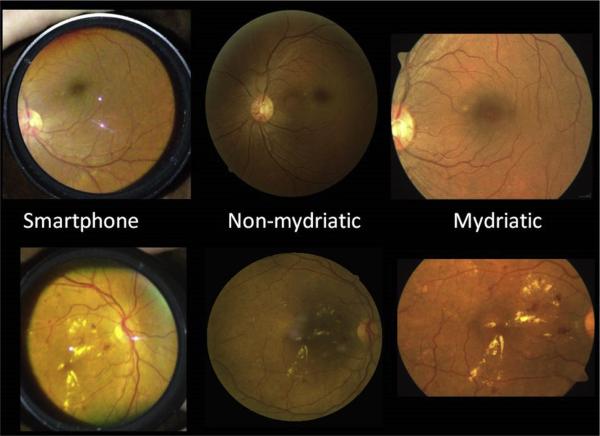
Purpose: We compared smartphone fundus photography, nonmydriatic fundus photography, and 7-field mydriatic fundus photography for their abilities to detect and grade diabetic retinopathy (DR).
Design: This was a prospective, comparative study of 3 photography modalities.
Participants: Diabetic patients (n = 300) were recruited at the ophthalmology clinic of a tertiary diabetes care center in Chennai, India.
Methods: Patients underwent photography by all 3 modalities, and photographs were evaluated by 2 retina specialists.
Main outcome measures: The sensitivity and specificity in the detection of DR for both smartphone and nonmydriatic photography were determined by comparison with the standard method, 7-field mydriatic fundus photography.
Results: The sensitivity and specificity of smartphone fundus photography, compared with 7-field mydriatic fundus photography, for the detection of any DR were 50% (95% confidence interval [CI], 43-56) and 94% (95% CI, 92-97), respectively, and of nonmydriatic fundus photography were 81% (95% CI, 75-86) and 94% (95% CI, 92-96%), respectively. The sensitivity and specificity of smartphone fundus photography for the detection of vision-threatening DR were 59% (95% CI, 46-72) and 100% (95% CI, 99-100), respectively, and of nonmydriatic fundus photography were 54% (95% CI, 40-67) and 99% (95% CI, 98-100), respectively.
Conclusions: Smartphone and nonmydriatic fundus photography are each able to detect DR and sight-threatening disease. However, the nonmydriatic camera is more sensitive at detecting DR than the smartphone. At this time, the benefits of the smartphone (connectivity, portability, and reduced cost) are not offset by the lack of sufficient sensitivity for detection of DR in most clinical circumstances.
Copyright © 2015 American Academy of Ophthalmology. Published by Elsevier Inc. All rights reserved.
Figures
References
-
- IDF . Diabetes Atlas. 6th ed. Brussels: International Diabetes Federation; 2013. [November 18, 2013]. Available at: http://www.idf.org/sites/default/files/EN_6E_Atlas_Full.pdf.
-
- Whiting DR, Guariguata L, Weil C, Shaw J. IDF diabetes atlas: global estimates of the prevalence of diabetes for 2011 and 2030. Diabetes Res Clin Pract. 2011;94:311–21. - PubMed
-
- Anjana RM, Pradeepa R, Deepa M, et al. Prevalence of diabetes and prediabetes (impaired fasting glucose and/or impaired glucose tolerance) in urban and rural India: phase I results of the Indian Council of Medical Research-INdia DIABetes (ICMRINDIAB) study. Diabetologia. 2011;54:3022–7. - PubMed
Publication types
MeSH terms
Substances
Grants and funding
LinkOut - more resources
Full Text Sources
Other Literature Sources
Medical
