Review
. 2015 Aug;16(8):469-86.
doi: 10.1038/nrn3978.

Immune mediators in the brain and peripheral tissues in autism spectrum disorder

Affiliations
Review

Immune mediators in the brain and peripheral tissues in autism spectrum disorder

Myka L Estes et al. Nat Rev Neurosci. 2015 Aug.

Abstract

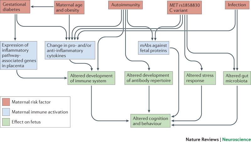
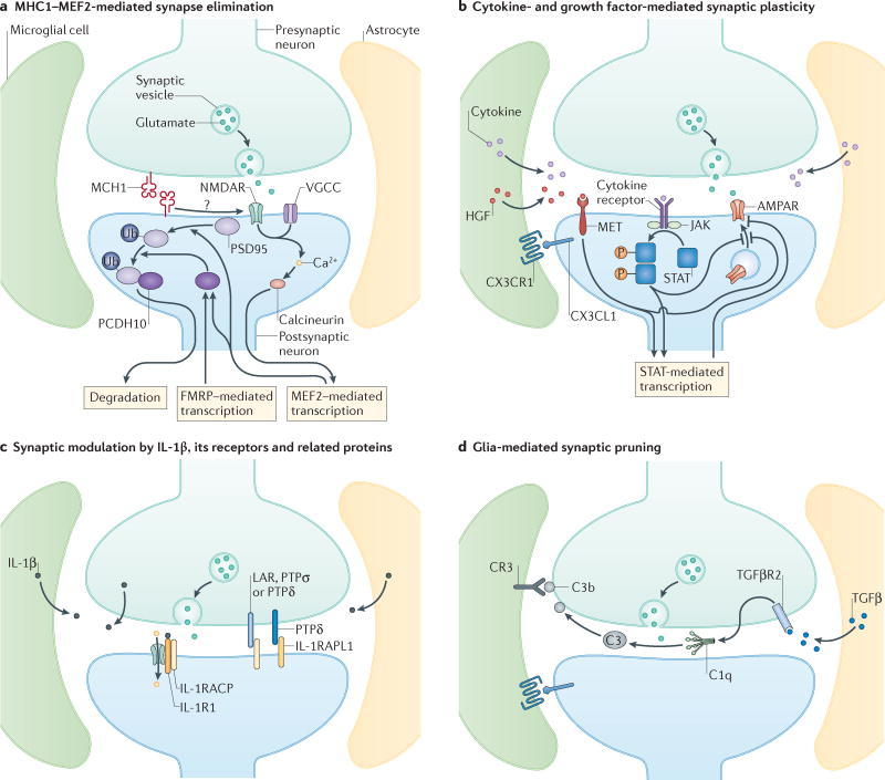
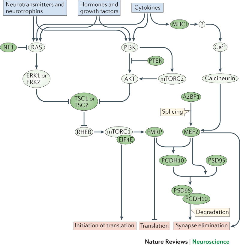
Increasing evidence points to a central role for immune dysregulation in autism spectrum disorder (ASD). Several ASD risk genes encode components of the immune system and many maternal immune system-related risk factors--including autoimmunity, infection and fetal reactive antibodies--are associated with ASD. In addition, there is evidence of ongoing immune dysregulation in individuals with ASD and in animal models of this disorder. Recently, several molecular signalling pathways--including pathways downstream of cytokines, the receptor MET, major histocompatibility complex class I molecules, microglia and complement factors--have been identified that link immune activation to ASD phenotypes. Together, these findings indicate that the immune system is a point of convergence for multiple ASD-related genetic and environmental risk factors.


Conflict of interest statement

Competing interests statement: The authors declare no competing interests.

Figures

Figure 1
Figure 1. ASD risk factors during pregnancy converge on maternal immune system activation
Maternal autoimmunity, infection during pregnancy, maternal age and obesity, gestational diabetes, and maternal MET variant rs1858830 ‘C’ allele are all associated with a higher incidence of ASD. These risk factors (red boxes) cause maternal immune activation (MIA) (blue boxes), which manifests as changes in the maternal peripheral cytokine milieu, generation of IgG maternal autoantibodies (mAbs) that are reactive to fetal proteins and activation of inflammatory pathway genes within the placenta. Based on findings in animal models, MIA is sufficient to induce long-lasting changes in brain development, gut microbiota, immune and endocrine systems of the developing fetus (green boxes).
Figure 2
Figure 2. Immune molecules at glutamatergic synapses
At the ‘quad-partite’ synapse, pre- and postsynaptic neurons, astrocytes and microglia communicate using immune mediators, many of which are altered in individuals with ASD. Each panel represents molecular pathways that are used by immune molecules at the synapse to regulate synapse formation and/or plasticity. a | MHCI molecules are located at synapses, where they act through calcineurin to activate MEF2 transcription factors to negatively regulate synapse strength and density. The MHCI-dependent activation of MEF2 requires calcium influx through NMDARs and voltage-gated calcium channels (VGCCs). MEF2 acts in concert with FMRP to stimulate the ubiquitination of PSD-95 and increased association with protocadherin 10, which then chaperons PSD-95 to proteosomes. b | In general, cytokines released by astrocytes, microglia, and/or neurons bind to their specific receptors and activate JAK/STAT which regulates signalling at the synapse and alters transcription, leading to negative regulation of AMPA receptor (AMPAR) expression either through inhibiting new insertion or increasing internalization. Growth factors, especially HGF, are also thought to be secreted from glial cells into the synaptic cleft, where they bind and activate the MET receptor, which negatively regulates AMPAR expression possibly through STAT-mediated transcription. Chemokines are also presented by neurons and bind to receptors on glial cells, depicted here for the interaction between CX3C chemokine receptor 1 (CX3CR1) on microglia and its ligand, CX3CL1, secreted by neurons or expressed in a tethered form on the neuronal cell surface. CX3CR1-CX3CL1 signaling is required for the migration of sufficient numbers of microglia into the brain in early development, and synaptic plasticity under physiological conditions. c | IL-1β exerts distinct effects at the synapse. Binding to IL-1R1 recruits the IL-1 accessory receptor (IL-1AcP), which increases NMDAR signaling. Unbound IL-1RAcP acts as a trans-synaptic adhesion molecule through its interactions with pre-synaptic PTPσ, PTPδ and LAR. The IL-1R accessory-like receptor 1 (IL-1RAPL1) also acts as a synaptic organizer binding to pre-synaptic PTPδ. These trans-synaptic interactions exert multiple effects on synapse formation and plasticity. d | Astrocyte-secreted TGF-β binds to neuronal TGFβRII (placed here presynaptically due to findings at the neuromuscular junction), which induces neuronal secretion of the complement protein C1q. C1q initiates the complement cascade leading to cleavage of C3 into C3b, which binds to synaptic surfaces. Microglial expressed complement receptor 3 recognizes tagged synapses and initiates synaptic pruning at a subset of synapses. CX3CR1-CX3CL1 is also required for microglia-mediated synaptic pruning in early development, and spine elimination and formation in mature circuits. While currently unknown, local neuron-microglia signaling through CX3CR1-CX3CL1 may serve as an instructive signal for complement-mediated synaptic pruning.
Figure 3
Figure 3. Synaptic immune signaling converges on mTOR
Immune and neuronal receptor signaling activate molecular pathways, which feed into the mTOR pathway and activate MEF2-dependent transcriptional regulation. These central signalling pathways may become dysregulated through genetic mutations or environmental exposures associated with ASD and thereby alter neural development and function. mTOR activity regulates numerous processes including protein synthesis, mitochondrial function, lipid synthesis, cell growth and proliferation, synaptic plasticity, neurogenesis, neuronal cell death, ion channel expression and cytoskeletal dynamics. Importantly, mTORC1 regulates the synthesis of glutamatergic receptors and protein products, including SHANK, SAPAP, neuroligins, AMPA and NMDA receptor subunits, many of which are genetically associated with ASD. Mutations in the genes that cause most of the syndromic forms of ASD—FMR1, NF1, PTEN, and TCS1/2—disrupt components of the mTOR signaling pathway. The MEF2 transcription factor is also implicated in ASD. MEF2C haploinsufficiency syndrome is characterized by ASD-like behaviors, perhaps through the function of MEF2 in regulating transcription during synapse formation and elimination. MEF2 also likely regulates the expression of cytokine receptors in a positive or negative feedback loop, since the promoters of some cytokine receptors (like Il1rapl1, for example) contain a MEF2 binding motif. MEF2 is also a target of the splicing regulator A2BP1—the central gene in a synaptic module identified in a transcriptome analysis of brain tissue from individuals with ASD. Green fill indicates protein products of genes associated with ASD.

References

    1. King BH, Navot N, Bernier R, Webb SJ. Update on diagnostic classification in autism. Curr Opin Psychiatry. 2014;27:105–109. - PMC - PubMed
    1. CDC. Prevalence of Autism Spectrum Disorder Among Children Aged 8 Years--Autism and Developmental Disabilities Monitoring Network, 11 Sites, United States, 2010. In: Baio J, editor. Morbidity and Mortality Weekly Report. Vol. 63. Naitonal Center on Birth Defects and Developmental Disabilities, CDC; 2014. pp. 1–21. - PubMed
    1. Hertz-Picciotto I, Delwiche L. The rise in autism and the role of age at diagnosis. Epidemiology. 2009;20:84–90. - PMC - PubMed
    1. Hallmayer J, et al. Genetic heritability and shared environmental factors among twin pairs with autism. Arch Gen Psychiatry. 2011;68:1095–1102. - PMC - PubMed
    1. Rosenberg RE, et al. Characteristics and concordance of autism spectrum disorders among 277 twin pairs. Arch Pediatr Adolesc Med. 2009;163:907–914. - PubMed

Publication types