. 2016 Mar 31;35(13):1703-15.
doi: 10.1038/onc.2015.235. Epub 2015 Jul 20.

RNA-binding protein HuR sequesters microRNA-21 to prevent translation repression of proinflammatory tumor suppressor gene programmed cell death 4

Affiliations

RNA-binding protein HuR sequesters microRNA-21 to prevent translation repression of proinflammatory tumor suppressor gene programmed cell death 4

D K Poria et al. Oncogene. .

Abstract

Translation control of proinflammatory genes has a crucial role in regulating the inflammatory response and preventing chronic inflammation, including a transition to cancer. The proinflammatory tumor suppressor protein programmed cell death 4 (PDCD4) is important for maintaining the balance between inflammation and tumorigenesis. PDCD4 messenger RNA translation is inhibited by the oncogenic microRNA, miR-21. AU-rich element-binding protein HuR was found to interact with the PDCD4 3'-untranslated region (UTR) and prevent miR-21-mediated repression of PDCD4 translation. Cells stably expressing miR-21 showed higher proliferation and reduced apoptosis, which was reversed by HuR expression. Inflammatory stimulus caused nuclear-cytoplasmic relocalization of HuR, reversing the translation repression of PDCD4. Unprecedentedly, HuR was also found to bind to miR-21 directly, preventing its interaction with the PDCD4 3'-UTR, thereby preventing the translation repression of PDCD4. This suggests that HuR might act as a 'miRNA sponge' to regulate miRNA-mediated translation regulation under conditions of stress-induced nuclear-cytoplasmic translocation of HuR, which would allow fine-tuned gene expression in complex regulatory environments.


Figures
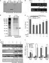
Figure 1
Figure 1
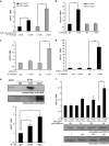
HuR binds to the PDCD4 mRNA 3′-UTR and reverses miR-21-mediated translation repression. (a) MCF7 cell lysates were immunoprecipitated with anti-HuR antibody or control IgG. RNA associated with the immunoprecipitate was subjected to RT–PCR using PDCD4 or GAPDH primers. (b) 32P-UTP-labeled PDCD4 3′-UTR RNA was incubated with MCF7 S10 cytoplasmic lysate, UV-crosslinked, digested with RNase-A. The RNase A digested reactions were immunoprecipitated with anti-HuR antibody or control IgG, and resolved on SDS-12% PAGE. The band corresponding to HuR is indicated by arrow. (c) Immunoblots of lysates of MCF7 cells transfected with two increasing concentrations of pSUP-miR-21 and co-transfected with two increasing concentrations of pCI-neo-myc-HuR together with the higher amount of pSUP-miR-21, probed with anti-PDCD4, anti-myc, anti-HuR and anti-GAPDH antibodies. The two bands in the anti-HuR immunoblot represent endogenous HuR and exogenously expressed myc-tagged HuR. Densitometric quantitation of PDCD4 protein bands, normalized to GAPDH, is given. (d) RT–PCR of total RNA isolated from MCF7 cells transfected with miR-21 and HuR-expressing plasmids with miR-21 and u6B snRNA primers. Densitometric quantitation of miR-21 PCR products, normalized to U6B snRNA, is given. (e) MCF7 cells transfected with Fluc-PDCD4 3′-UTR reporter gene construct and pCMV-Rluc were co-transfected with two increasing concentrations of pSUP-miR-21 and of three increasing concentrations of pCI-neo-myc-HuR in presence of the higher amount of pSUP-miR-21. Fluc values are normalized to Rluc values as transfection control. (f) MCF7 cells were either untransfected (-miR-21/-HuR) or transfected with pSUP-miR-21 together with (+miR-21/+HuR) or without pCI-neo-myc-HuR (+miR-21/-HuR) or with pCI-neo-myc-HuR alone (-miR-21/+HuR). Cell lysates were immunoprecipitated with anti-HuR antibody, anti-Ago2 antibody and control IgG. RNA associated with the immunoprecipitates was subjected to qRT–PCR using PDCD4 and GAPDH primers, and PDCD4 mRNA levels were normalized to GAPDH mRNA levels. Mean±s.d. from three experiments are represented in all graphs. * signifies a P-value⩽0.05 and ** signifies a P-value⩽0.01 (paired one-tailed t-test).
Figure 2
Figure 2
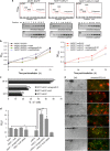
Derepression of PDCD4 translation by HuR counteracts miR-21-mediated increase in cell proliferation and decrease in apoptosis. (a) Ribosomal fractions from MCF7-EGFP, MCF7-miR-21 and MCF7-miR-21 cell line transfected with pCI-neo-myc-HuR were analysed by 10–50% sucrose density gradient fractionation. Ribosomal RNA content, measured at 254 nm, is plotted against fraction numbers. RNA isolated from selected fractions were analyzed by semiquantative RT–PCR using PDCD4 and GAPDH primers. (b) MCF7-EGFP and MCF7-miR-21 cell lines transfected with pCI-neo-myc-HuR or antagomiR against miR-21 or siRNA against HuR were allowed to grow post transfection and MTT assay was performed at various time points. OD595 readings are plotted. (c) MCF7-miR-21 cell line was transfected with pCI-neo-myc-HuR or antagomiR-21 and 48 h post transfection, DNA content of cells was analyzed by PI staining followed by flow cytometry. (d) MCF7-EGFP and MCF7-miR-21 cell lines transfected with pCI-neo-myc-HuR or an antagomiR against miR-21 or siRNA against HuR were serum-starved for 48 h post transfection. Caspase activation was measured using a caspase 3/7 assay using a luminescent substrate. RLU values are normalized to that of MCF7-EGFP cells, taken as 1. (e) MCF7 cells were either mock transfected, transfected with pSUP-miR-21 alone or together with pCI-neo-myc-HuR or HuR siRNA or with antagomiR-21 or HuR alone and then serum starved for 48 h to induce apoptosis. Cells were stained with AnexinV-FITC and PI to detect apoptosis. Mean±s.d. from three experiments done in duplicate are represented in all graphs. * signifies a P-value⩽0.05 and ** signifies a P-value⩽0.01 (t-test) in all graphs.
Figure 3
Figure 3
LPS treatment causes nuclear-cytoplasmic relocalization of HuR and prevents miR-21-mediated repression of PDCD4. (a) MCF7 cells were treated with LPS, and immunofluorescence of cells collected at various time points was observed using anti-HuR primary and AlexaFluor568-conjugated secondary antibodies. Nucleus was visualized using DAPI staining. Right panel shows merge of the DIC and fluorescent images. (b) Lysates of cells treated with LPS for 4 h were immunoprecipitated with anti-HuR antibody and control IgG. RNA associated with the immunoprecipitates was subjected to qRT–PCR using PDCD4 and GAPDH-specific primers, and PDCD4 mRNA levels were normalized to GAPDH mRNA levels. The data represent fold excess of normalized PDCD4 mRNA in HuR immunoprecipitate over IgG immunoprecipitate. Mean±s.d. from three experiments are represented. * signifies a P-value⩽0.05 and ** signifies a P-value⩽0.01 (t-test) (c). MCF-EGFP, MCF7-miR-21 and MCF7-miR-21 cell line transfected with HuR siRNA were treated with LPS for 4 h and cell lysates were immunoblotted using anti-PDCD4, anti-HuR and anti-β-actin antibodies. Densitometric quantitation of PDCD4 protein bands, normalized to β-actin, is given.
Figure 4
Figure 4
HuR acts in trans to prevent miR-21 mediated translation repression of PDCD4. (a) Schematic diagram of full-length (FL) PDCD4 3′-UTR and various deletions from the 5′- and 3′-ends. The miR-21 target site (nt 228–249) as well as the HuR-binding sites, as determined from the RNA-protein interaction studies (below), are indicated. (b) 32P-UTP labeled full-length and various deletion mutants of PDCD4 3′-UTR RNA were incubated with purified HuR protein, UV-crosslinked, digested with RNase A and resolved on 10% SDS–PAGE. (c) Reporter gene constructs containing full-length and various deletion mutants of PDCD4 3′-UTR were transfected into MCF cells together with pSUP-miR-21 or pCI-Neo-myc-HuR or both. (d) Reporter gene constructs containing full-length or various mutants (HuR binding and miR-21 binding) of the PDCD4 3′-UTR were co-transfected with pSUP-miR-21 vector and treated with LPS for 4 h. Mean±s.d. of Fluc/Rluc values, normalized to control, from three independent experiments done in duplicate are represented in all graphs. * signifies a P-value⩽0.05 and ** signifies a P-value⩽0.01 (t-test) in all graphs.
Figure 5
Figure 5
HuR binds directly with miR-21. (a) Sequence of miR-21 (guide strand) and target site in PDCD4 3′-UTR with the ARE within the seed sequence of miR-21 indicated. (b) Cy5-labeled fluorescent RNA corresponding to miR-21, miR-125b, ARE-mutated miR-21, a non-specific RNA of equivalent length were incubated with purified HuR protein and the RNA-protein complex resolved by native PAGE. miR-21 was also incubated with purified BSA followed by native PAGE. (c) Isothermal Titration Calorimetry (ITC) was performed using in vitro transcribed miR-21 RNA as analyte and purified HuR protein as titrant. Heat change on injection of titrant was measured (upper panel) and heat change per mole of titrant was plotted against the molar ratio of titrant to analyte. (d) Filter binding assay using 32P-ATP end-labeled RNA corresponding to miR-21, ARE-mutant miR-21, double-stranded miR-21, miR-125b and a non-specific RNA of equivalent length and purified HuR protein. Only single-stranded miR-21 (blue line) showed significant binding. miR-125b and non-specific RNA did not show any binding at all. The miR-21 binding curve was fitted using a polynomial function and the Kd was computed from the curve. Mean±s.d. from three experiments are represented.
Figure 6
Figure 6
HuR binds to miR-21 in cells independently of PDCD4 mRNA. (a) Lysates from miR-21 and EGFP-expressing cell lines in which HuR protein was overexpressed were immunoprecipitated with anti-HuR and control IgG antibodies. qRT–PCR was done with immunoprecipitated RNA using miR-21 and U6B snRNA-specific primers. (b) Lysates from miR-21 and EGFP-expressing cell lines were immunoprecipitated with anti-HuR and control IgG antibodies followed by qRT–PCR using PDCD4 and GAPDH-specific primers. (c) MCF7 cells were treated with LPS for 4 h, followed by immunoprecipitation with anti-HuR and control IgG antibodies and qRT–PCR using miR-21 and U6B snRNA-specific primers. (d) Lysates from MCF7 cells in which PDCD4 3′-UTR RNA has been overexpressed were immunoprecipitated with anti-HuR and control IgG antibodies followed by qRT–PCR using miR-21 and U6B-specific primers. The data show fold difference in miR-21 levels (normalized to U6B snRNA levels) in the HuR immunoprecipitates compared with IgG immunoprecipitates in panels a, c and d and PDCD4 mRNA (normalized to GAPDH levels) in panel b. (e) Lysates from MCF7 cells were immunoprecipitated with anti-HuR and anti-Ago2 antibodies and the immunoprecipitates resolved on SDS–PAGE and blotted with anti-HuR and anti-Ago2 antibodies (upper panel). Quantative RT–PCR was performed with RNA isolated from the immunoprecipitates using miR-21 and U6B snRNA-specific primers. The data show fold difference in miR-21 levels (normalized to U6B snRNA levels) in the HuR and Ago2 immunoprecipitates compared with IgG immunoprecipitates (lower panel). (f) MCF7 cells were treated with LPS for 0–4 h, followed by immunoprecipitation with anti-HuR and control IgG antibodies and qRT–PCR using miR-21 and U6B snRNA-specific primers. Fold difference in miR-21 level (normalized to U6B level) in HuR immunoprecipitate compared with IgG immunoprecipitate is shown. The cell lysates were immunoblotted with anti-PDCD4 and GAPDH antibodies (lower panel). Densitometric quantitation of PDCD4 protein bands, normalized to GAPDH, is given. Mean±s.d. from three experiments are represented in all graphs. * signifies a P-value⩽0.05 and ** signifies a P-value⩽0.01 (t-test) in all graphs.
Figure 7
Figure 7
Integration of miR-21 and HuR-mediated regulation and model of the ‘miRNA sponge‘. (a) Intersection of mRNAs having HuR and miRNA-binding sites with predicted miR-21 target mRNAs. The intersection of 109 mRNAs represent the co-regulon of HuR and miR-21. (b) Pie-chart showing distribution of various AREs and HuR-binding sites in miRNA sequences from miRbase. Total number of miRNAs with ARE sequences or HuR-binding sites is 672 and total number of AREs and HuR-binding sites is 1245. (c) Frequency distribution showing number of AREs per miRNA sequence. (d) Proposed model showing dynamic equilibrium between miR-21 and HuR binding to PDCD4 mRNA and ‘miRNA sponge‘ function of HuR. High level of miR-21 allows miRNA-RISC complex to be loaded on PDCD4 mRNA 3′-UTR and prevents HuR binding to the 3′-UTR, causing translation repression. High level of HuR in the cytoplasm sequesters miR-21, and causes conformational change in the PDCD4 3′-UTR, causing more of miR-21 to dissociate from PDCD4 mRNA 3′-UTR and RISC complex, leading to translation derepression.

References

    1. Lankat-Buttgereit B, Göke R. The tumour suppressor Pdcd4: recent advances in the elucidation of function and regulation. Biol Cell 2009; 101: 309–317. - PubMed
    1. Hilliard A, Hilliard B, Zheng S-J, Sun H, Miwa T, Song W et al. Translational regulation of autoimmune inflammation and lymphoma genesis by programmed cell death 4. J Immunol 2006; 177: 8095–8102. - PubMed
    1. Biyanee A, Ohnheiser J, Singh P, Klempnauer K-H. A novel mechanism for the control of translation of specific mRNAs by tumor suppressor protein Pdcd4: inhibition of translation elongation. Oncogene 2014; 34: 1384–1392. - PubMed
    1. Anderson P. Post-transcriptional regulons coordinate the initiation and resolution of inflammation. Nat Rev Immunol 2010; 10: 24–35. - PubMed
    1. Gebauer F, Hentze MW. Molecular mechanisms of translational control. Nat Rev Mol Cell Biol 2004; 5: 827–835. - PMC - PubMed

Publication types

MeSH terms