

3-D volumetric MRI evaluation of the placenta in fetuses with complex congenital heart disease
- PMID: 26190037
- PMCID: PMC4554892
- DOI: 10.1016/j.placenta.2015.06.013
3-D volumetric MRI evaluation of the placenta in fetuses with complex congenital heart disease
Abstract
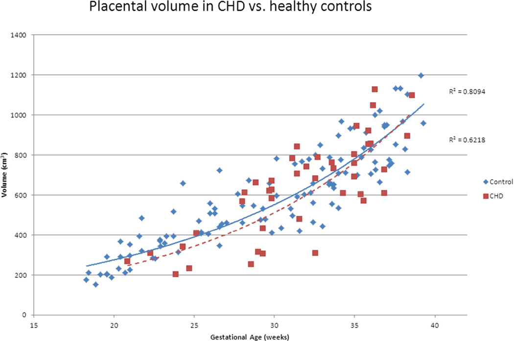
Introduction: Placental insufficiency remains a common cause of perinatal mortality and neurodevelopmental morbidity. Congenital heart disease (CHD) in the fetus and its relationship to placental function is unknown. This study explores placental health and its relationship to neonatal outcomes by comparing placental volumes in healthy pregnancies and pregnancies complicated by CHD using in vivo three-dimensional MRI studies.
Methods: In a prospective observational study, pregnant women greater than 18 weeks gestation with normal pregnancies or pregnancies complicated by CHD were recruited and underwent fetal MR imaging. The placenta was manually outlined and the volume was calculated in cm(3). Brain volume was also calculated and clinical data were also collected. Relationships, including interactive effects, between placental and fetal growth, including brain growth, were evaluated using longitudinal multiple linear regression analysis.
Results: 135 women underwent fetal MRI between 18 and 39 weeks gestation (mean 31.6 ± 4.4). Placental volume increased exponentially with gestational age (p = 0.041). Placental volume was positively associated with birth weight (p < 0.001) and increased more steeply with birth weight in CHD-affected fetuses (p = 0.046). Total brain and cerebral volumes were smaller in the CHD group (p < 0.001), but brainstem volume (p < 0.001) was larger. Placental volumes were not associated with brain volumes.
Discussion: Impaired placental growth in CHD is associated with gestational age and birth weight at delivery. Abnormalities in placental development may contribute to the significant morbidity in this high-risk population. Assessment of placental volume by MRI allows for in vivo assessments of placental development.
Keywords: Congenital heart disease; Fetal MRI; Placenta; Volumetric assessment.
Copyright © 2015 Elsevier Ltd. All rights reserved.
Conflict of interest statement
Nickie Andescavage: I have no conflicts of interest to disclose.
Alexa Yarish: I have no conflicts of interest to disclose.
Mary Donofrio: I have no conflicts of interest to disclose.
Dorothy Bulas: I have no conflicts of interest to disclose.
Iordanis Evangelou: I have no conflicts of interest to disclose.
Gilbert Vezina: I have no conflicts of interest to disclose.
Robert McCarter: I have no conflicts of interest to disclose.
Adre DuPlessis: I have no conflicts of interest to disclose.
Catherine Limperopoulos: I have no conflicts of interest to disclose.
Figures


References
-
- Gagnon R. Placental insufficiency and its consequences. Eur J Obstet Gynecol Reprod Biol. 2003 Sep 22;110(Suppl 1):S99–S107. - PubMed
-
- Redline RW. Disorders of placental circulation and the fetal brain. Clin Perinatol. 2009 Sep;36(3):549–559. - PubMed
-
- Jurewicz J, Polanska K, Hanke W. Chemical exposure early in life and the neurodevelopment of children--an overview of current epidemiological evidence. Ann Agric Environ Med. 20(3):465–486. - PubMed
-
- O'Donnell K, O'Connor TG, Glover V. Prenatal stress and neurodevelopment of the child: focus on the HPA axis and role of the placenta. Dev Neurosci. 2009;31(4):285–292. - PubMed
-
- Pawluski JL, Brummelte S, Barha CK, Crozier TM, Galea LA. Effects of steroid hormones on neurogenesis in the hippocampus of the adult female rodent during the estrous cycle, pregnancy, lactation and aging. Front Neuroendocrinol. 2009 Aug;30(3):343–357. - PubMed
Publication types
MeSH terms
Grants and funding
LinkOut - more resources
Full Text Sources
Other Literature Sources
Medical
