. 2015 Jul 27;34(2):229-41.
doi: 10.1016/j.devcel.2015.06.021. Epub 2015 Jul 16.

A synthetic niche for nephron progenitor cells

Affiliations

A synthetic niche for nephron progenitor cells

Aaron C Brown et al. Dev Cell. .

Abstract

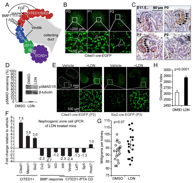
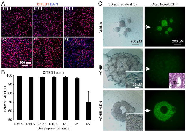
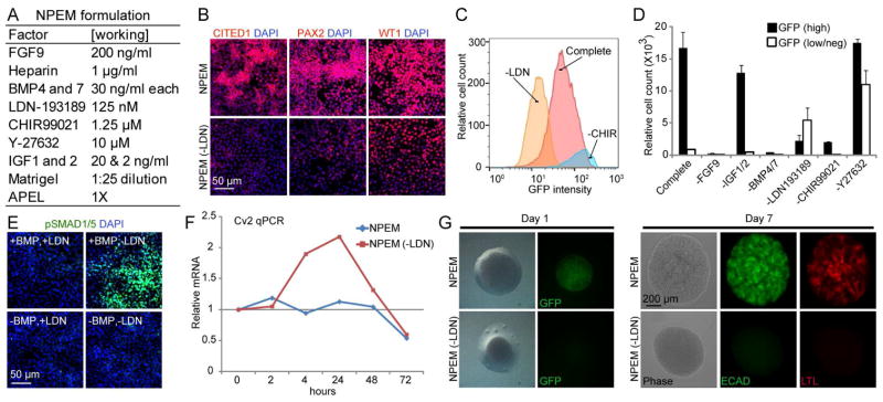
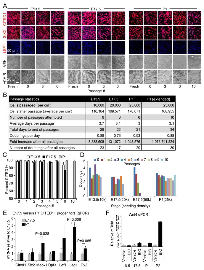
FGF, BMP, and WNT balance embryonic nephron progenitor cell (NPC) renewal and differentiation. By modulating these pathways, we have created an in vitro niche in which NPCs from embryonic kidneys or derived from human embryonic stem cells can be propagated. NPC cultures expanded up to one billion-fold in this environment can be induced to form tubules expressing nephron differentiation markers. Single-cell culture reveals phenotypic variability within the early CITED1-expressing NPC compartment, indicating that it is a mixture of cells with varying progenitor potential. Furthermore, we find that the developmental age of NPCs does not correlate with propagation capacity, indicating that cessation of nephrogenesis is related to factors other than an intrinsic clock. This in vitro nephron progenitor niche will have important applications for expansion of cells for engraftment and will facilitate investigation of mechanisms that determine the balance between renewal and differentiation in these cells.


Figures

Figure 1
Figure 1. SMAD inhibition with LDN-193189 retains NPCs in the CITED1 compartment in vivo
(A) Schematic of cap mesenchyme compartments and key signaling pathways required for their maintenance and differentiation. (B) Cited1creERT2-EGFP kidneys harvested at postnatal stages. GFP expression in cap mesenchymes (c). (C) Immunostaining of pSMAD1/5 (black arrows) in kidney sections isolated from E17.5 to P1. Cap mesenchymes are outlined with dotted black lines. (D) pSMAD1/5 immunoblot of NZCs after intraperitoneal injection of P0 pups with either vehicle or 3 mg/kg of LDN twice daily until P2. Percent remaining after LDN treatment quantified by densitometry and normalized to β-tubulin in graph. NZCs were isolated from 4 kidney pairs per treatment group and pooled. (E) Fluorescent imaging of kidneys from Cited1 or Six2 EGFP mice in vehicle and LDN treated animals. Representative image from 4 kidney pairs per group shown. (F) Cap mesenchyme marker analysis of isolated NZCs by qPCR. Data represent the mean ±SD of qPCR technical replicates from 5 (DMSO) and 6 (LDN) pooled kidney pairs. (G) Distribution of kidney weights from P0 mice treated for 2 days with DMSO or LDN and harvested at 2 weeks. Error bars represent the mean ±SE (standard error) of weights and P-value is derived from the Student’s t-test. (H) Relative number of glomeruli counted per kidney (glomerular index). Kidneys from 7 mice per group were serially sectioned and a section was counted every 100 μM. Error bars represent the mean ±SE and P-value is derived from the Student’s t-test.
Figure 2
Figure 2. SMAD inhibition with LDN-193189 retains NPCs in the CITED1 compartment in vitro
(A) CITED1 immunostaining of freshly plated monolayer cultures of purified progenitors isolated at developmental time points. (B) Quantitation of CITED1+ progenitor purity from images shown in (A). Error bars represent average values ±SD calculated from individual culture well replicates. (C) Stereo microscopy and GFP expression of purified CITED1+ progenitors in aggregate culture isolated from Cited1creERT2-EGFP kidneys and treated with CHIR (3 μM) and LDN (75 nM). Insets (stereo and H&E – 50 μM) show tubules (T) with lumens after CHIR only treatment.
Figure 3
Figure 3. SMAD inhibition maintains nephron progenitor potential
(A) NPEM formulation. (B) Immunostaining of CITED1+ progenitors expanded in NPEM for 3 days in the presence or absence of LDN. (C) Flow cytometry histogram of CITED1+ progenitors isolated from Cited1creERT2-EGFP mice propagated in complete NPEM or in the absence of the indicated factors. (D) Quantitation of GFP intensity (GFP (high) or GFP (low/neg)) by flow cytometry to quantify progenitor cell state in the absence of individual factors. ±SD calculated from culture well replicates shown. (E) pSMAD1/5 immunostaining of CITED1+ progenitors grown in NPEM with or without LDN or BMP. (F) Cv2 expression in CITED1+ progenitors grown in NPEM in the presence and absence of LDN over 72 hours. (G) Aggregate culture of CITED1+ progenitors isolated from Cited1creERT2-EGFP mice that were initially expanded for 3 days in monolayer in the presence or absence of LDN. See also Figures S1 and S2.
Figure 4
Figure 4. FGF, BMP and WNT expand functionally competent CITED1+ NPCs
(A) Immunostaining of CITED1+ progenitors expanded in NPEM. DAPI shown in blue. Bottom two panels show stereomicroscopy of aggregate cultures demonstrating formation of tubule-like structures with CHIR treatment. VEH indicates vehicle control. (B) Passage statistics for CITED1+ progenitors from each developmental stage. E13.5 and E17.5 progenitors were passaged 6 times, whereas P1 progenitors were cultured to passage 10 (P1 extended). (C) Percentage CITED1+ progenitors after each passage. Error bars represent mean ±SD from immunofluorescence images for each passage. (D) Number of CITED1+ progenitor doublings after each passage for the seeding densities indicated (k = x1000). (E) Expression of cap mesenchyme transcripts in freshly isolated CITED1+ progenitors. (F) Wnt4 expression in CITED1+ progenitors cultured in keratinocyte basal medium and treated with BIO (0.5 μM) for 6 hours. Average values ± SE in (E) and (F) shown. See also Figures S3 and S4.
Figure 5
Figure 5. Expanded NPCs retain differentiation potential
(A) Time course images of expanded CITED1+ progenitors differentiated in aggregate culture with CHIR (3 μM). (B) Confocal microscopy of ECAD+ and LTL+ tubules from expanded CITED1+ progenitors differentiated in aggregate culture. LTL+ tubules are outlined with dotted white lines. (C) Contiguous LTL+/ECAD+ positive tubules differentiated in aggregate cultures with CHIR. White arrows indicate connecting junction. (D) E17.5 kidney section (left panel) showing distinct proximal (cyan arrowhead), distal (red arrowhead) and an undetermined ECAD+ nephron tubule (green arrowhead) that is not ECAD+ collecting duct (right panel, red arrow). (E) Expanded E17.5 progenitors express distal tubule markers ECAD and NCC (red arrowhead) when treated in aggregate culture with CHIR (day 1–7) and LDN (day 3–7). Green arrowhead shows adjacent ECAD+/NCC− tubule.
Figure 6
Figure 6. NPEM supports clonal expansion of functional NPCs from a heterogeneous CITED1+ pool
(A) SIX2 immunostaining of isolated NZCs and CITED1+ progenitors (pass 2) expanded in NPEM. (B) Stereo microscopy and immunostaining of single cell derived colonies obtained from NZCs seeded in NPEM. (C) Graphical representation of cell doublings in colonies seeded from a single CITED1+ progenitor. (D) Number of cells recovered (black bars, left y-axis) and percent CITED1+ (white boxes, right y-axis) of single cell seeded colonies after passage 1. Error bars represent mean ±SD (standard deviation) for three quantitated images. (E) Phase contrast (left) and LTL immunostain (right) of 24 clones (from (D)) differentiated with CHIR (3 μM) in aggregate culture. (F) Stereo and confocal microscopy of an aggregate derived from a single CITED1+ progenitor after propagation in NPEM for 23 days through 2 passages. See also Figure S5.
Figure 7
Figure 7. hESC derived NPCs expanded in NPEM medium retain their capacity for organotypic epithelial differentiation
(A) CITED1 and PAX2 immunostaining of cells differentiated for 10 days from H9 hESCs using conditions reported previously by Takasato et al. (2014). (B) CITED1 and PAX2 immunostaining of cells differentiated using the Takasato et al. (2014) procedure and expanded in NPEM. (C) PAX2 immunostaining of ES cell derived progenitors expanded in NPEM with and without LDN treatment at passage 1. (D) Stereomicroscopy of cells differentiated using the Takasato et al. (2014) procedure and transferred to aggregate cultures containing CHIR. White boxes circle areas of tubulogenesis. (E) Stereo microscopy of cells differentiated using the Takasato et al. (2014) procedure, expanded in NPEM and transferred to aggregate cultures containing CHIR. LTL immunostaining (boxed regions, insets) shown in red. (F) H&E staining of CHIR treated aggregate cultures containing cells expanded in NPEM (pass 2) show extensive formation of tubules (T) with lumens (arrows). (G) LTL and ECAD immunostaining of CHIR treated aggregate cultures containing cells expanded in NPEM (pass 2). (H) LTL and PAX8 immunostaining of CHIR treated aggregate cultures containing cells expanded in NPEM (pass 2). See also Figures S6 and S7.

References

    1. Abdel-Kader K, Unruh ML, Weisbord SD. Symptom burden, depression, and quality of life in chronic and end-stage kidney disease. Clin J Am Soc Nephrol. 2009;4:1057–1064. - PMC - PubMed
    1. Amit M, Carpenter MK, Inokuma MS, Chiu CP, Harris CP, Waknitz MA, Itskovitz-Eldor J, Thomson JA. Clonally derived human embryonic stem cell lines maintain pluripotency and proliferative potential for prolonged periods of culture. Dev Biol. 2000;227:271–278. - PubMed
    1. Bach LA, Hale LJ. Insulin-like growth factors and kidney disease. Am J Kidney Dis. 2015;65:327–336. - PubMed
    1. Barak H, Huh SH, Chen S, Jeanpierre C, Martinovic J, Parisot M, Bole-Feysot C, Nitschke P, Salomon R, Antignac C, et al. FGF9 and FGF20 maintain the stemness of nephron progenitors in mice and man. Dev Cell. 2012;22:1191–1207. - PMC - PubMed
    1. Blank U, Brown A, Adams DC, Karolak MJ, Oxburgh L. BMP7 promotes proliferation of nephron progenitor cells via a JNK-dependent mechanism. Development. 2009;136:3557–3566. - PMC - PubMed

Publication types

MeSH terms