. 2015 Aug 10;34(3):267-82.
doi: 10.1016/j.devcel.2015.05.025. Epub 2015 Jul 16.

Regeneration of Sensory Hair Cells Requires Localized Interactions between the Notch and Wnt Pathways

Affiliations

Regeneration of Sensory Hair Cells Requires Localized Interactions between the Notch and Wnt Pathways

Andrés Romero-Carvajal et al. Dev Cell. .

Abstract

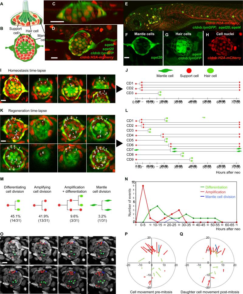
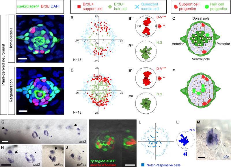
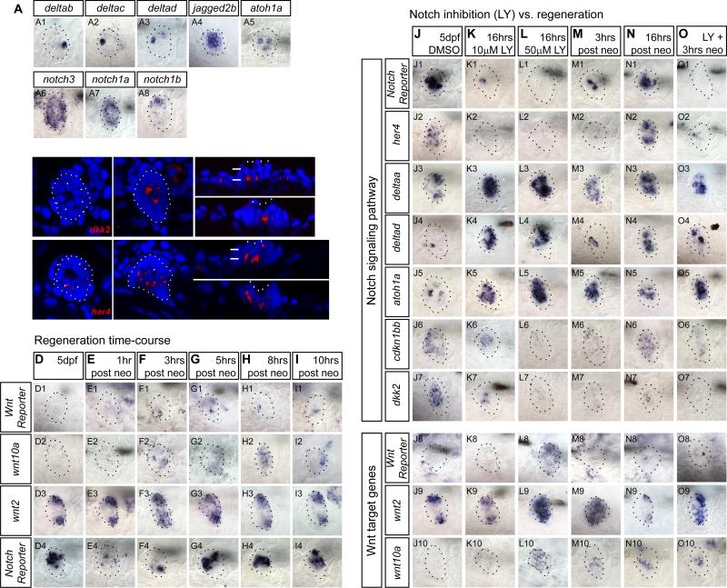
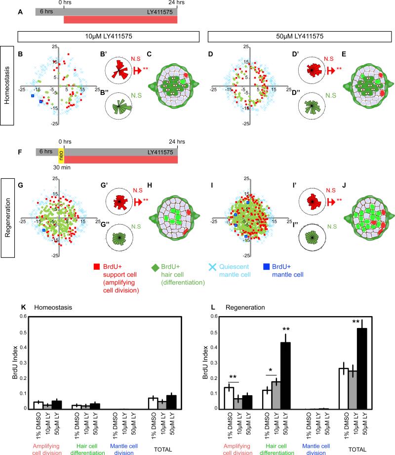
In vertebrates, mechano-electrical transduction of sound is accomplished by sensory hair cells. Whereas mammalian hair cells are not replaced when lost, in fish they constantly renew and regenerate after injury. In vivo tracking and cell fate analyses of all dividing cells during lateral line hair cell regeneration revealed that support and hair cell progenitors localize to distinct tissue compartments. Importantly, we find that the balance between self-renewal and differentiation in these compartments is controlled by spatially restricted Notch signaling and its inhibition of Wnt-induced proliferation. The ability to simultaneously study and manipulate individual cell behaviors and multiple pathways in vivo transforms the lateral line into a powerful paradigm to mechanistically dissect sensory organ regeneration. The striking similarities to other vertebrate stem cell compartments uniquely place zebrafish to help elucidate why mammals possess such low capacity to regenerate hair cells.


Figures

Figure 1
Figure 1. Support cells (SCs) are multipotent progenitors
(A) Horizontal and (B) lateral views of a neuromast (NM). (C-H) Quadruple transgenic larvae express the mantle cell (MC) marker sqet20 (F, green), the hair cell (HC) marker sqet4 (G, cytoplasmic green), the cell membrane marker cldnb:lynGFP (G) and the nuclear maker cldnb:H2A-mCherry (H). (I) Still images of a time-lapse of a homeostatic NM (Movie S1). Split images show different focal planes. Numbers in NMs label the progenitors shown in (J). Time = hours : minutes. (J) Lineage analysis of the mitotic events in (I) and Movie S1. (K) Time-lapse of a regenerating NM (Movie S2B). CD1 is shown in Movie S2C. (L) Lineage analysis in a regenerating NM (Figure 1K; Movie S2). (M) SCs self-renew or differentiate into two hair cells: Quantification of lineages of three time-lapse movies of regenerating NMs from Figures S1F-S1H. (N) Proliferation dynamics during regeneration. Amplifying divisions occur first (p<0.0001, Fisher's exact test). (O) Proliferating cells and their progeny do not actively move in a regenerating NM. Lineages from Figure 1L are color-coded: red: amplifying cell divisions, green: differentiation, blue: MC divisions (Movie S3). mCherry nuclei are in grey. (P) Vectors show directions and distances of cell displacement before mitosis (metaphase) for every cell division recorded during the first 24hrs in Figures S1F-S1H). Central HC progenitors are not displaced. (Q) Vectors show cell displacements of one of the daughter SCs back to their original positions. Displacements for P and Q are quantified in Figure S1I. Scale bars = 10μm. See also Figure S1, Movies S1-S3.
Figure 2
Figure 2. Support cell amplification is restricted to polar compartments during homeostasis and regeneration
(A, D) 24hr BrdU incorporation in primI-derived NMs during homeostasis and regeneration. Scale bar = 10μm. (B, E) Amplifying cell divisions are clustered in the D/V compartments of NMs. BrdU plots show the positions of BrdU+ nuclei of 18 NMs superimposed on the same XY plane. Red squares indicate amplifying divisions; green diamonds indicate BrdU+ cells that differentiated into sqet4+ hair cells. Blue crosses indicate quiescent mantle cells. Axes are in pixels. (B’, B”, E’, E”) Rose diagrams for the angular position of BrdU+ SCs (red), BrdU+ HC (green). Bipolar clustering (D-V) and directional bias to the posterior (red arrow) were statistically analyzed using the Binomial test (***p<0.001). (C, F) Number and location of progenitors that divide within 24hrs. (G-J) wnt2 and deltaa are expressed in poles. Arrowheads label primI-derived NMs. Asterisks label primII-derived NMs. Dashed lines outline NMs. Scale bar in (G) =100μm; in (H)=60μm. (K-K’) EGFP driven by the Notch reporter Tg(Tp1bglob:eGFP) occurs in central cells beneath the red HCs labeled with Tg(atoh1a:dTomato). The Atoh1a reporter is mosaic, leaving some cells unlabeled. (K’) Orthogonal view of K. Scale bar = 10 μm (L-L’) Superimposed EGFP+ Notch reporter cells of 15 NMs (blue squares) are biased toward the anterior side of the NM. (see rose diagram). (M) In situ hybridization of egfp mRNA in Tg(Tp1bglob:eGFP). Scale bar = 10 μm. See also Figure S2.
Figure 3
Figure 3. Downregulation of Notch mimics expression changes during regeneration
(A) Notch ligand and receptor expression in 5dpf neuromasts. (B-C) dkk2 and her4 are expressed in SCs below HCs as shown by confocal imaging of the in situ hybridization signal. B and B’, C and C’ are different focal planes. B” and B’”, C” and C’” are orthogonal views. White arrows point at HCs. (D-I) Time course of mRNA expression of the Wnt reporter Tg(6xTcf/LefBS-miniP:d2EGFP), the Wnt targets wnt10a and wnt2, and the Notch reporter Tg(Tp1bglob:EGFP) at different time points after neomycin treatment. Notch is downregulated first, followed by the activation of Wnt signaling. (Row 2) wnt10a is inactive in control neuromasts but is activated 3-8hrs after hair cell death. (Row 3) wnt2 is present in the poles of homeostatic neuromasts and is upregulated 3h after HC death. (Row 4) mRNA of the Notch reporter shows that Notch downregulation occurs 1-3hrs after hair cell death. (J-O) Notch inhibition with LY411575 mimics expression changes observed during the first 16hrs of regeneration. Larvae were pre-treated with LY or DMSO for 6hrs before starting the time-course (Figures 4A and 4F). (J-K) Lower doses of LY (10μM) downregulate the Notch target her4 (K2) and the Notch reporter mRNA (K1), the cell cycle inhibitor cdkn1bb (K6), and the Wnt inhibitor dkk2 (K7). Also, 10μM LY activates the HC differentiation markers deltad (K4) and atoh1a (K5) and the polar marker delta (K3). (L) After 50μM LY, dkk2 is absent (L7) and wnt2 and wnt10a are activated (L8-L10). (M-N) deltaa (M3), deltad (M4), and atoh1a (M5) are upregulated after HC death. The Notch reporter (N1), her4 (N2), wnt2 (N9), wnt10a (N10) and cdkn1bb (N7) show that Notch downregulation is transient and is reactivated 16hrs after HC death. (O) Notch downregulation during regeneration mimics changes in expressionduring regeneration. Larvae were pre-treated 6hrs in 50μM LY before neo.
Figure 4
Figure 4. Notch inhibits proliferation and differentiation in a dose-dependent manner
(A, F) sqet4;sqet20 larvae were treated for 30hrs with the γ-secretase inhibitor LY411575 (LY). After 6hrs in drug, BrdU was added for 24hrs. DMSO treated controls shown in Figures 2B and 2E. (B-E) Notch inhibition disrupts the proliferative compartments. After 10μM LY, amplifying cell divisions are no longer clustered in the dorso-ventral poles. (G-J) During regeneration, Notch inhibition enhances differentiating divisions in the central region of the NM and in the normally quiescent anterior and posterior compartments. (K) Notch inhibition does not affect proliferation rates (BrdU index) during homeostasis. (L) During regeneration 10μM LY does not affect total proliferation rates but induces differentiation at the expense of amplifying cell divisions. 50μM LY induces hyper-proliferation and an increase in differentiation. Error bars show the 95% confidence interval (CI).
Figure 5
Figure 5. Notch signaling inhibits proliferation via Wnt inhibition and via a Wnt-independent mechanism during homeostasis and regeneration
(A) Heat-shock protocol to experimentally induce hs:nicd or hs:dkk2 expression in regenerating NMs. Larva required 1hr-heat-shock pulses at least 12hrs before HC ablation in order to activate expression of the tagged reporter (c-Myc-tag or RFP respectively, not shown). (B-E) BrdU plots for primI-derived regenerating NMs in sibling, Wnt activated (using the GSK3β inhibitor 1-Azakenpaullone, AZK) and Notch activated Tg(hsp70l:Gal4); Tg(UAS:myc-Notch1a-intra) transgenic larvae, referred to as (hs:nicd). All larvae carry the sqet4 transgene. (B) In DMSO-treated, heat-shocked (hs) siblings amplifying cell divisions occur in the D-V poles and differentiating divisions in the center. (C) AZK (3μM) increases SC proliferation in the D-V poles. (D) Notch activation in hs:nicd larvae disrupts the proliferative compartments and reduces total proliferation. (E) Activation of Wnt with AZK in hs:nicd larvae restores the clustering of SC amplification and proliferation rates. (F-I) BrdU plots for primI-derived regenerating NMs in sibling or Wnt-inhibited Tg(hsp70l:Gal4); Tg(UAS:dkk2-RFP);sqet4, called hs:dkk2, larvae. (G) Wnt inhibition in hs:dkk2 larvae depletes amplifying cell divisions and reduces differentiating divisions in the center. (H) Notch inhibition disrupts polar compartments but maintains amplifying divisions and increases differentiating divisions in the center. (I) Combined Notch and Wnt inhibition (hs:dkk2 + LY) depletes amplifying divisions in the poles but leaves differentiating divisions unaffected. (J-K) BrdU indexes of amplifying, differentiating and total divisions after individual and combinatorial manipulations of the Wnt and Notch pathways. Error bar = 95% CI. See also Figure S3.
Figure 6
Figure 6. Wnt controls proliferation in the poles but does not affect hair cell differentiation
(A) Notch pathway genes are active during homeostasis, whereas Wnt targets are absent, with the exception of wnt2 (A8). (B) Notch inhibition causes activation of the Wnt reporter Tg(6xTcf/LefBS-miniP:d2EGFP) (B7) and Wnt targets wnt2 (B8) and wnt10a (B9). (C) AZK-induced Wnt activation has no effect on HC differentiation markers, such as atoh1a (C4), Notch pathway genes (C1-2), or dkk2 (C6). (D) LY and AZK combined phenocopy the effects of LY alone. (E) hs:dkk2 does not affect the expression of Notch pathway genes (F) LY-induced upregulation of Wnt target genes is reversed by hs:dkk2 induction. (G) Increased Notch signaling in hs:nicd larvae enhances Notch reporter expression. Only 20% of neuromast cells express nicd (data not shown). (H) hs:nicd inhibits the AZK-induced activation of wnt10a (H9) and the Wnt reporter (H7). (I-Q) BrdU plots for primI-derived homeostatic and regenerating sqet4+ NMs. Larvae were treated with DMSO, AZK, 50μM LY or AZK+LY as in Figures 3A and 3F. (I, N) In homeostatic and regenerating NMs amplifying cell divisions are clustered in the poles. During regeneration, centrally located SCs divide and differentiate. (J, O) AZK enhances SC amplification in the polar compartments without affecting HC differentiation. (K, P) LY enhances HC differentiation and disrupts the polar compartments. (L, Q) Combined Wnt activation and Notch inhibition disrupts the polar compartments and randomizes amplification and differentiation. (M, R) BrdU indexes of amplifying, differentiating and total divisions after single and combinatorial manipulations of the Wnt and Notch pathways. Error bar = 95% CI. See also Figure S4.
Figure 7
Figure 7. Mantle cells are quiescent stem cells
(A) Protocol that transforms most SCs into hair cells, followed by neo treatment to test the MC response. (B-G) BrdU incorporation in primI-derived sqet20 NMs at different time points. Scale bar = 10μm. (H) sqet20+ MCs are reduced 10hrs after the second neo treatment but recover by 24hrs. (I) MC BrdU index (No. of BrdU+, sqet20+ cells / total No. of sqet20+ cells). The proliferation of MCs significantly increases between 10-36hrs after the second neo treatment. (J) In Tg(cldnb:mKO2-zCdt1) cldnb drives the Cdt1-tagged mKO2 fluorescent protein in NMs. The Cdt1 ubiquitination domain forces degradation of mKO2 once DNA replication begins. (K) The quiescent state of MCs was analyzed 24hrs after the second neo treatment. (L-L’) mKO2-zCdt1 expression is strong in MCs (O). (M,M’) Treating embryos twice with neo does not affect the quiescent state of MCs. (N-O) Depletion of SCs by LY in sqet20;sqet4 larvae causes some MCs to lose mKO2-zCdt1 expression suggesting that they re-entered the cell cycle. Error bar = 95% CI. See also Figure S5.
Figure 8
Figure 8. Model of the molecular control of cell behaviors during regeneration
Notch signaling controls tissue homeostasis in the NM by restricting proliferation and differentiation through Wnt-dependent and -independent mechanisms. During regeneration Notch is transiently downregulated activating Wnt and proliferation in the center and D-V poles. (A) SC amplification (red nuclei) occurs in the D-V compartments (red cytoplasm) next to peripheral, green MCs. Amplifying cells express wnt2 and deltaa. HC differentiation occurs in the central, Notch+ domain (yellow). (B) Wnt/Notch signaling interactions. In the center (outlined in yellow), Notch inhibits differentiation by inhibiting atoh1a and delta ligands. Notch inhibits proliferation possibly via cdkn1bb and also by inhibiting Wnt signaling through the activation of dkk2. Wnt signaling activates proliferation of HC progenitors in the center and non-cell autonomously of SC progenitors in the poles. The mechanisms by which Wnt initiates proliferation in the poles and the roles of wnt10a and wnt2 have yet to be discovered. Red lines show inhibition, blue arrows indicate activation and dashed arrows show indirect, non-cell-autonomous activation of proliferation.

Similar articles

Cited by

References

    1. Alunni A, Krecsmarik M, Bosco A, Galant S, Pan L, Moens CB, Bally-Cuif L. Notch3 signaling gates cell cycle entry and limits neural stem cell amplification in the adult pallium. Development. 2013;140:3335–3347. - PMC - PubMed
    1. Balak KJ, Corwin JT, Jones JE. Regenerated hair cells can originate from supporting cell progeny: evidence from phototoxicity and laser ablation experiments in the lateral line system. J Neurosci. 1990;10:2502–2512. - PMC - PubMed
    1. Burns JC, Corwin JT. Responses to cell loss become restricted as the supporting cells in mammalian vestibular organs grow thick junctional actin bands that develop high stability. J Neurosci. 2014;34:1998–2011. - PMC - PubMed
    1. Chapouton P, Skupien P, Hesl B, Coolen M, Moore JC, Madelaine R, Kremmer E, Faus-Kessler T, Blader P, Lawson ND, et al. Notch activity levels control the balance between quiescence and recruitment of adult neural stem cells. J Neurosci. 2010;30:7961–7974. - PMC - PubMed
    1. Chen P, Zindy F, Abdala C, Liu F, Li X, Roussel MF, Segil N. Progressive hearing loss in mice lacking the cyclin-dependent kinase inhibitor Ink4d. Nat Cell Biol. 2003;5:422–426. - PubMed

Publication types

MeSH terms

LinkOut - more resources