. 2015 Sep 7;15(17):3521-9.
doi: 10.1039/c5lc00507h. Epub 2015 Jul 20.

Microfluidic device to control interstitial flow-mediated homotypic and heterotypic cellular communication

Affiliations

Microfluidic device to control interstitial flow-mediated homotypic and heterotypic cellular communication

Luis F Alonzo et al. Lab Chip. .

Abstract

Tissue engineering can potentially recreate in vivo cellular microenvironments in vitro for an array of applications such as biological inquiry and drug discovery. However, the majority of current in vitro systems still neglect many biological, chemical, and mechanical cues that are known to impact cellular functions such as proliferation, migration, and differentiation. To address this gap, we have developed a novel microfluidic device that precisely controls the spatial and temporal interactions between adjacent three-dimensional cellular environments. The device consists of four interconnected microtissue compartments (~0.1 mm(3)) arranged in a square. The top and bottom pairs of compartments can be sequentially loaded with discrete cellularized hydrogels creating the opportunity to investigate homotypic (left to right or x-direction) and heterotypic (top to bottom or y-direction) cell-cell communication. A controlled hydrostatic pressure difference across the tissue compartments in both x and y direction induces interstitial flow and modulates communication via soluble factors. To validate the biological significance of this novel platform, we examined the role of stromal cells in the process of vasculogenesis. Our device confirms previous observations that soluble mediators derived from normal human lung fibroblasts (NHLFs) are necessary to form a vascular network derived from endothelial colony forming cell-derived endothelial cells (ECFC-ECs). We conclude that this platform could be used to study important physiological and pathological processes that rely on homotypic and heterotypic cell-cell communication.


Figures
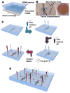
Figure 1
Figure 1. Four-chambered microfluidic device to study interstitial flow-driven communication between discrete microenvironments
(A) An illustration of the components of the microfluidic device. PDMS enclosed chambers are obtained by irreversibly plasma bonding the PDMS device to a thin sheet of PDMS and a glass coverslip. (B) A macroscopic view of the device shows two distinct tissue compartments – where the cell containing hydrogels are housed - connected in parallel, as well as the adjacent microfluidic lines which allow for the introduction of cell culture media. (C) Schematic of the device's loading process. Two distinct cell containing hydrogels may be manually injected sequentially into their respective tissue compartments using a micropipetter. After the hydrogels have polymerized, cell culture media can be similarly injected into the adjacent microfluidic lines. The pipette tips used to inject the media are then used to create a hydrostatic pressure gradient across the tissue compartments by adjusting the volume of media in each of the tips every ~24 hours for the remainder of the experiment. (D) Each custom made platform is designed to hold four separate devices.
Figure 2
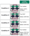
Figure 2. Cellular seeding pattern and interstitial flow experimental conditions
The microfluidic device is amenable to diverse experimental culturing conditions due to the inherent ability to control the cellular composition within each tissue compartment (green and blue channels) and the communication between the compartments through the regulation of interstitial flow direction. For the purpose of validating the device, this study examined the role of stromal cells in vessel network formation; therefore, ECFC-ECs were 1) co-cultured within the same tissue chamber with NHLFs, 2) co-cultured in the adjacent tissue chamber separately connected to NHLFs, 3) cultured in the absence of NHLFs, or 4) cultured in the presence of NHLF pre-conditioned media only. Two types of cellular communications are possible: heterotypic and homotypic cell-cell communication. Heterotypic cellular communication between the two discrete compartments in parallel (green and blue channels) can be achieved by adjusting the volumes of the pipette tips to obtain an interstitial flow pattern in the transverse direction (y-axis). Similarly, a homotypic cellular communication between the two diamond-shaped compartments in series (green only or blue only channels) can be achieved by adjusting the volumes of the pipette tips to obtain an interstitial flow pattern in the longitudinal direction (x-axis).
Figure 3
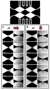
Figure 3. Finite element simulations demonstrate control of pressure distribution and associated interstitial flow distribution within the microfluidic device
(A) A continuous 3D environment can be achieved through the sequential loading of the tissue compartments (TRITC and FITC labeled) which allows for the uninterrupted communication between adjacent compartments. (B–D) Heterotypic communication between the two compartments in parallel can be achieved by setting a pressure difference (ΔP) between the top microfluidic channel and the bottom microfluidic channel (Pα = Pα′ > Pβ = Pβ′), which results in a transverse (y-axis) interstitial convective flow pattern. Theoretical fluid flow velocities along the length of the two compartments aligned with the direction of flow is shown (white dotted line). (E–G) Homotypic communication between the diamond-shaped compartments in series can be achieved by setting a ΔP between the left sides of the top/bottom and the right sides of the top/bottom of the microfluidic lines (Pα = Pβ > Pα′ = Pβ′), which results in a longitudinal (x-axis) interstitial convective flow pattern. Theoretical fluid flow velocities along the length of the two compartments aligned with the direction of flow is shown (white dotted line). Scale bar: 30 um.
Figure 4
Figure 4. Fluorescence recovery after photobleaching (FRAP) validates simulated interstitial flow velocity results
FRAP was used to determine the local flow velocities at two regions within the tissue compartments (#1, #2) where the COMSOL simulations yielded the most extreme interstitial flow velocities. The black circles indicate the originally bleached area, and arrows indicate the direction of the bulk interstitial convective flow. (A) In the case of longitudinal interstitial flow (x-axis), the flow velocity was measured to be 69 μm/s at the junctions which connect the two diamond-shaped compartments in series (#1), while the velocity at the junctions which connect the two tissue compartments in parallel (#2) was negligible. (B) In contrast, when a transverse interstital flow (y-axis) was applied, the flow velocity at spot #2 was measured to be 8 μm/s, while it remained negligible at spot #1. Scale bars: 50 um.
Figure 5
Figure 5. Formation of 3D in vitro interconnected vessel networks depends on interstitial flow-driven communication between ECFC-ECs and NHLFs
Qualitative confirmation of vessel network formation after 1 week of culture was conducted via fluorescently labeling ECFC-ECs with an anti-human CD31 antibody. (A–B) Significant vessel networks developed when ECFC-ECs and NHLFs were co-cultured within the same tissue compartment (condition 1) regardless of the direction of the interstitial convective flow. (C–D) However, under the same interstitial flow conditions and in the absence of NHLFs (condition 2), ECFC-ECs failed to form significant vessel networks. (F,H) Similarly, when NHLFs were cultured in a separate compartment or when NHLF conditioned media was introduced, but the interstitial flow direction was arranged to restrict ECFC-ECs' exposure to NHLF soluble mediators, no significant vessel network formation occurred. (E–G) Significant vessel network formation can be rescued when the interstitial flow direction is setup to allow for ECFC-ECs to be exposed to NHLF soluble mediators from adjacent NHLFs or pre-conditioned media. (I–K) Qualitative observations were confirmed through the quantitative analysis of standard indices measured from the resulting vessel networks. Total vessel length and number of branches peaked for cellular condition 1, significantly different from conditions 2, 3 and 4 under in both interstitial flow directions (*, p<0.05). In addition, parameters show significant difference between the two interstitial flow directions for conditions 3 and 4 (i.e. ECFC-EC exposure to NHLF soluble mediators; #, p<0.05). Scale bar: 100 um.

References

    1. Pampaloni F, Reynaud EG, Stelzer EHK. Nat Rev Mol Cell Biol. 2007;8:839–845. - PubMed
    1. Tung YC, Hsiao AY, Allen SG, Torisawa Y, Ho M, Takayama S. Analyst. 2011;136:473–8. - PMC - PubMed
    1. Weigelt B, Lo AT, Park CC, Gray JW, Bissell MJ. Breast Cancer Res Treat. 2010;122:35–43. - PMC - PubMed
    1. Yamada KM, Cukierman E. Cell. 2007;130:601–10. - PubMed
    1. Chen G, Lv Y, Guo P, Lin C, Zhang X, Yang L, Xu Z. Curr Stem Cell Res Ther. 2013;8:313–23. - PubMed

Publication types

MeSH terms