

Identification of interleukin-26 in the dromedary camel (Camelus dromedarius): Evidence of alternative splicing and isolation of novel splice variants
- PMID: 26190308
- PMCID: PMC7112506
- DOI: 10.1016/j.molimm.2015.06.022
Identification of interleukin-26 in the dromedary camel (Camelus dromedarius): Evidence of alternative splicing and isolation of novel splice variants
Abstract
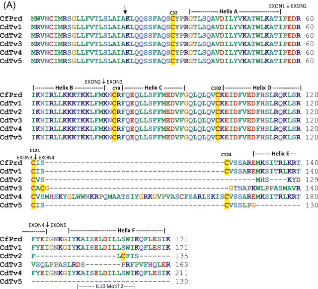
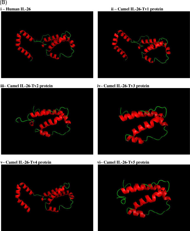
Interleukin-26 (IL-26) is a member of the IL-10 family of cytokines. Though conserved across vertebrates, the IL-26 gene is functionally inactivated in a few mammals like rat, mouse and horse. We report here the identification, isolation and cloning of the cDNA of IL-26 from the dromedary camel. The camel cDNA contains a 516 bp open reading frame encoding a 171 amino acid precursor protein, including a 21 amino acid signal peptide. Sequence analysis revealed high similarity with other mammalian IL-26 homologs and the conservation of IL-10 cytokine family domain structure including key amino acid residues. We also report the identification and cloning of four novel transcript variants produced by alternative splicing at the Exon 3-Exon 4 regions of the gene. Three of the alternative splice variants had premature termination codons and are predicted to code for truncated proteins. The transcript variant 4 (Tv4) having an insertion of an extra 120 bp nucleotides in the ORF was predicted to encode a full length protein product with 40 extra amino acid residues. The mRNA transcripts of all the variants were identified in lymph node, where as fewer variants were observed in other tissues like blood, liver and kidney. The expression of Tv2 and Tv3 were found to be up regulated in mitogen induced camel peripheral blood mononuclear cells. IL-26-Tv2 expression was also induced in camel fibroblast cells infected with Camel pox virus in-vitro. The identification of the transcript variants of IL-26 from the dromedary camel is the first report of alternative splicing for IL-26 in a species in which the gene has not been inactivated.
Keywords: Alternative splicing; Camelids; Cytokine; IL-10; IL-26; Virus.
Copyright © 2015 Elsevier Ltd. All rights reserved.
Figures


Similar articles
-
Comparison of virokine from camel pseudocowpoxvirus (PCPV) with interleukin 10 of the Dromedary camel (Camelus dromedarius).Cytokine. 2013 Feb;61(2):356-9. doi: 10.1016/j.cyto.2012.12.008. Epub 2013 Jan 8. Cytokine. 2013. PMID: 23306428
-
The main WAP isoform usually found in camel milk arises from the usage of an improbable intron cryptic splice site in the precursor to mRNA in which a GC-AG intron occurs.BMC Genet. 2019 Jan 29;20(1):14. doi: 10.1186/s12863-018-0704-x. BMC Genet. 2019. PMID: 30696406 Free PMC article.
-
Cloning and sequence analysis of IL-2, IL-4 and IFN-γ from Indian Dromedary camels (Camelus dromedarius).Res Vet Sci. 2012 Jun;92(3):420-6. doi: 10.1016/j.rvsc.2011.03.028. Epub 2011 May 6. Res Vet Sci. 2012. PMID: 21529863
-
Role and Molecular Mechanisms of Alternative Splicing of Th2-Cytokines IL-4 and IL-5 in Atopic Bronchial Asthma.Biochemistry (Mosc). 2023 Oct;88(10):1608-1621. doi: 10.1134/S0006297923100152. Biochemistry (Mosc). 2023. PMID: 38105028 Review.
-
Alternative splicing: new insights from global analyses.Cell. 2006 Jul 14;126(1):37-47. doi: 10.1016/j.cell.2006.06.023. Cell. 2006. PMID: 16839875 Review.
Cited by
-
Host/genetic factors associated with COVID-19 call for precision medicine.Precis Clin Med. 2020 Jul 21;3(3):228-234. doi: 10.1093/pcmedi/pbaa026. eCollection 2020 Sep. Precis Clin Med. 2020. PMID: 35960669 Free PMC article.
-
Camelid type I interferons: Identification and functional characterization of interferon alpha from the dromedary camel (Camelus dromedarius).Mol Immunol. 2020 Mar;119:132-143. doi: 10.1016/j.molimm.2020.01.020. Epub 2020 Jan 31. Mol Immunol. 2020. PMID: 32014632 Free PMC article.
-
Beta interferons from the extant camelids: Unique among eutherian mammals.Dev Comp Immunol. 2022 Aug;133:104443. doi: 10.1016/j.dci.2022.104443. Epub 2022 May 12. Dev Comp Immunol. 2022. PMID: 35568245 Free PMC article.
-
Refining the Camelus dromedarius Myostatin Gene Polymorphism through Worldwide Whole-Genome Sequencing.Animals (Basel). 2022 Aug 14;12(16):2068. doi: 10.3390/ani12162068. Animals (Basel). 2022. PMID: 36009658 Free PMC article.
-
α3-Deletion Isoform of HLA-A11 Modulates Cytotoxicity of NK Cells: Correlations with HIV-1 Infection of Cells.J Immunol. 2017 Sep 15;199(6):2030-2042. doi: 10.4049/jimmunol.1602183. Epub 2017 Aug 7. J Immunol. 2017. PMID: 28784847 Free PMC article.
References
-
- Allen M., Pratscher B., Roka F., Krepler C., Wacheck V., Schöfer C., Pehamberger H., Müller M., Lucas T. Loss of novel mda-7 splice variant (mda-7s) expression is associated with metastatic melanoma. J. Invest. Dermatol. 2004;123:583–588. - PubMed
-
- Biasini M., Bienert S., Waterhouse A., Arnold K., Stude G., Schmidt T., Kiefer F., Cassarino T.G., Bertoni M., Bordoli L., Schwede T. SWISS-MODEL: modelling protein tertiary and quaternary structure using evolutionary information. Nucleic Acids Res. 2014;42(W1):W252–W258. doi: 10.1093/nar/gku340. - DOI - PMC - PubMed
-
- Boue S., Letunic I., Bork P. Alternative splicing and evolution. Bioessays. 2003;25:1031–1034. - PubMed
MeSH terms
Substances
LinkOut - more resources
Full Text Sources
Other Literature Sources
