

Primary nodal peripheral T-cell lymphomas: diagnosis and therapeutic considerations
- PMID: 26190436
- PMCID: PMC4519704
- DOI: 10.1016/j.bjhh.2015.03.017
Primary nodal peripheral T-cell lymphomas: diagnosis and therapeutic considerations
Abstract
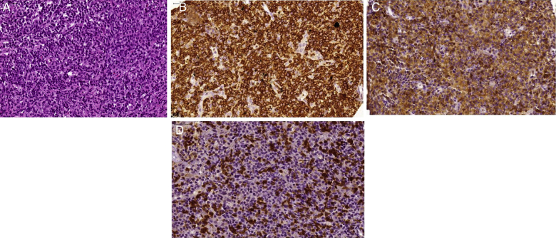
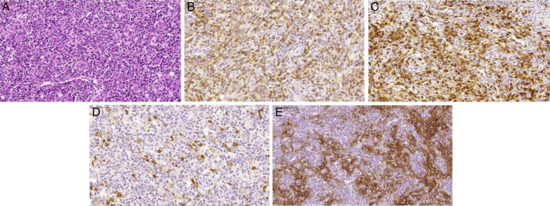
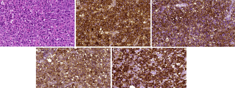
Nodal peripheral T-cell lymphomas are a rare group of neoplasms derived from post-thymic and activated T lymphocytes. A review of scientific articles listed in PubMed, Lilacs, and the Cochrane Library databases was performed using the term "peripheral T-cell lymphomas". According to the World Health Organization classification of hematopoietic tissue tumors, this group of neoplasms consists of peripheral T-cell lymphoma not otherwise specified (PTCL-NOS), angioimmunoblastic T-cell lymphoma (AITL), anaplastic large cell lymphoma-anaplastic lymphoma kinase positive (ALCL-ALK(+)), and a provisional entity called anaplastic large cell lymphoma-anaplastic lymphoma kinase negative (ALCL-ALK(-)). Because the treatment and prognoses of these neoplasms involve different principles, it is essential to distinguish each one by its clinical, immunophenotypic, genetic, and molecular features. Except for anaplastic large cell lymphoma-anaplastic lymphoma kinase positive, which has no adverse international prognostic index, the prognosis of nodal peripheral T-cell lymphomas is worse than that of aggressive B-cell lymphomas. Chemotherapy based on anthracyclines provides poor outcomes because these neoplasms frequently have multidrug-resistant phenotypes. Based on this, the current tendency is to use intensified cyclophosphamide, doxorubicin, vincristine, prednisolone (CHOP) regimens with the addition of new drugs, and autologous hematopoietic stem cell transplantation. This paper describes the clinical features and diagnostic methods, and proposes a therapeutic algorithm for nodal peripheral T-cell lymphoma patients.
Keywords: Antineoplastic combined chemotherapy protocols; Diseases classification; Immunophenotyping; T-cell Lymphoma; World Health Organization.
Copyright © 2015 Associação Brasileira de Hematologia, Hemoterapia e Terapia Celular. Published by Elsevier Editora Ltda. All rights reserved.
Figures
References
-
- Jaffe E.S. Pathobiology of peripheral T-cell lymphomas. Hematol Am Soc Hematol Educ Prog. 2006:317–322. - PubMed
-
- Vose J., Armitage J., Weisenburger D. International peripheral T-cell and natural killer lymphoma study: pathology findings and clinical outcomes. J Clin Oncol. 2008;26(25):4124–4130. - PubMed
-
- Dogan A., Feldman A F. T- and NK-cell lymphoma update. Diagn Histopathol. 2009;16(2):99–110.
-
- Rizvi M.A., Evens A.M., Tallman M.S., Nelson B.P., Rosen S.T. T-cell non-Hodgkin lymphoma. Blood. 2006;107(4):1255–1264. - PubMed
LinkOut - more resources
Full Text Sources
Other Literature Sources
Medical
Research Materials
