. 2015 Oct;135(2):309-22.
doi: 10.1111/jnc.13250. Epub 2015 Aug 31.

Interferon gamma induces protective non-canonical signaling pathways in primary neurons

Affiliations

Interferon gamma induces protective non-canonical signaling pathways in primary neurons

Lauren A O'Donnell et al. J Neurochem. 2015 Oct.

Abstract

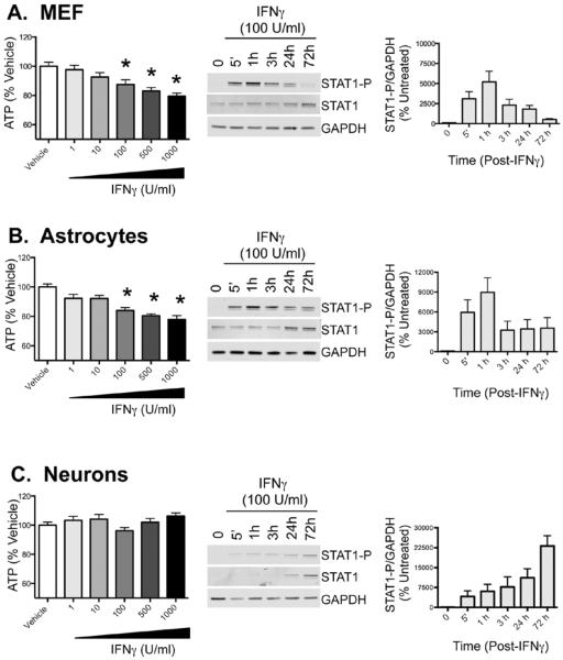
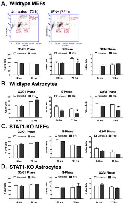
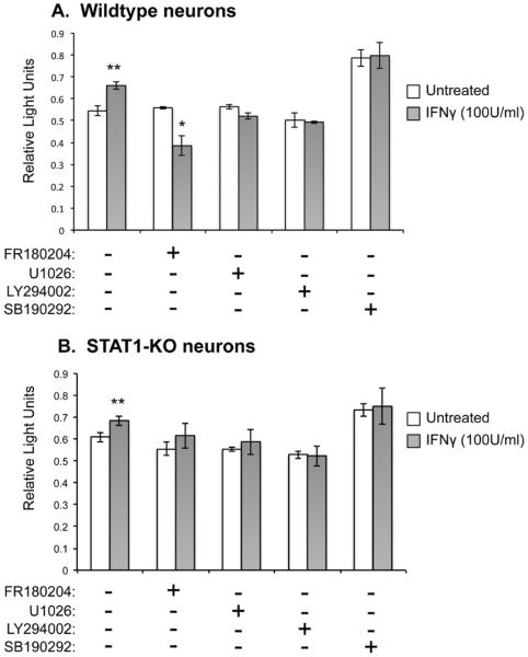
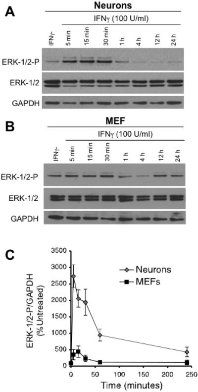
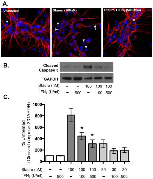
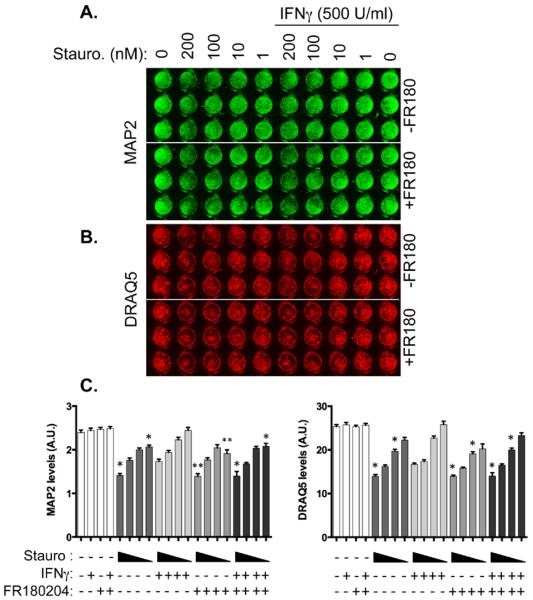
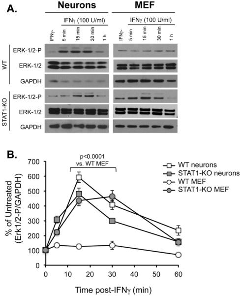
The signal transduction molecule, Stat1, is critical for the expression of type I and II interferon (IFN)-responsive genes in most cells; however, we previously showed that primary hippocampal mouse neurons express low basal Stat1, with delayed and attenuated expression of IFN-responsive genes. Moreover, IFNγ-dependent resolution of a neurotropic viral challenge in permissive mice is Stat1-independent. Here, we show that exogenous IFNγ has no deleterious impact on neuronal viability, and staurosporine-induced apoptosis in neurons is significantly blunted by the addition of IFNγ, suggesting that IFNγ confers a pro-survival signal in neurons. To identify the pathways induced by IFNγ in neurons, the activation of alternative signal transducers associated with IFNγ signaling was assessed. Rapid and pronounced activation of extracellular signal regulated kinase (Erk1/2) was observed in neurons, compared to a modest response in fibroblasts. Moreover, the absence of Stat1 in primary fibroblasts led to enhanced Erk activation following IFNγ addition, implying that the cell-specific availability of signal transducers can diversify the cellular response following IFN engagement.

Keywords: Erk-1/2; Interferon gamma; Stat1; cytokines; hippocampus; neuron.


Figures

Figure 1
Figure 1. IFNγ reduces ATP production in primary fibroblasts and astrocytes, but not in primary neurons
Primary mouse embryonic fibroblasts (MEFs) (A), cortical astrocytes (B) and hippocampal neurons (C) were explanted from E16 mouse embryos. All cell types were treated with vehicle control (PBS) or IFNγ at indicated concentrations (1–1000U/ml). ATP levels were measured using CellTiter-Glo assay 72h post treatment. Fluorescence values were plotted as a percentage of PBS-treated controls. Shown are the mean + S.E.M. from 3–5 experiments. Western blots for phosphorylated Stat1 (Stat1-P), total Stat1, and GAPDH as a loading control were performed over the 72h time course. Representative blots are shown in the middle column, and quantitation of the western blots is shown in right column with Stat1-P signal normalized to GAPDH levels (n=3). Each graph includes cells from 3 biological replicates from independent dissections, and statistical analysis was applied by 1-way ANOVA with Bonferroni multiple comparison test (* p<0.05 vehicle control treated cells).
Figure 2
Figure 2. IFNγ inhibits cell cycle progression in fibroblasts and astrocytes in a Stat1-dependent manner
Primary wild type fibroblasts (A) and astrocytes (B) were treated with IFNγ (100 U/ml) for 24 or 72h. Stages of the cell cycle were measured by incorporation of BrdU into newly-synthesized DNA and by 7-AAD, which stained total DNA. Samples were then assayed by flow cytometry. Representative scatter plots and gates for fibroblasts (MEFs) at 72h post-IFNγ treatment are shown in (A). Stat1-KO fibroblasts (C) and astrocytes (D) were also assayed by the same approach. Experiments with cells from individual dissections were averaged (n=3–4) and plotted with SEM. Statistical significance was assessed via paired t-test (* p<0.01).
Figure 3
Figure 3. Blockade of Erk1/2 in neurons negates the IFNγ-mediated protection against Stat1-mediated cytotoxicity
Primary wildtype (A) and Stat1-KO (B) hippocampal neurons were pre-treated with inhibitors FR180204 (Erk1/2 inhibitor), U1026 (Mek1/2 inhibitor), LY294002 (PI3-Kinase inhibitor), and SB190292 (p38-Mapk inhibitor) (10μM). After 1h, IFNγ (100 U/ml) was applied to the cultures in the presence of the inhibitors and incubated for 72h. Cultures were assayed by MTS assay as a measure of cell survival. Statistical analysis was applied by paired t-test (* p<0.05 vs. corresponding untreated; # p<0.01 vs. corresponding untreated; n=2).
Figure 4
Figure 4. IFNγ activates Erk1/2 in primary neurons but not in fibroblasts
(A) and (B) Hippocampal neurons and fibroblasts (MEFs) explanted from mouse embryos were treated with IFNγ (100 U/ml). Cells were lysed at indicated times post treatment and subjected to Western blot for Erk1/2, phosphorylated Erk1/2 (Erk1/2-P), and GAPDH as a loading control. C. Western blots from A and B were quantified by densitometry using ImageJ software. Erk1/2 and p-Erk1/2 signal was normalized to GAPDH as a loading control. Statistical analysis was determined using two-way ANOVA (n = 3, p < 0.002).
Figure 5
Figure 5. IFNγ blocks apoptotic signaling in neurons upon staurosporine exposure
Cultured hippocampal neurons were treated with IFNγ at the indicated concentrations (100 or 500 U/ml). After 30 min of pretreatment with IFNγ, staurosporine (30 or 100nM) was added to the cultures for 72 h. A. Cells were fixed and stained for MAP2 (red) as a marker for neurons and Hoechst (blue) for cell nuclei. Arrowheads indicate pyknotic nuclei and arrows indicate beaded dendrites. B. Cells were lysed in protein solubilization buffer and analyzed by Western blot with antibodies against cleaved caspase 3 and GAPDH as a loading control. C. Western blots were quantified by densitometry using ImageJ software. Cleaved caspase 3 signal was normalized to GAPDH as a loading control and statistical analysis was determined using a paired t test (n = 4, *p < 0.04 versus signal in untreated control).
Figure 6
Figure 6. Inhibition of Erk1/2 abrogates IFNγ-mediated protection against staurosporine
Primary hippocampal cultures were treated with the Erk inhibitor FR180204 for 30 min prior to addition of IFNγ (500 U/ml). One hour after IFNγ treatment, cells were treated with a range of staurosporine concentrations (200, 100, 10, and 1 nM) for 72h. Cultures were stained for MAP2 as a neuronal marker (green; A) and DRAQ5 as a label for DNA (red, B). Results were quantified in C. Graphs show the average signal intensity from three biological replicates of independent dissections (n = 5 plates) with SEM. Triangles below the graph indicate decreasing concentrations of staurosporine (200–1 nM). Statistical analysis was applied via one-way ANOVA with multiple comparisons (* p<0.05 versus IFNγ + equivalent staurosporine concentration, ** p<0.01 versus IFNγ + equivalent staurosporine concentration.)
Figure 7
Figure 7. Enhanced Erk1/2 activation in Stat1 KO fibroblasts
Wildtype and Stat1-KO hippocampal neurons and MEFs were treated with IFNγ (100 U/ml). Whole-cell lysates were harvested post treatment at times indicated and subjected to Western blot analysis for phospho-Erk1/2 (Erk-1/2/-P), total Erk-1/2, and GPADH as a loading control (A). The signal of both Erk bands was analyzed together by densitometry on ImageJ software, and p-Erk1/2 signal was normalized to GAPDH as a loading control (B). Statistical analysis was applied using two-way ANOVA with Tukey's post hoc test (n=3).

References

    1. Aksamitiene E, Kholodenko BN, Kolch W, Hoek JB, Kiyatkin A. PI3K/Akt-sensitive MEK-independent compensatory circuit of ERK activation in ER-positive PI3K-mutant T47D breast cancer cells. Cell Signal. 2010;22:1369–1378. - PMC - PubMed
    1. Amadoro G, Ciotti MT, Costanzi M, Cestari V, Calissano P, Canu N. NMDA receptor mediates tau-induced neurotoxicity by calpain and ERK/MAPK activation. Proceedings of the National Academy of Sciences of the United States of America. 2006;103:2892–2897. - PMC - PubMed
    1. Anderson CN, Tolkovsky AM. A role for MAPK/ERK in sympathetic neuron survival: protection against a p53-dependent, JNK-independent induction of apoptosis by cytosine arabinoside. J Neurosci. 1999;19:664–673. - PMC - PubMed
    1. Banker G. a. G., K. Culturing Nerve Cells. A Bradford Book; 1998.
    1. Bapat S, Verkleij A, Post JA. Peroxynitrite activates mitogen-activated protein kinase (MAPK) via a MEK-independent pathway: a role for protein kinase C. FEBS letters. 2001;499:21–26. - PubMed

Publication types

MeSH terms