Review
. 2015 Sep 1;22(3):381-97.
doi: 10.1016/j.cmet.2015.06.010. Epub 2015 Jul 16.

Calcium Homeostasis and Organelle Function in the Pathogenesis of Obesity and Diabetes

Affiliations
Review

Calcium Homeostasis and Organelle Function in the Pathogenesis of Obesity and Diabetes

Ana Paula Arruda et al. Cell Metab. .

Abstract

A number of chronic metabolic pathologies, including obesity, diabetes, cardiovascular disease, asthma, and cancer, cluster together to present the greatest threat to human health. As research in this field has advanced, it has become clear that unresolved metabolic inflammation, organelle dysfunction, and other cellular and metabolic stresses underlie the development of these chronic metabolic diseases. However, the relationship between these systems and pathological mechanisms is poorly understood. Here we discuss the role of cellular Ca(2+) homeostasis as a critical mechanism integrating the myriad of cellular and subcellular dysfunctional networks found in metabolic tissues such as liver and adipose tissue in the context of metabolic disease, particularly in obesity and diabetes.


Conflict of interest statement

The authors have no conflicts of interest related to the contents of this manuscript.

Figures

Figure 1
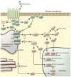
Figure 1. Ca2+ homeostasis in the ER
Right: tool-kit of Ca2+ handling proteins. Ligand binding of GPCRs leads to the biosynthesis of the intracellular messengers such as IP3, which diffuses into the cell and binds to IP3R, stimulating Ca2+ release from the ER. Ca2+ channels activated by IP3 release Ca2+ from ER to the cytosol. RyR also releases Ca2+ from ER to the cytosol. SERCA (Ca2+-ATPase) pumps Ca2+ from cytosol to the ER lumen at the expense of ATP hydrolysis. STIM is an ER membrane protein that possesses 2 EF hand domains. Under basal conditions, Ca2+ is bound to these domains and the protein remains in its monomeric form. After Ca2+ release from ER, Ca2+ dissociates from the of STIM proteins leading to oligomerization and trafficking of STIM through the ER membrane toward the plasma membrane contact sites. This localization, allows STIM to couple with the plasma membrane Ca2+ channel Orai and Ca2+ enters the cell from the extracellular space, a process known as capacitive or store operator Ca2+ entry (SOCE). SERCA colocalizes with STIM/Orai complex, and is thought to pump Ca2+ directly from cytosol to ER lumen. Left: ER protein folding machinery: During glycoprotein biosynthesis, the translation of nascent polypeptides is followed by their translocation of the peptides through the SEC61 pore. Sugar molecules are added to the polypeptides that are recognized by lectin chaperones such as calnexin (CNX) and calretculin (CRT), which function in complex with ERp57 and ERp72. Additional chaperone assistance is provided by the ATP-driven chaperone BiP (GRP78). Calnexin and calreticulin also bind Ca2+ with high capacity, functioning as the main Ca2+ buffers in the ER lumen. Ca2+ depletion leads to accumulation of misfolded proteins and the activation of the unfolded protein response (UPR), mediated by three canonical ER luminal sensors, IRE1, PERK, and ATF6, which are inactive and bound to BiP in the absence of stress. GPCR: G protein–coupled receptor. IP3: inositol triphosphate. RyR: Ryanodine receptor. STIM1: stroma interacting protein.
Figure 2
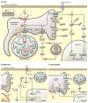
Figure 2. Components of the mitochondria associated ER membranes (MAMs) and Ca2+ homeostasis in the mitochondria (A)
Electron microscopic images exemplifying the close contacts between smooth ER and mitochondria in mouse liver cells. Dense areas show the proteinaceous bridges formed between the two membranes. (B) The MAM connection is mediated by protein tethers such as mitofusin2 (MFN2) and stabilized by proteins such as PACS-2 bound to ER-resident calnexin (CNX). The MAMs are enriched in proteins that regulate Ca2+ transport from ER to the mitochondria such as IP3R, connected to the mitochondria anion transporter (VDAC) by the chaperone GRP75. Through these structures Ca2+ leaves the ER and enters the mitochondria matrix via the mitochondria Ca2+ uniporter (MCU) located in the IMM. MCU is regulated by a series of binding partners such as MICU1 and MICU2, MICUR1 and EMRE. MICU1 functions as a gatekeeper for MCU-mediated Ca2+ uptake. MICU2 seems to inhibit MCU function. EMRE is essential for the interaction between MCU and the MICU proteins and is indispensable for MCU-mediated Ca2+ transport. The exit pathway for mitochondrial Ca2+ is mediated by the NCXL antiporter that exchanges Na+/Ca2+. MAMs are also enriched in key enzymes of lipid biosynthesis to facilitate the conversion of PA to PE and PC, as described in the text. (C) Within the mitochondria, Ca2+ regulates the activity of matrix and citric acid cycle enzymes such as pyruvate dehydrogenase phosphatase (PDP1), α-ketoglutarate dehydrogenase (aKGDH), and isocitrate dehydrogenase (IDH). This culminates in increased NADH production that feeds the respiratory chain to produce ATP. (D) Mitochondrial Ca2+ overload leads to increased mitochondrial ROS production and the opening the mitochondrial permeability transition pore (PTP). The opening of the PTP leads to a collapse of membrane potential and mitochondrial swelling, with consequent loss of nucleotides and cytochrome C (CytoC), and is directly linked with apoptotic cell death. Apoptosis is regulated by the BCL2 class of proteins such as BID, BAX and BAD. Under normal conditions, the proapoptotic protein BAX is in the cytosol. However, under apoptotic condition this protein interacts with BAD or BID located in the OMM, stimulating the permeabilization of the OMM.
Figure 3
Figure 3. Intracellular Ca2+ signaling
GPCR activation by hormones can induce Ca2+ release by activating phospholipase C (PLC) leading to the hydrolysis of phosphatidylinositol 4,5 bisphosphate (PIP2) to produce the intracellular messengers IP3 and DAG. IP3 diffuses in to the cell and binds to IP3R, stimulating Ca2+ release from the ER. Alternatively, GPCR activation leads to the production of cAMP, which activates the protein kinase A (PKA), enabling it to phosphorylate IP3R and induce its activity. In the cytosol the two main Ca2+ sensors involved in signal transduction are CaM kinase (CamK) and Calcineurin (CLN). CaMKs have been shown to directly phosphorylate many targets including the inflammatory proteins JNK and p38 and transcription factors including FOXO and CREB. CLN is activated by sustained high Ca2+ levels and regulates the activity of at least three important transcription pathways involving Nuclear factor of activated T-cells (NFAT) and CREB-regulated transcription coactivator 2 (CRTC2).
Figure 4
Figure 4. Dysfunction of Ca2+ homeostasis in metabolic disease
(A) In the liver of obese humans and animal models, the UPR is activated and the phosphorylation of IRE1 (inositol required 1), PERK (PKR-like endoplasmic reticulum localized kinase), and eIF2α are increased. However, the levels of sXBP1 and ATF6 (activating transcription factor 6) activity are decreased in obesity in the liver (similar to beta cells). Due to changes in ER membrane composition (increased ratio of PC/PE) the transport of Ca2+ from cytosol to ER lumen by SERCA is impaired, leading to decreased Ca2+ levels in ER. Also, in the liver of obese animals IP3R1 expression and activity are increased, which contributes both to decreased levels of ER Ca2+ and increased cytosolic and mitochondrial Ca2+. In the mitochondria, Ca2+ overload correlates with increased oxidative stress (ROS) and decreased oxidative function. Increased cytosolic Ca2+ also affects several cellular pathways such as autophagosome accumulation, and activates proteases such as calpain that cleaves and degrades ATG7. Both phenomena culminate in decreased autophagy, as seen in liver in obesity. Increased cytosolic Ca2+ also activates calcineurin leading to dephosphorylation of CRTC2 and its nuclear translocation, and activates CaMK to phosphorylate FOXO, with a direct impact on gluconeogenesis. CaMK also phosphorylates p38 and JNK leading to increased inflammation. In both adipocytes (B) and macrophages (C) of obese animal models, the UPR is activated and the phosphorylation of IRE1, PERK, ATF6 and eIF2α are increased. (B) In hypertrophic adipocytes there is increased deposition of Ca2+ surrounding the lipid droplets and increased cytosolic Ca2+ levels. NFAT transcription factors isoforms 2 and 4 are induced in obese adipose tissue driving expression of inflammatory genes. TRPV4 (Transient receptor potential cation channel subfamily V member 4) is a Ca2+-permeable ion channel that was first identified as an osmolality sensor. In adipocytes, it acts as a negative regulator of PGC1α and a positive regulator of inflammatory genes and proteins and JNK activity. These effects are related to the activation of ERK1/2. (C) Macrophages derived from obese animals show increased IP3R1 and ERO1α activity, which leads to higher Ca2+ release from ER to cytosol. In addition reduced levels of SERCA2b mRNA and protein expression is observed in this condition. Rise in cytosolic Ca2+ and mitochondrial oxidative stress have been shown to engage the NLRP3 inflammasome pathway. In addition, the alterations in macrophage activity in obesity have been shown to activate CaMKK (Calcium/calmodulin-dependent protein kinase kinase 2), the deletion of which leads to impaired cytokine secretion and phagocytosis and induces morphological changes.

Similar articles

Cited by

References

    1. Amaya MJ, Nathanson MH. Calcium signaling in the liver. Comprehensive Physiology. 2013;3:515–539. - PMC - PubMed
    1. Anderson KA, Lin F, Ribar TJ, Stevens RD, Muehlbauer MJ, Newgard CB, Means AR. Deletion of CaMKK2 from the liver lowers blood glucose and improves whole-body glucose tolerance in the mouse. Mol Endocrinol. 2012;26:281–291. - PMC - PubMed
    1. Arruda AP, Pers BM, Parlakgul G, Guney E, Inouye K, Hotamisligil GS. Chronic enrichment of hepatic endoplasmic reticulum-mitochondria contact leads to mitochondrial dysfunction in obesity. Nature medicine. 2014;20:1427–1435. - PMC - PubMed
    1. Baines CP, Kaiser RA, Purcell NH, Blair NS, Osinska H, Hambleton MA, Brunskill EW, Sayen MR, Gottlieb RA, Dorn GW, et al. Loss of cyclophilin D reveals a critical role for mitochondrial permeability transition in cell death. Nature. 2005;434:658–662. - PubMed
    1. Bartelt A, Heeren J. Adipose tissue browning and metabolic health. Nature reviews Endocrinology. 2014;10:24–36. - PubMed

Publication types

MeSH terms