

Renal transplant vascular complications: the role of Doppler ultrasound
- PMID: 26191097
- PMCID: PMC4504861
- DOI: 10.1007/s40477-014-0085-6
Renal transplant vascular complications: the role of Doppler ultrasound
Abstract
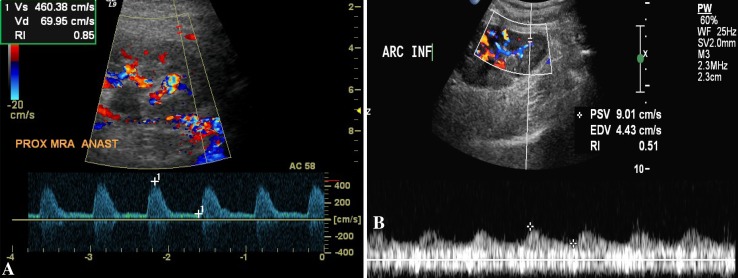
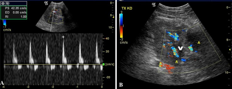
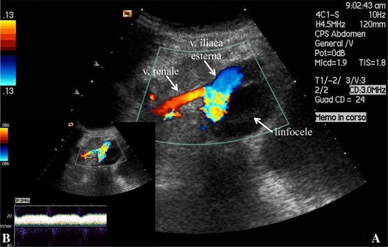
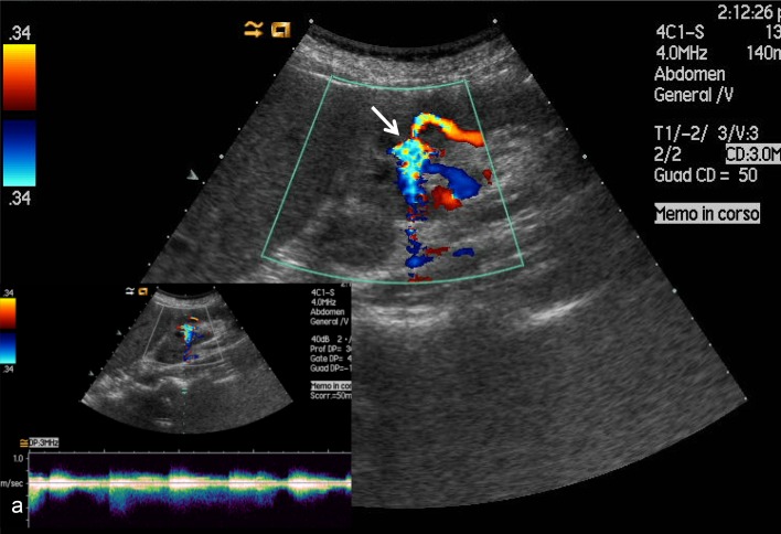
Improvements in the care of kidney transplant recipients and advances in immunosuppressive therapy have reduced the incidence of graft rejection. As a result, other types of kidney transplant complications, such as surgical, urologic, parenchymal, and vascular complications, have become more common. Although vascular complications account for only 5-10 % of all post-transplant complications, they are a frequent cause of graft loss. Ultrasonography, both in B-mode and with Doppler ultrasound, is a fundamental tool in the differential diagnosis of renal allograft dysfunction. Doppler ultrasound is highly specific in cases of transplanted renal artery stenosis, pseudoaneurysms, arteriovenous fistulas, and thrombosis with complete or partial artery or vein occlusion. A single measurements of color Doppler indexes display high diagnostic accuracy and in particular cases are more useful during the post-transplantation follow-up period. More recent techniques, such as contrast-enhanced ultrasound, undoubtedly increase the accuracy of ultrasonography in the diagnosis of vascular complications involving the transplanted kidney.
La progressiva riduzione dell’incidenza del rigetto ha reso più frequenti le complicanze urologiche, chirurgiche, parenchimali e vascolari. Queste ultime, pur rappresentando soltanto il 5–10 % di tutte le complicanze post-trapianto, sono frequente causa di perdita del graft. L’esame ultrasonografico, sia in B-mode che con l’ausilio del color Doppler, è fondamentale nella diagnosi differenziale delle cause che possono innescare una disfunzione del graft. Sebbene sia ormai indiscussa la sua utilità nella diagnosi di complicanze parenchimali, chirurgiche e urologiche, non è ancora consolidato il suo ruolo in caso di complicanze a carico dell’asse vascolare renale. L’ecocolor-Doppler, in particolare, possiede una specificità tale da poter essere considerato uno strumento diagnostico nella maggior parte delle complicanze vascolari del rene trapiantato, sia acute (occlusione parziale o totale dei vasi renali) che croniche (stenosi dell’arteria renale, pseudo aneurisma e fistola artero-venosa) Gli indici color-Doppler possiedono, infatti, una alta accuratezza diagnostica nella loro singola determinazione, risultando in casi particolari più utili nel follow-up. L’utilizzo di tecniche più moderne, come il mezzo di contrasto ecografico, consente indubbiamente di aumentare l’accuratezza diagnostica dell’esame ultrasonografico nel caso delle complicanze vascolari del rene trapiantato.
Keywords: Doppler ultrasound; Renal transplant; Ultrasonography; Vascular complications.
Figures
References
-
- Granata A, Floccari F, Lentini P, Vittoria S, Di Pietro F, Zamboli P, et al. Vascular complications following kidney transplant: the role of color-Doppler imaging. G Ital Nefrol. 2012;29(S57):S99–S105. - PubMed
Publication types
LinkOut - more resources
Full Text Sources
Other Literature Sources
Research Materials
