

doi: 10.1007/s40477-015-0164-3.
eCollection 2015 Jun.
Volumetric blood flow measurement using Doppler ultrasound: concerns about the technique
Affiliations
- PMID: 26191112
- PMCID: PMC4504867
- DOI: 10.1007/s40477-015-0164-3
Item in Clipboard
Volumetric blood flow measurement using Doppler ultrasound: concerns about the technique
J Ultrasound.
.
No abstract available
Figures
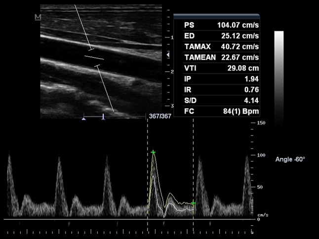
Spectral Doppler of the common carotid artery (CCA), obtained with pulsed-wave Doppler. Sample volume encompasses the whole vessel diameter. Time-averaged maximum velocity [TAMAX (also called TAP), represented with a continuous yellow line delineating the maximum velocities of the spectrum occurring along the cardiac cycle] and time-averaged mean velocity (TAMEAN, represented with a continuous white line delineating the mean velocities of the spectrum occurring along the cardiac cycle) were obtained. Of note, TAMAX and TAMEAN values are different, with TAMEAN lower than TAMAX. Cross-sectional area was estimated in 0.28 cm2, and the resultant volume flow measurements were 684 ml/min using TAMAX and 376 ml/min with TAMEAN
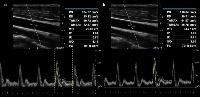
Doppler ultrasound of the CCA, with sample volume encompassing the whole diameter of the vessel (a) and with the sample volume in the center of the vessel (b). Broadening of the Doppler spectrum is demonstrated (a), indicating the recording of all range of velocities. TAMEAN is also lower in a as expected
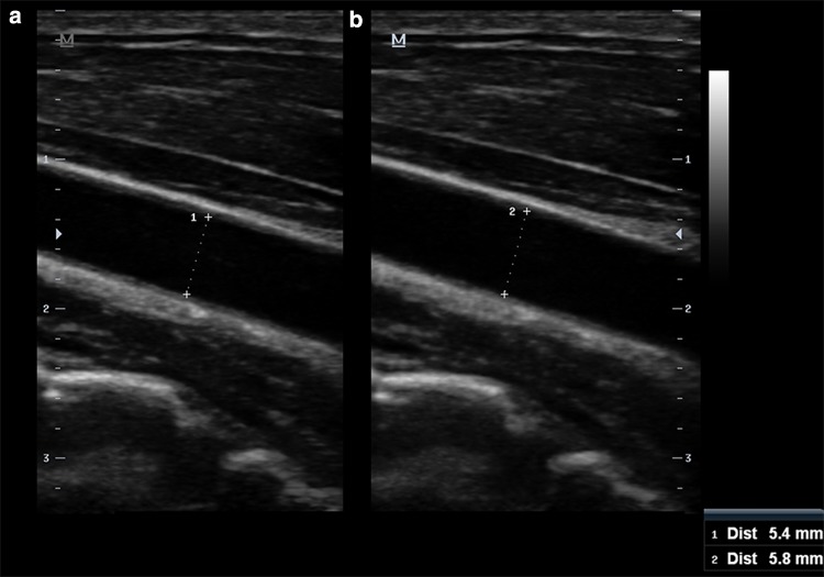
Measurement of the diameter of the CCA. a Diastolic frame; b systolic frame. The systolic diameter is larger in comparison with the diastolic diameter. Note the effect in calculated area of 0.23 cm2 (a) and 0.26 cm2 (b)
References
-
- Daigle RJ. Arterial hemodynamics, anatomy and physiology. In: Daigle RJ, editor. Techniques in non-invasive vascular diagnosis: an encyclopedia of vascular testing. 3. Littleton: Summer Publishing; 2008. pp. 141–158.
LinkOut - more resources
Full Text Sources
Other Literature Sources
