

Up-regulation of microRNA-15b correlates with unfavorable prognosis and malignant progression of human glioma
- PMID: 26191187
- PMCID: PMC4503059
Up-regulation of microRNA-15b correlates with unfavorable prognosis and malignant progression of human glioma
Abstract
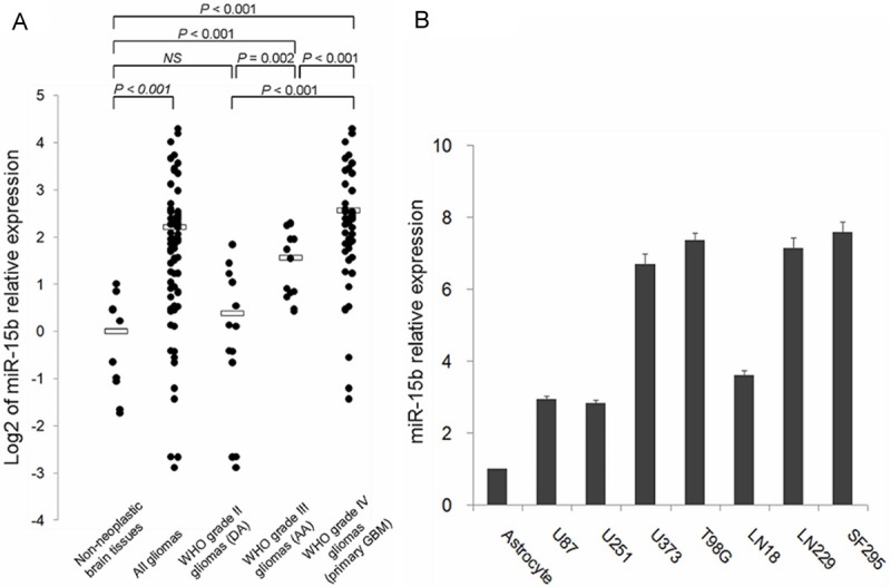
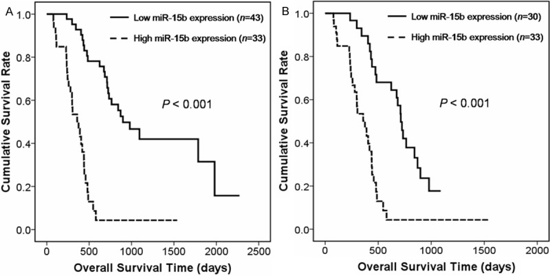
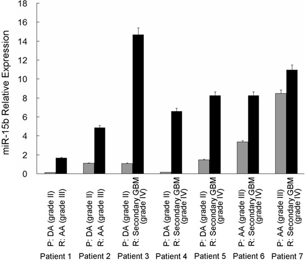
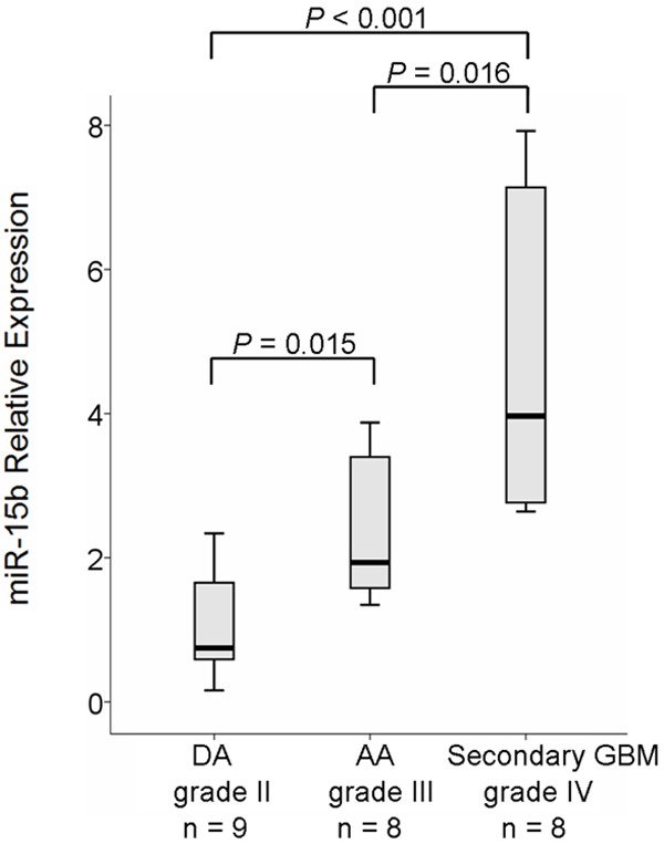
Recent studies have demonstrated that microRNA-15b (miR-15b) regulates cell cycle progression, proliferationnd apoptosis in glioma cells by targeting Cyclins. However, the clinical significance of miR-15b in human glioma remains unclear. Therefore, the aim of this study was to investigate the significance of miR-15b expression in diagnosis, prognosis and malignant progression of glioma. Quantitative real-time reverse transcriptive-PCR (qRT-PCR) was performed to examine miR-15b expression levels in 76 glioma tissues (13 grade II, 13 grade III and 50 grade IV gliomas) and seven glioma cell lines, as well as 10 non-neoplastic brain tissues and human astrocyte as control. MiR-15b showed significant increased expression in high-grade gliomas (P≤0.001) and glioma cells (fold change 2.8-7.6) relative to non-neoplastic brains and astrocyte, respectively. Additionally, high miR-15b expression was significantly associated with advanced WHO grade (P≤0.001), advanced patient age (P≤0.001) and low Karnofsky performance score (KPS, P≤0.001). Furthermore, Kaplan-Meier survival analysis and Cox regression analysis showed that patients with high miR-15b expression had significantly poor overall survival rate (P≤0.001) and miR-15b expression was an independent prognosis-predicting factor for glioma patients (P≤0.001; risk ratio=5.6), respectively. Moreover, miR-15b expression was examined in seven independent patients with primary grade II or III gliomas that spontaneously progressed to grade III or IV gliomas. Statistically significant higher expression (P=0.01) in the recurrent tumor compared with the corresponding primary tumor was observed in all of the seven patients. Our results suggest that miR-15b may be a prognostic predictor and be involved in malignant progression of glioma.
Keywords: glioma; malignant progression; miRNA-15b; microRNA; prognosis; up-regulation.
Figures
References
-
- Ohgaki H, Kleihues P. Epidemiology and etiology of gliomas. Acta Neuropathol. 2005;109:93–108. - PubMed
-
- Verhaak RG, Hoadley KA, Purdom E, Wang V, Qi Y, Wilkerson MD, Miller CR, Ding L, Golub T, Mesirov JP, Alexe G, Lawrence M, O’Kelly M, Tamayo P, Weir BA, Gabriel S, Winckler W, Gupta S, Jakkula L, Feiler HS, Hodgson JG, James CD, Sarkaria JN, Brennan C, Kahn A, Spellman PT, Wilson RK, Speed TP, Gray JW, Meyerson M, Getz G, Perou CM, Hayers DN Cancer Genome Atlas Research Network. Integrated genomic analysis identifies clinically relevant subtypes of glioblastoma characterized by abnormalities in PDGFRA, IDH1, EGFR and NF1. Cancer Cell. 2010;17:98–110. - PMC - PubMed
-
- Houshmehr H, Weisenberger DJ, Diefes K, Phillips HS, Pujara K, Berman BP, Pan F, Pelloski CE, Sulman EP, Bhat KP, Verhaak RG, Hoadley KA, Hayes DN, Perou CM, Schmidt HK, Ding L, Wilson RK, Van Den Berg D, Shen H, Bengtsson H, Neuvial P, Cope LM, Buckley J, Herman JG, Baylin SB, Laird PW, Aldape K Cancer Genome Atlas Research Network. Identification of a CpG island methlator phenotype that defines a distinct subgroup of glioma. Cancer Cell. 2010;17:510–22. - PMC - PubMed
-
- Inui M, Martello G, Piccolo S. MicroRNA control of signal transduction. Nat Rev Mol Cell Biol. 2010;11:252–63. - PubMed
Publication types
MeSH terms
Substances
LinkOut - more resources
Full Text Sources
Medical