

Adenoid cystic carcinoma of the right main bronchus showing squamous differentiation and mimicking mucoepidermoid carcinoma: a case report
- PMID: 26191305
- PMCID: PMC4503176
Adenoid cystic carcinoma of the right main bronchus showing squamous differentiation and mimicking mucoepidermoid carcinoma: a case report
Abstract
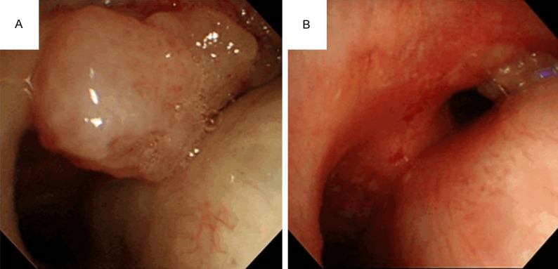
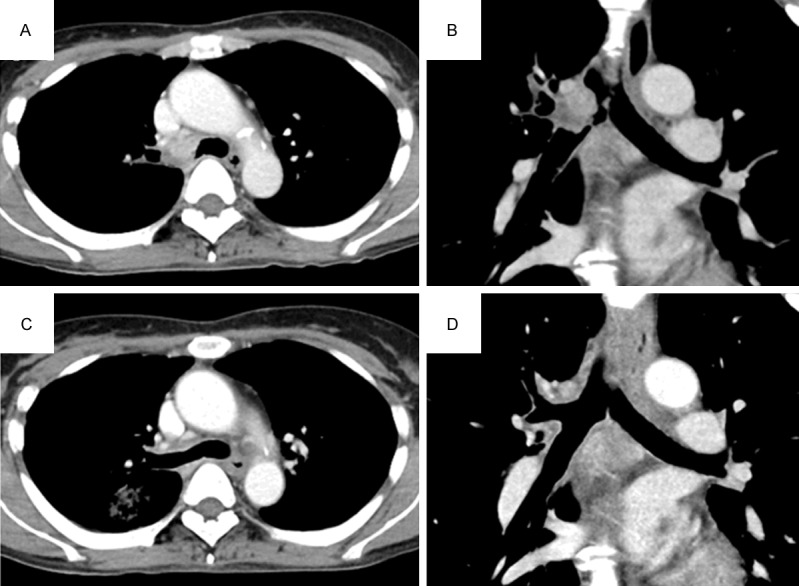
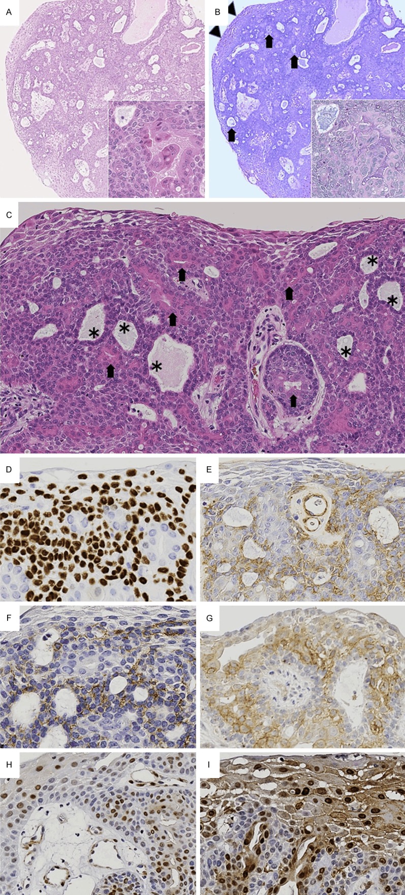
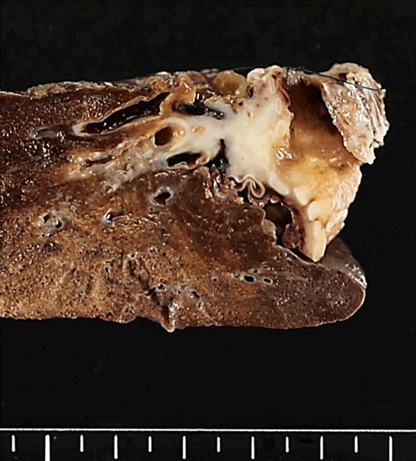
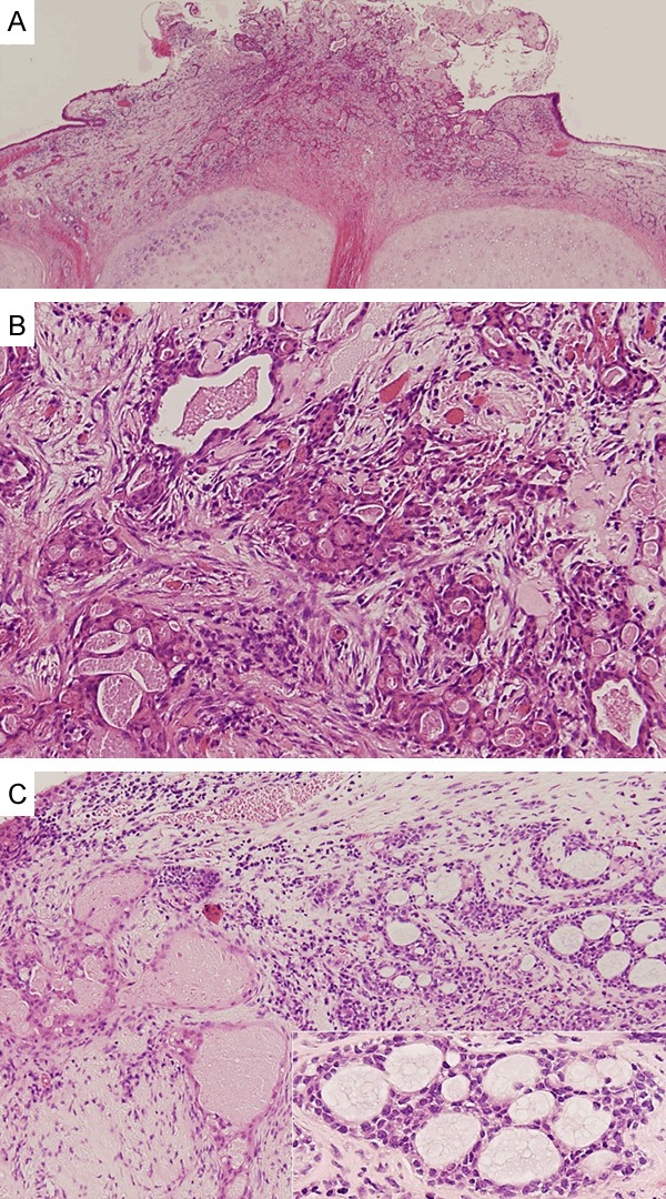
Complete dissection of tracheobronchial adenoid cystic carcinoma (TACC) by surgery alone is sometimes difficult and has a greater propensity than tracheobronchial mucoepidermoid carcinoma (TMEC) for its surgical margin to become positive. In addition, TACC is more likely to present distant metastases than TMEC. Considering these facts, TACC and TMEC should be differentiated based on histopathological examination of biopsy specimens. Herein, we present a case of 54-year-old woman with a tumor in the right main bronchus, whose biopsy specimen was difficult to diagnose as TACC or TMEC. The specimen from the rounded protrusion of the tumor showed squamous differentiation, along with the presence of glandular and basaloid cells, making morphological examination alone ineffective in rendering a definite diagnosis. Thus, the addition of immunohistochemical analysis, αSMA and CD43 expression in basaloid cells and c-kit expression in glandular cells, was useful for accurately diagnosing TACC in this case. The squamous component was considered to be neoplastic because of its increased expression of cyclin D1 and overexpression of p16. The surgically resected specimen contained typical morphology of ACC, and the diagnosis of TACC was definitely confirmed.
Keywords: Bronchus; adenoid cystic carcinoma; immunohistochemistry; mucoepidermoid carcinoma; squamous differentiation.
Figures
Similar articles
-
Treatment outcomes of patients with tracheobronchial mucoepidermoid carcinoma compared with those with adenoid cystic carcinoma.Eur J Surg Oncol. 2020 Oct;46(10 Pt A):1888-1895. doi: 10.1016/j.ejso.2020.04.020. Epub 2020 May 5. Eur J Surg Oncol. 2020. PMID: 32418755
-
Immunohistochemical study of basaloid squamous cell carcinoma, adenoid cystic and mucoepidermoid carcinoma in the upper aerodigestive tract.Anticancer Res. 2000 Mar-Apr;20(2B):1205-11. Anticancer Res. 2000. PMID: 10810423
-
Adenoid cystic carcinoma of the trachea and bronchus--a clinicopathologic study with DNA flow cytometric analysis and oncogene expression.Eur J Cardiothorac Surg. 2002 Oct;22(4):621-5. doi: 10.1016/s1010-7940(02)00406-2. Eur J Cardiothorac Surg. 2002. PMID: 12297183
-
Basaloid-squamous cell carcinoma of the bronchus. Report of a case with review of the literature.Arch Pathol Lab Med. 1995 Dec;119(12):1167-70. Arch Pathol Lab Med. 1995. PMID: 7503668 Review.
-
[Solid variant of mammary adenoid cystic carcinoma with basaloid features: a clinicopathologic and immunohistochemical study].Zhonghua Bing Li Xue Za Zhi. 2012 Dec;41(12):803-7. doi: 10.3760/cma.j.issn.0529-5807.2012.12.003. Zhonghua Bing Li Xue Za Zhi. 2012. PMID: 23324227 Review. Chinese.
References
-
- Xu LT, Sun ZF, Li ZJ, Wu LH, Zhang ZY, Yu XQ. Clinical and pathologic characteristics in patients with tracheobronchial tumor: report of 50 patients. Ann Thorac Surg. 1987;43:276–278. - PubMed
-
- Kwak SH, Lee KS, Chung MJ, Jeong YJ, Kim GY, Kwon OJ. Adenoid cystic carcinoma of the airways: helical CT and histopathologic correlation. AJR Am J Roentgenol. 2004;183:277–281. - PubMed
-
- Zhu F, Liu Z, Hou Y, He D, Ge X, Bai C, Jiang L, Li S. Primary salivary gland-type lung cancer: clinicopathological analysis of 88 cases from China. J Thorac Oncol. 2013;8:1578–1584. - PubMed
-
- Li X, Zhang W, Wu X, Sun C, Chen M, Zeng Q. Mucoepidermoid carcinoma of the lung: common findings and unusual appearances on CT. Clin Imaging. 2012;36:8–13. - PubMed
Publication types
MeSH terms
Substances
LinkOut - more resources
Full Text Sources
Research Materials