. 2015 Jun 26;4(6):e38.
doi: 10.1038/cti.2015.10. eCollection 2015 Jun.

B cell-derived circulating granzyme B is a feature of acute infectious mononucleosis

Affiliations

B cell-derived circulating granzyme B is a feature of acute infectious mononucleosis

Magdalena Hagn et al. Clin Transl Immunology. .

Abstract

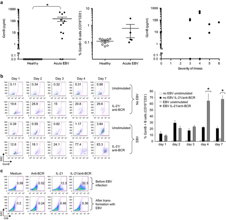
Granzyme B (GzmB) is a serine protease best known for inducing target cell apoptosis when released by cytotoxic T lymphocytes (CTLs) or natural killer cells with pore-forming perforin. As a result, GzmB detected in the serum of virus-infected individuals has typically been attributed to these sources. Here, we show that patients with recently diagnosed infectious mononucleosis caused by Epstein-Barr virus (EBV) have high circulating levels of GzmB that may be derived from infected B cells early in course of disease. We recently reported that human B cells from healthy donors secrete active GzmB when stimulated in vitro through B-cell receptor (BCR) ligation and interleukin (IL)-21. We found that infecting B cells with EBV greatly amplified GzmB secretion in response to the same stimuli, but the expression was terminated once the infection had become latent. Our results represent a rare instance of GzmB expression by non-CTL/natural killer cells in the context of infection with a human pathogen.


Figures

Figure 1
Figure 1
Patients with freshly diagnosed IM show elevated serum levels of granzyme B, likely due to large secretion from B cells during acute, but not chronic infection with EBV. After obtaining informed consent, peripheral blood and serum was taken from healthy donors or patients with recently diagnosed IM, as determined by detection of anti-VCA-IgM, or age-matched EBV+ healthy controls (n=12 each group). (a) Left panel shows results of serum levels of GzmB in each donor, as determined by cytometric bead array (BD Biosciences, San Jose, CA, USA). Horizontal lines represent mean levels in each group±s.e.m. *Indicates two-sided P-value <0.0001 for the difference between patients and healthy individuals by Mann–Whitney U test. Middle panel shows percentages of GzmB+ B cells in patients and healthy controls as determined by intracellular flow cytometry (mean±s.e.m.). Cells were harvested following 4 h incubation with Brefeldin A (1 μg ml−1), fixed, permeabilized, and GzmB expression was assessed using a PE-labelled GzmB antibody (1:400, Sanquin, Amsterdam, The Netherlands). Cell-surface markers were analysed using anti-CD3 and anti-CD19 (BD Biosciences). Right panel shows the severity of illness index in each patient compared with GzmB levels. (b, c) PBMC from healthy donors were isolated and either infected with the B95.8 strain of EBV or left untreated. After infection, cells were plated in 48-well plates at 1 × 10⁶ cells per ml and stimulated for 16 h with or without human recombinant IL-21 (50 ng ml−1, Invitrogen, Carlsbad, CA, USA) and 6.5 μg ml−1 anti-BCR (affinity purified rabbit F(ab')2 against human IgA+IgG+IgM (H+L); Jackson ImmunoResearch Laboratories, West Grove, PA, USA) on the time points indicated. Next, cells were harvested on indicated time points after infection, and GzmB expression was assessed by intracellular flow cytometry. (b) Left panel: dot plots show percentages of GzmB+ CD19+ CD3 B cells. Right panel: bar graphs depict summarized results of five independent experiments. Error bars show±s.e.m., *Indicates P<0.01 by Student's t-test. (c) Dot plots show GzmB+ B cells from freshly isolated B cells from a healthy volunteer (purity >99%, upper panel, negative selection, Miltenyi Biotech, Bergisch Gladbach, Germany) or after full in vitro transformation with the B95.8 strain of EBV (4–5 weeks after infection). B cells were stimulated for 16 h with either IL-21 or anti-BCR or both. Successful EBV transformation was determined by observation of distinct morphological feature changes by microscopy and by expression of EBNA-2 protein in B cells by western immunoblot (not shown).

References

    1. Luzuriaga K, Sullivan JL. Infectious mononucleosis. N Engl J Med. 2010;362:1993–2000. - PubMed
    1. Sutton VR, Wowk ME, Cancilla M, Trapani JA. Caspase activation by granzyme B is indirect, and caspase autoprocessing requires the release of proapoptotic mitochondrial factors. Immunity. 2003;18:319–329. - PubMed
    1. Rickinson AB, Moss DJ. Human cytotoxic T lymphocyte responses to Epstein-Barr virus infection. Annu Rev Immunol. 1997;15:405–431. - PubMed
    1. Hagn M, Schwesinger E, Ebel V, Sontheimer K, Maier J, Beyer T, et al. Human B cells secrete granzyme B when recognizing viral antigens in the context of the acute phase cytokine IL-21. J Immunol. 2009;183:1838–1845. - PubMed
    1. Hagn M, Sontheimer K, Dahlke K, Brueggemann S, Kaltenmeier C, Beyer T, et al. Human B cells differentiate into granzyme B-secreting cytotoxic B lymphocytes upon incomplete T-cell help. Immunol Cell Biol. 2012;90:457–467. - PubMed

LinkOut - more resources