. 2015 Jul 20;10(7):e0133263.
doi: 10.1371/journal.pone.0133263. eCollection 2015.

The Mitochondrial Peptidase Pitrilysin Degrades Islet Amyloid Polypeptide in Beta-Cells

Affiliations

The Mitochondrial Peptidase Pitrilysin Degrades Islet Amyloid Polypeptide in Beta-Cells

Hanjun Guan et al. PLoS One. .

Abstract

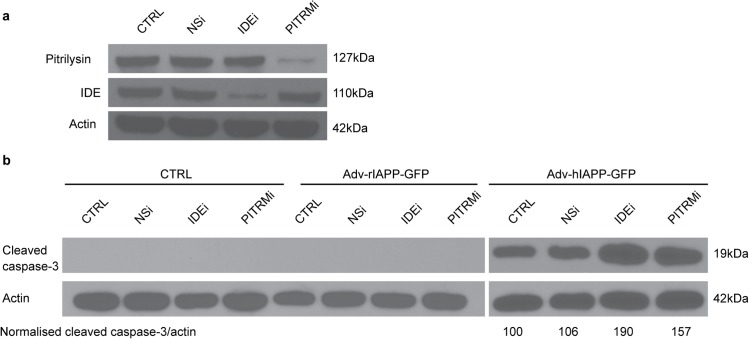
Amyloid formation and mitochondrial dysfunction are characteristics of type 2 diabetes. The major peptide constituent of the amyloid deposits in type 2 diabetes is islet amyloid polypeptide (IAPP). In this study, we found that pitrilysin, a zinc metallopeptidase of the inverzincin family, degrades monomeric, but not oligomeric, islet amyloid polypeptide in vitro. In insulinoma cells when pitrilysin expression was decreased to 5% of normal levels, there was a 60% increase in islet amyloid polypeptide-induced apoptosis. In contrast, overexpression of pitrilysin protects insulinoma cells from human islet amyloid polypeptide-induced apoptosis. Since pitrilysin is a mitochondrial protein, we used immunofluorescence staining of pancreases from human IAPP transgenic mice and Western blot analysis of IAPP in isolated mitochondria from insulinoma cells to provide evidence for a putative intramitochondrial pool of IAPP. These results suggest that pitrilysin regulates islet amyloid polypeptide in beta cells and suggest the presence of an intramitochondrial pool of islet amyloid polypeptide involved in beta-cell apoptosis.


Conflict of interest statement

Competing Interests: The authors have declared that no competing interests exist.

Figures

Fig 1
Fig 1. Degradation of monomeric hIAPP by pitrilysin.
hIAPP (20 μM) was incubated with 40 nM pitrilysin at 37°C with the reaction followed by HPLC. Peaks were collected and identified by mass spectral analysis. a. HPLC chromatograms of hIAPP degradation by pitrilysin at the indicated times. * substrate impurity; # buffer contamination; and a-m hIAPP cleavage products by pitrilysin. b. Immunodepletion of pitrilysin reduces hIAPP degradation activity. Purified pitrilysin was incubated with a polyclonal rabbit anti-pitrilysin antibody, normal rabbit IgG, or PBS followed by incubation with protein A/G-Sepharose beads. The beads were spun down and the supernatants incubated with hIAPP. The hIAPP remaining was detected by HPLC as described in Methods. c. Schematic of the cleavage sites on hIAPP. Thick arrows represent initial cleavages on hIAPP by pitrilysin and thin arrows represent secondary cleavages.
Fig 2
Fig 2. Pitrilysin does not degrade oligomeric hIAPP in vitro.
a. Oligomers of hIAPP were generated from hIAPP incubated with rat cardiac myocytes and then incubated with pitrilysin (400 nM) at 37°C for 1 hr. Reactions were analyzed by Western blot analysis using mouse monoclonal anti-human amylin antibody E-5. b. Western blot of hIAPP oligomers generated from HIP rat cardiac myocytes incubated with pitrilysin. c. hIAPP were preincubated at 37°C to induce oligomer formation and then incubated with or without 40 nM pitrilysin at 37°C for 1hr. Reactions were analyzed by Western blots using rabbit anti-IAPP that recognizes monomeric IAPP or rabbit anti-oligomer antibody A11. rIAPP (20 μM) was treated at the same condition as a negative control.
Fig 3
Fig 3. Pitrilysin expression in pancreatic beta-cells.
a. INS 832/13 cells were transfected with pcDNA3.1-pitrilysin-flag. Forty-eight hrs. after transfection, cells were fixed and stained for flag tagged pitrilysin and mitochondrial MDH. b. The mitochondrial location of pitrilysin in human islets evidenced by colocalization of pitrilysin with the mitochondrial marker, voltage-dependent anion channel. c. Western blot analysis of pitrilysin in extracts of human islets and subjected to Western blot analysis with polyclonal rabbit anti-pitrilysin antibody. Lane 1: human islet lysate (10 μg); Lane 2: 60 ng recombinant pitrilysin.
Fig 4
Fig 4. Knockdown of pitrilysin in INS cells exacerbates hIAPP-induced apoptosis.
Stably transfected INS 832/13 cells expressing an shRNA specific to rat pitrilysin (PITRMi), rat IDE (IDEi) or non-silencing control shRNA (NSi) were selected using 10 μg/ml puromycin. Stable cells were then transduced with Adv-prepro-hIAPP-GFP (MOI = 100) and apoptosis determined by Western blot analysis of cleaved caspase-3. Band densities were analyzed using NIH Image J software. The ratios of cleaved caspase-3 to actin intensities are indicated (n = 3). a. Pitrilysin in INS832/13 cells is reduced ~95% by lenti-PITRMi. b. An approximate 60% increase (162.1 ± 13.2% of control, p<0.01) in apoptosis induced by hIAPP in pitrilysin shRNA treated cells compared to the lenti-NSi control or parental INS 832/13 cells. shRNA treatment itself did not induce apoptosis.
Fig 5
Fig 5. Overexpression of pitrilysin protects INS cells from hIAPP-induced toxicity.
INS 832/13 cells were transduced with lentivirus carrying human pitrilysin (PITRM), an inactive mutant (PITRMx), or empty lentiviral vector. Stable cell lines were isolated and then transduced with Adv-prepro-hIAPP-GFP. Apoptosis was determined at 48 hrs. after transduction as in Fig 4 with the ratios of cleaved caspase-3 to actin intensities indicated (n = 3). a. Pitrilysin protein levels were increased by 80% (PITRMx) and by 60% (PITRM) in the stable cell lines. b. An ~30% decrease (68.7 ± 10.3% of control, p<0.05) in apoptosis induced by hIAPP was observed in pitrilysin overexpressing cells compared to empty lentiviral vector control, inactive pitrilysin (PITRMx) overexpressing cells, or parental INS 832/13 cells. Treatment with Adv-prepro-rIAPP-GFP did not cause apoptosis.
Fig 6
Fig 6. Mitochondrial localization of hIAPP.
a. INS 832/13 cells were transfected with pCMV6-XL5-prepro-hIAPP. Forty-eight hrs. after transfection, cells were fixed and stained for hIAPP (mouse anti-hIAPP) and mitochondrial malate dehydrogenase. b. hIAPP/mMDH and insulin/mMDH immunocytochemistry performed on paraffin sections of pancreas from hIAPP transgenic mice. Staining was visualized with FITC conjugated goat anti-mouse and cy3 conjugated goat anti-rabbit secondary antibodies. Confocal images were taken as described in Methods and merged with Adobe Photoshop software. Scale bar: 10μm. Yellow color denotes colocalization.
Fig 7
Fig 7. Detection of IAPP in the mitochondrial fraction from INS cells.
a. Protein components of subcellular fractions prepared from INS cells were immunoblotted with anti-IAPP and antibodies to a cytosolic marker (cytosolic thiolase), an ER marker (calnexin), and a mitochondrial markers (mitochondrial malate dehydrogenase, mMDH). H: homogenate; C: cytosol; Mi: Microsomal fraction; Mc: crude mitochondrial fraction; Mp: purified mitochondrial fraction. b. Purified mitochondria from INS cells were incubated with trypsin (10 μg/ml) in the presence or absence of Triton X-100 (0.01%) at 37°C for 30min. The reactions were stopped with 50 μg/ml of trypsin inhibitor and subjected to SDS-PAGE and Western blot analysis. IAPP in mitochondria is insensitive to trypsin without prior Triton X-100 permeabilization suggesting an intramitochondrial location of IAPP.

Similar articles

Cited by

References

    1. Butler AE, Janson J, Bonner-Weir S, Ritzel R, Rizza RA, Butler PC. Beta-cell deficit and increased beta-cell apoptosis in humans with type 2 diabetes. Diabetes. 2003;52(1):102–10. . - PubMed
    1. Johnson KH, O'Brien TD, Betsholtz C, Westermark P. Islet amyloid polypeptide: mechanisms of amyloidogenesis in the pancreatic islets and potential roles in diabetes mellitus. Lab Invest. 1992;66(5):522–35. . - PubMed
    1. Haataja L, Gurlo T, Huang CJ, Butler PC. Islet amyloid in type 2 diabetes, and the toxic oligomer hypothesis. Endocr Rev. 2008;29(3):303–16. 10.1210/er.2007-0037 . - DOI - PMC - PubMed
    1. Kahn SE, Andrikopoulos S, Verchere CB. Islet amyloid: a long-recognized but underappreciated pathological feature of type 2 diabetes. Diabetes. 1999;48(2):241–53. . - PubMed
    1. Zraika S, Hull RL, Verchere CB, Clark A, Potter KJ, Fraser PE, et al. Toxic oligomers and islet beta cell death: guilty by association or convicted by circumstantial evidence? Diabetologia. 2010;53(6):1046–56. 10.1007/s00125-010-1671-6 . - DOI - PMC - PubMed

Publication types

LinkOut - more resources