. 2015 Jul 20:5:12287.
doi: 10.1038/srep12287.

Opportunistic pathogen Candida albicans elicits a temporal response in primary human mast cells

Affiliations

Opportunistic pathogen Candida albicans elicits a temporal response in primary human mast cells

José Pedro Lopes et al. Sci Rep. .

Abstract

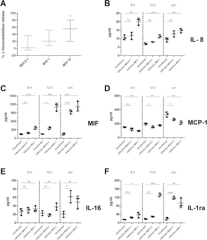
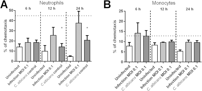
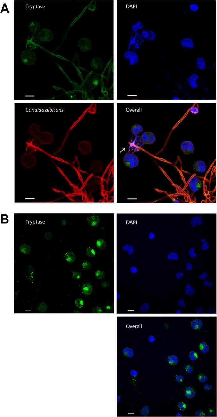
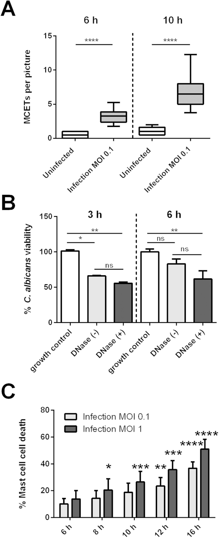
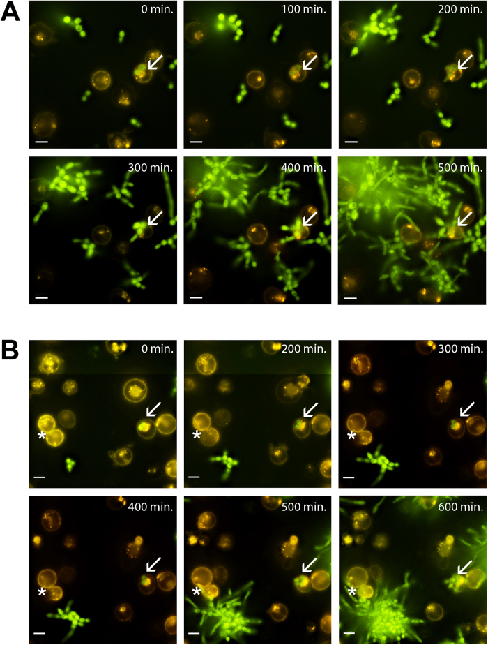
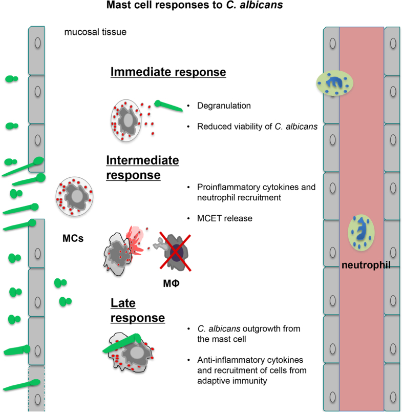
Immunosuppressed patients are frequently afflicted with severe mycoses caused by opportunistic fungal pathogens. Besides being a commensal, colonizing predominantly skin and mucosal surfaces, Candida albicans is the most common human fungal pathogen. Mast cells are present in tissues prone to fungal colonization being expectedly among the first immune cells to get into contact with C. albicans. However, mast cell-fungus interaction remains a neglected area of study. Here we show that human mast cells mounted specific responses towards C. albicans. Collectively, mast cell responses included the launch of initial, intermediate and late phase components determined by the secretion of granular proteins and cytokines. Initially mast cells reduced fungal viability and occasionally internalized yeasts. C. albicans could evade ingestion by intracellular growth leading to cellular death. Furthermore, secreted factors in the supernatants of infected cells recruited neutrophils, but not monocytes. Late stages were marked by the release of cytokines that are known to be anti-inflammatory suggesting a modulation of initial responses. C. albicans-infected mast cells formed extracellular DNA traps, which ensnared but did not kill the fungus. Our results suggest that mast cells serve as tissue sentinels modulating antifungal immune responses during C. albicans infection. Consequently, these findings open new doors for understanding fungal pathogenicity.


Figures

Figure 1
Figure 1. C. albicans induced mast cell degranulation and cytokine release in a MOI-dependent manner.
(A) HMC-1 cells were infected with opsonized C. albicans yeasts (MOI 0.1, 1 and 10) for 1 hour, after which ß-hexosaminidase release was measured from supernatants of infection. (BF) Shown are 5 cytokines at 6, 12 and 24 h post infection that were released differentially from different supernatants of mast cells infected with C. albicans (MOI 0.1 and 1) or of mast cells left uninfected. ß–hexosaminidase percentage release was defined by the amount of ß-hexosaminidase release from infected cell divided by spontaneous ß-hexosaminidase release from uninfected cells (% of ß –hexosaminidase release/% of ß –hexosaminidase control). Significance for (A–F) was analysed by Tukey one-way ANOVA *P ≤ 0.05. Data are presented as means of n = 4 (4) ± SD (ß-hexosaminidase release assay) and n = 3 (3) ± SD (Cytokine Multiplex).
Figure 2
Figure 2. Neutrophils but not monocytes differentially migrated in response to supernatants from mast cells infected with C. albicans.
Supernatants collected from C. albicans-infected mast cells (MOI 0.1) at 6 h, 12 h and overnight infection were used as chemoattractants to neutrophils and monocytes in a transwell system. End-point cell migration was plotted per condition, per time as ratio of migrated cells using as 100% control cells added to the lower compartment without inserted transwell system. (A) Neutrophil migration is increased over time towards supernatants of infection but not to C. albicans and HMC-1 alone (controls). (B) Monocytes show no significant chemoattraction towards supernatants of infected mast cells. Variations between neutrophil or monocyte migration over time towards supernatants of infection and C. albicans control were analysed for statistical significance using a one-way ANOVA with Tukey post-test. As positive control for migration we used fMLP causing chemotaxis significantly above background of approximately 45% after 30 min for neutrophils and 13% after 90 min for monocytes. These values are indicated as a horizontal, dashed line in the graphs of the figure. Data are presented as means of n = 5 (3) ± SD.
Figure 3
Figure 3. C. albicans induced MCETs in a time-dependent manner.
Mast cells were infected for 6 h with C. albicans with an MOI 0.1 (A) or left uninfected (B). Shown are representative micrographs of indirect immunofluorescence from fixed and permeabilized samples with DNA (blue), mast cell tryptase (green) as well as C. albicans (red) stained samples. MCETs were identified by co-localization of extracellular laminar DNA with tryptase immunostaining (arrows). Scale bars, 10 μm.
Figure 4
Figure 4. Quantification of cellular death and antifungal activity of mast cells.
(A) The graph depicts the relative amount of MCETs per micrograph of C. albicans-infected mast cells (MOI 0.1) as compared to the uninfected mast cell control at two different time points. At both time points analysed (6 h and 10 h) the variation between MCETs compared to uninfected samples was analysed for statistical significance. (B) The graph represents the viability of fungal cells after normalization to the biological replicates 100% growth control. C. albicans viability is reduced in mast cell infection (MOI 1) up to 3 h. (C) C. albicans-induced mast cell death in a time and dose-dependent manner as determined with Sytox green. The Y-axis represents the relative amount of dead cells after normalization to the mast cell lysis control. (A) Significance was analysed by t-test and by Tukey one-way ANOVA (B) *P ≤ 0.05. (B) Data are presented as means of at least six replicates (A) and n = 3 (6) ± SD. (C) For cell death significance was analysed by Bonferroni two-way ANOVA *P ≤ 0.05 comparing to the mast cell uninfected control at each time point. Data represents n= 4 (5) ±SD.
Figure 5
Figure 5. C. albicans internalization is followed by fungal outgrowth.
Shown are images of the indicated time points for C. albicans infected mast cells (MOI 0.1). (A) Arrows show an intracellular yeast cell (GFP-expressing C. albicans strain CAI4 pENO1-GFP-CyC1t) replicating inside the mast cell (orange, membrane stain DiI) finally rupturing the plasma membrane as determined by loss of signal. An extracellular C. albicans hyphal tip growing towards a mast cell nudged the host cell and induced collapse of the plasma membrane (*). Complete movies are available as Movie S2 and S3 in the supplemental material. Scale bar, 10 μm.
Figure 6
Figure 6. Summary of mast cell and C. albicans interaction.
Our findings indicate that mast cells specifically respond to C. albicans by degranulation, secretion of cytokines and chemokines, internalization of C. albicans and the release of MCETs. Interestingly, the processes seem to be organised and thus can be divided into three time periods: Initial (up to 3 h), intermediate (3 h to 12 h), and late responses (>12 h).

References

    1. Mikulska M., Del Bono V., Ratto S. & Viscoli C. Occurrence, presentation and treatment of candidemia. Expert Rev Clin Immunol 8, 755–765, 10.1586/eci.12.52 (2012). - DOI - PubMed
    1. Patterson T. F. Advances and challenges in management of invasive mycoses. Lancet 366, 1013–1025, 10.1016/S0140-6736(05)67381-3 (2005). - DOI - PubMed
    1. Montravers P. et al. A multicentre study of antifungal strategies and outcome of Candida spp. peritonitis in intensive-care units. Clin Microbiol Infect 17, 1061–1067, 10.1111/j.1469-0691.2010.03360.x (2011). - DOI - PubMed
    1. Bassetti M. et al. Epidemiology, species distribution, antifungal susceptibility, and outcome of candidemia across five sites in Italy and Spain. J Clin Microbiol 51, 4167–4172, 10.1128/JCM.01998-13 (2013). - DOI - PMC - PubMed
    1. Abraham S. N. & St John A. L. Mast cell-orchestrated immunity to pathogens. Nat Rev Immunol 10, 440–452, 10.1038/nri2782 (2010). - DOI - PMC - PubMed

Publication types

MeSH terms

LinkOut - more resources