. 2015 Oct 15;192(8):998-1008.
doi: 10.1164/rccm.201410-1820OC.

The Causal Role of IL-4 and IL-13 in Schistosoma mansoni Pulmonary Hypertension

Affiliations

The Causal Role of IL-4 and IL-13 in Schistosoma mansoni Pulmonary Hypertension

Rahul Kumar et al. Am J Respir Crit Care Med. .

Abstract

Rationale: The etiology of schistosomiasis-associated pulmonary arterial hypertension (PAH), a major cause of PAH worldwide, is poorly understood. Schistosoma mansoni exposure results in prototypical type-2 inflammation. Furthermore, transforming growth factor (TGF)-β signaling is required for experimental pulmonary hypertension (PH) caused by Schistosoma exposure.

Objectives: We hypothesized type-2 inflammation driven by IL-4 and IL-13 is necessary for Schistosoma-induced TGF-β-dependent vascular remodeling.

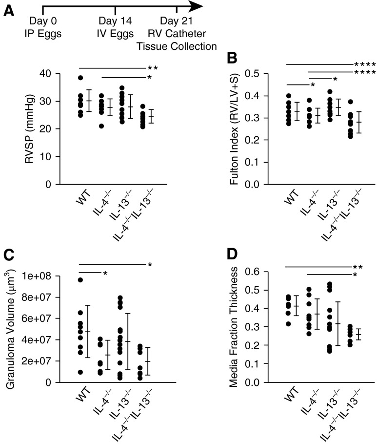
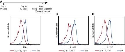
Methods: Wild-type, IL-4(-/-), IL-13(-/-), and IL-4(-/-)IL-13(-/-) mice (C57BL6/J background) were intraperitoneally sensitized and intravenously challenged with S. mansoni eggs to induce experimental PH. Right ventricular catheterization was then performed, followed by quantitative analysis of the lung tissue. Lung tissue from patients with schistosomiasis-associated and connective tissue disease-associated PAH was also systematically analyzed.

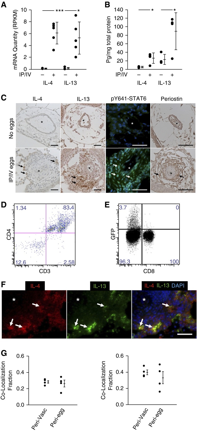
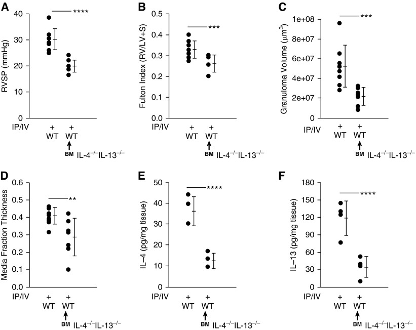
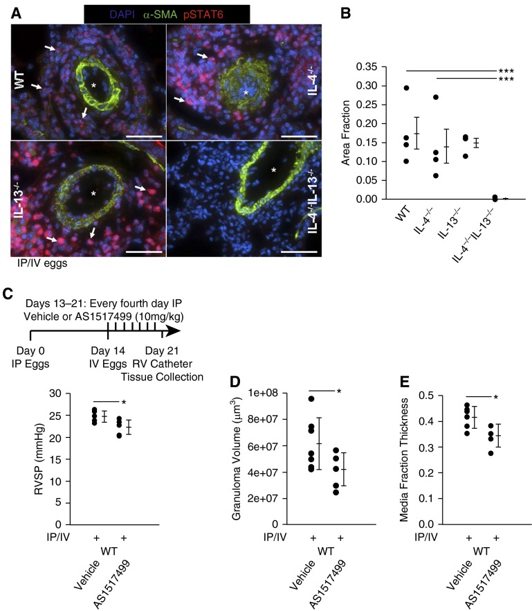
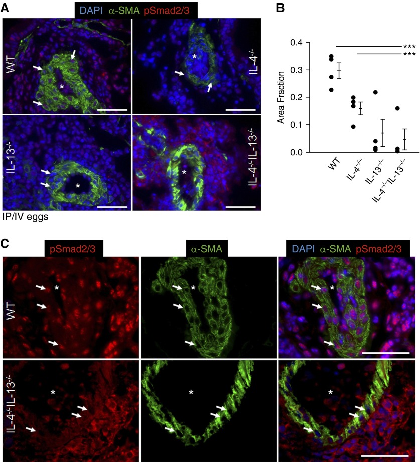
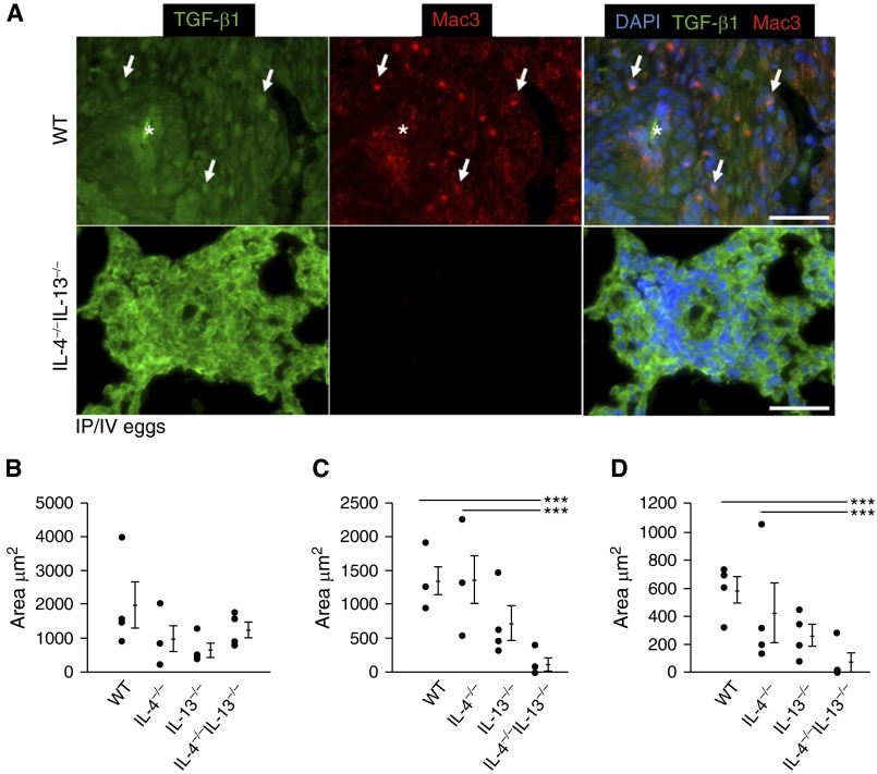
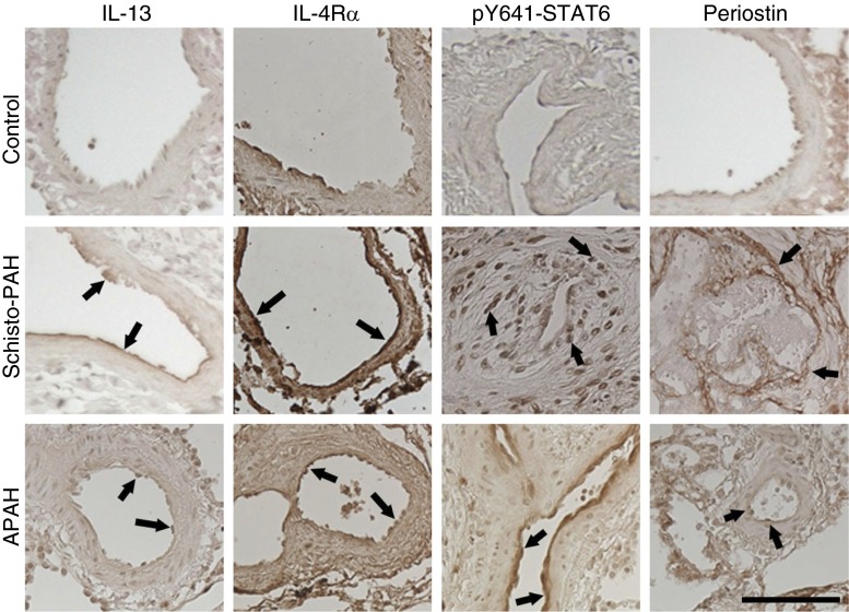
Measurements and main results: Mice with experimental Schistosoma-induced PH had evidence of increased IL-4 and IL-13 signaling. IL-4(-/-)IL-13(-/-) mice, but not single knockout IL-4(-/-) or IL-13(-/-) mice, were protected from Schistosoma-induced PH, with decreased right ventricular pressures, pulmonary vascular remodeling, and right ventricular hypertrophy. IL-4(-/-)IL-13(-/-) mice had less pulmonary vascular phospho-signal transducer and activator of transcription 6 (STAT6) and phospho-Smad2/3 activity, potentially caused by decreased TGF-β activation by macrophages. In vivo treatment with a STAT6 inhibitor and IL-4(-/-)IL-13(-/-) bone marrow transplantation also protected against Schistosoma-PH. Lung tissue from patients with schistosomiasis-associated and connective tissue disease-associated PAH had evidence of type-2 inflammation.

Conclusions: Combined IL-4 and IL-13 deficiency is required for protection against TGF-β-induced pulmonary vascular disease after Schistosoma exposure, and targeted inhibition of this pathway is a potential novel therapeutic approach for patients with schistosomiasis-associated PAH.

Keywords: Th2 cells; pulmonary hypertension; schistosomiasis; transforming growth factor-β.


Figures

Figure 1.
Figure 1.
Schistosoma-exposed mouse tissue demonstrates evidence of Th2 signaling. (A) Whole-lung quantity of IL-4 and IL-13 messenger RNA (mRNA) transcripts (n = 5 samples per group; RPKM; mean ± SD; t test, *P < 0.05; ***P < 0.005). (B) IL-4 and IL-13 whole-lung ELISA (mean ± SD; n = 4 samples per group; t test, *P < 0.05). (C) Immunostaining for IL-4, IL-13, phospho–signal transducer and activator of transcription factor 6 (STAT6) and periostin in Schistosoma-exposed and unexposed mouse lung tissue (asterisks, vessel lumens; arrows, representative positive stained cells; scale bars, 50 μm). (D) Flow cytometry analysis of GFP+ cells from intraperitoneal and intravenous (IP/IV) egg–exposed IL-4GFP mice for CD3+ and CD4+ cells. (E) Percentage of CD3+CD4+ and CD3+CD8+ cells in IP/IV egg–exposed mice positive for IL-4GFP (flow studies repeated at least three times). (F and G) Qualitative (F) and quantitative (G) colocalization of IL-4 and IL-13 using double immunoflorescence analysis (G, left, fraction of IL-4–positive area that colocalizes with IL-13; G, right, fraction of IL-13–positive area that colocalizes with IL-4; asterisk, vessel lumens; arrows, representative double-positive cells; scale bar, 50 μm; plots mean ± SD; n = 4 samples per group; the groups are perivascular/adventitia and periegg/granuloma images). DAPI = 4′,6-diamidino-2-phenylindole; GFP = green fluorescent protein; RPKM = reads per kilobase per million mapped reads.
Figure 2.
Figure 2.
Mice exposed to Schistosoma mansoni eggs develop pulmonary hypertension and vascular remodeling, which is suppressed in the absence of IL-4 and IL-13. (A) Right ventricular systolic pressure (RVSP) of intraperitoneal and intravenous (IP/IV) egg–exposed mice (mean ± SD; n = 9–18 mice per group; analysis of variance (ANOVA), P = 0.01; post hoc Tukey test, *P < 0.05, **P < 0.01). (B) Fulton index (RV/[LV + S]) of IP/IV egg–exposed mice (mean ± SD; n = 9–18 mice per group; ANOVA, P = 0.001; post hoc Tukey test, *P < 0.05, ****P < 0.001). (C) Periegg granuloma volumes in IP/IV egg–exposed mice (mean ± SD; n = 9–18 mice per group; ANOVA, P = 0.03; post hoc Tukey test, *P < 0.05). (D) Quantitative fractional thickness of the pulmonary vascular media in IP/IV egg exposed mice (mean ± SD; n = 9–18 mice per group; ANOVA P = 0.007; post hoc Tukey test, *P < 0.05, **P < 0.01). LV = left ventricle; RV = right ventricle; S = systolic; WT = wild type.
Figure 3.
Figure 3.
Wild-type (WT) recipients of IL-4−/−IL-13−/− bone marrow (BM) showed decreased IL-4 and IL-13 levels and protection against pulmonary hypertension after exposure to Schistosoma mansoni eggs. WT and WT recipients of IL-4−/−IL-13−/− BM followed by intraperitoneal and intravenous (IP/IV) egg exposure. (A) Right ventricular systolic pressure (RVSP) (mean ± SD; n = 8–9 mice per group; t test, ****P < 0.001). (B) Fulton index (RV/[LV + S]) (mean ± SD; n = 8–9 mice per group; t test, ***P < 0.005). (C) Periegg granuloma volume (mean ± SD; n = 8–9 mice per group; t test, ***P < 0.005). (D) Quantitative fractional thickness of the pulmonary vascular media (mean ± SD; n = 8–9 mice per group; t test, **P < 0.01). (E) Concentration of whole-lung IL-4 (mean ± SD; n = 4 mice per group; t test, ****P < 0.001). (F) Concentration of whole-lung IL-13 (mean ± SD; n = 4 mice per group; t test, ****P < 0.001). LV = left ventricle; RV = right ventricle; S = systolic.
Figure 4.
Figure 4.
IL-4−/−IL-13−/− mice exposed to Schistosoma mansoni eggs have suppressed Th1 and Th17 responses. (A–C) Flow cytometric histograms of CD3+CD4+ populations from digested whole-lung tissue, individually gated for IFN-γ (A), IL-17A (B), and IL-17F (C) in intraperitoneal and intravenous (IP/IV) egg–exposed IL-4−/−IL-13−/− and wild-type (WT) mice (flow studies repeated at least three times).
Figure 5.
Figure 5.
Schistosoma-exposed IL-4−/−IL-13−/− mice have decreased phospho–signal transducer and activator of transcription factor 6 (STAT6), and STAT6 inhibition prevents Schistosoma-pulmonary hypertension. (A) Representative images showing representative immunostaining for phospho-Y641-STAT6 in Schistosoma-exposed wild-type (WT), IL-4−/−, IL-13−/−, and IL-4−/−IL-13−/− mice (images representative of n = 4/group; asterisks, vessel lumens; arrows, representative positive stained cells; scale bars, 50 μm). (B) Quantification of area staining positive for phospho-STAT6 in the pulmonary vascular adventitia in Schistosoma-exposed WT, IL-4−/−, IL-13−/−, and IL-4−/−IL-13−/− mice (mean ± SD; n = 4 specimens per group; analysis of variance, P = 0.009; post hoc Tukey test, ***P < 0.005). (C–E) IP/IV egg–exposed WT mice treated with the STAT6 inhibitor AS1517499 or equivalent volume of vehicle: right ventricular systolic pressure (RVSP) (mean ± SD; n = 7 mice per group; t test *P < 0.05) (C); granuloma volume (mean ± SD; n = 7 mice per group; t test *P < 0.05) (D); and quantitative fractional thickness of the pulmonary vascular media (mean ± SD; n = 7 mice per group; t test *P < 0.05) (E). α-SMA = α-smooth muscle actin; DAPI = 4′,6-diamidino-2-phenylindole; IP/IV = intraperitoneal and intravenous; RV = right ventricle.
Figure 6.
Figure 6.
Nuclear phospho-Smad2/3 in the media is decreased in IL-4−/−IL-13−/− Schistosoma-exposed mice. (A) Representative images showing immunostaining for phospho-Smad2/3 and α-smooth muscle actin (α-SMA) in Schistosoma-exposed wild-type (WT), IL-4−/−, IL-13−/−, and IL-4−/−IL-13−/− mice (images representative of n = 4/group; asterisks, vessel lumens; arrows, representative positive nuclear-stained cells; scale bars, 50 μm). (B) Quantification of the fraction of DAPI-positive pixels that also stain positive for phospho-Smad2/3 in the media of Schistosoma-exposed WT, IL-4−/−, IL-13−/−, and IL-4−/−IL-13−/− mice (mean ± SD; n = 4 specimens per group; analysis of variance, P = 0.002; post hoc Tukey test, ***P < 0.005). (C) Higher-magnification (×60 oil lens) representative images showing immunostaining for phospho-Smad2/3 and α-smooth muscle actin in Schistosoma-exposed WT and IL-4−/−IL-13−/− mice (asterisks, vessel lumens; arrows, representative media cells; scale bars, 50 μm). DAPI = 4′,6-diamidino-2-phenylindole; IP/IV = intraperitoneal and intravenous.
Figure 7.
Figure 7.
Macrophage density and transforming growth factor (TGF)-β1–macrophage colocalization is decreased in IL-4−/−IL-13−/− Schistosoma-exposed mice. (A) Representative images showing double immunofluorescence staining for TGF-β ligand and Mac3 (macrophage marker; images representative of n = 4/group; asterisks, vessel lumens; arrows, representative positive double-stained cells; scale bars, 50 μm). (B–D) Quantitative analysis of area in the adventitia that stains positive for TGF-β1 (mean ± SD; n = 4 specimens per group; analysis of variance [ANOVA], P = NS) (B), Mac3 (macrophages; mean ± SD; n = 4 specimens per group; ANOVA, P = 0.011; post hoc Tukey test, ***P < 0.005) (C), and colocalization of both TGF-β1 and macrophages (mean ± SD; n = 4 specimen per group; ANOVA, P = 0.011; post hoc Tukey test, ***P < 0.005) (D) in Schistosoma-exposed wild-type, IL-4−/−, IL-13−/−, and IL-4−/−IL-13−/− mice. DAPI = 4′,6-diamidino-2-phenylindole; IP/IV = intraperitoneal and intravenous; NS = not significant; WT = wild-type.
Figure 8.
Figure 8.
Human lung tissue immunostaining demonstrates increased Th2 signaling. Immunostaining for the type 2 signaling molecules IL-13, IL-4Rα, phospho–signal transducer and activator of transcription factor 6 (STAT6), and periostin in lung tissue of patients with Schistosoma–pulmonary arterial hypertension (Schisto-PAH) and connective tissue disease–associated PAH (APAH) compared with normal control subjects. Semiquantitative analysis is reported in Table E6. The images are representative of n = 5–11/group; arrows indicate representative positive vascular staining; scale bar, 100 μm.

Comment in

References

    1. Hovnanian A, Hoette S, Fernandes CJ, Jardim C, Souza R. Schistosomiasis associated pulmonary hypertension. Int J Clin Pract Suppl. 2010;(165):25–28. - PubMed
    1. Simonneau G, Gatzoulis MA, Adatia I, Celermajer D, Denton C, Ghofrani A, Gomez Sanchez MA, Krishna Kumar R, Landzberg M, Machado RF, et al. Updated clinical classification of pulmonary hypertension. J Am Coll Cardiol. 2013;62(25) Suppl:D34–D41. - PubMed
    1. Boros DL, Whitfield JR. Enhanced Th1 and dampened Th2 responses synergize to inhibit acute granulomatous and fibrotic responses in murine schistosomiasis mansoni. Infect Immun. 1999;67:1187–1193. - PMC - PubMed
    1. Burke ML, Jones MK, Gobert GN, Li YS, Ellis MK, McManus DP. Immunopathogenesis of human schistosomiasis. Parasite Immunol. 2009;31:163–176. - PubMed
    1. Jiang H, Harris MB, Rothman P. IL-4/IL-13 signaling beyond JAK/STAT. J Allergy Clin Immunol. 2000;105:1063–1070. - PubMed

Publication types

MeSH terms