. 2015 Sep;11(9):685-90.
doi: 10.1038/nchembio.1864. Epub 2015 Jul 20.

A biosynthetic pathway for a prominent class of microbiota-derived bile acids

Affiliations

A biosynthetic pathway for a prominent class of microbiota-derived bile acids

A Sloan Devlin et al. Nat Chem Biol. 2015 Sep.

Abstract

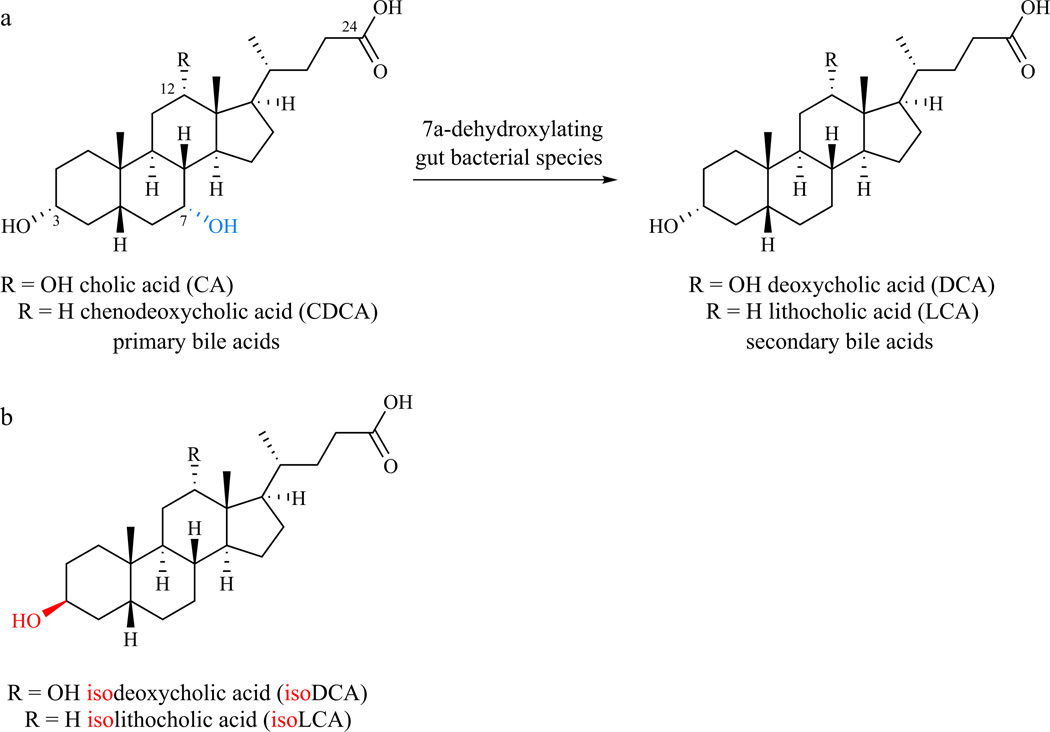
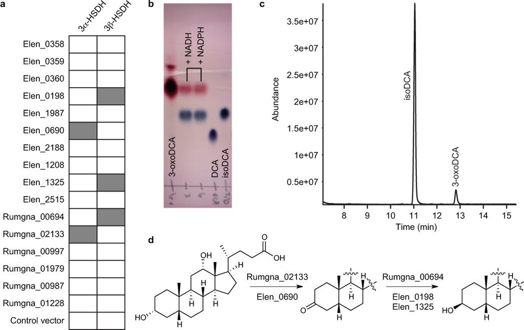
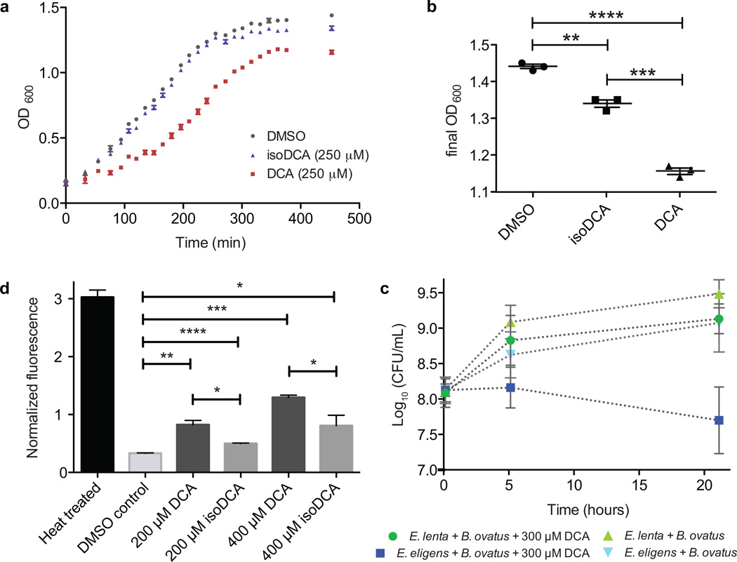
The gut bile acid pool is millimolar in concentration, varies widely in composition among individuals and is linked to metabolic disease and cancer. Although these molecules are derived almost exclusively from the microbiota, remarkably little is known about which bacterial species and genes are responsible for their biosynthesis. Here we report a biosynthetic pathway for the second most abundant class in the gut, 3β-hydroxy(iso)-bile acids, whose levels exceed 300 μM in some humans and are absent in others. We show, for the first time, that iso-bile acids are produced by Ruminococcus gnavus, a far more abundant commensal than previously known producers, and that the iso-bile acid pathway detoxifies deoxycholic acid and thus favors the growth of the keystone genus Bacteroides. By revealing the biosynthetic genes for an abundant class of bile acids, our work sets the stage for predicting and rationally altering the composition of the bile acid pool.


Figures

Figure 1
Figure 1. Chemical transformations of bile acids by gut bacteria
(a) Conversion of the primary bile acids CA and CDCA to the secondary bile acids DCA and LCA, respectively, involves the net reductive removal of the 7–OH group. (b) IsoDCA and isoLCA are 3β–OH epimers of DCA and LCA.
Figure 2
Figure 2. Elucidation of the biosynthetic pathway for isoDCA formation inEggerthella lentaand Ruminococcus gnavus
(a) Results of cell lysate assay in which candidate HSDH enzymes were expressed in E. coli and their ability to convert DCA to 3-oxoDCA (column 1) and 3-oxoDCA to isoDCA (column 2) was analyzed by TLC. Gray box: Product spot was present; white box: no product was observed. (b-c) Representative example showing enzymatic conversion by purified Rumgna_00694 of 3-oxoDCA into isoDCA by (b) TLC and (c) GC-MS. Retention times: isoDCA, 11.1 min; 3-oxoDCA, 12.9 min. In (c), NADPH was used as cofactor in the enzymatic reaction. (d) The biosynthetic pathway we elucidated for conversion of bile acids to iso bile acids involves the consecutive action of 3α- and 3β-hydroxysteroid dehydrogenases (HSDHs).
Figure 3
Figure 3. IsoDCA is less bacteriostatic than DCA
(a) B. ovatus growth rate in the presence of 250 µM DCA, 250 µM isoDCA, or DMSO control. Data shown are from one experiment with three technical replicates. Error bars represent standard deviation. (b) Saturated culture density of B. ovatus was lower in DCA-treated versus isoDCA-treated (P = 0.0002) or DMSO-treated cultures (P < 0.0001) (asterisks represent significant differences between groups, Welch’s t test, n = 3 per group). (c) Co-culture experiment showing the growth of B. ovatus in the presence of either the isoDCA producer E. lenta or the common gut commensal and non-producer Eubacterium eligens with or without 300 µM DCA (starting concentration). Data shown are from four biological replicates. Error bars represent standard deviation. (d) Cell membrane damage caused by exposure of actively dividing B. ovatus cells to either DCA or isoDCA was assessed using a propidium iodide (Pi) fluorescence assay. IsoDCA caused less membrane damage than DCA at both 200 µM (½ DCA MIC, P = 0.0158) and 400 µM (DCA MIC, P = 0.0367). Data shown are from three biological replicates. Error bars represent standard deviation. Asterisks represent significant differences between groups, Welch’s t test, n = 3 per group. (DMSO vs. 200 µM DCA, P = 0.0068; DMSO vs. 200 µM isoDCA, P <0.0001; DMSO vs. 400 µM DCA, P = 0.0004; DMSO vs. 400 µM isoDCA, P = 0.0435.)

References

    1. Ridlon J, Kang D, Hylemon P. Bile salt biotransformations by human intestinal bacteria. J. Lipid Res. 2006;47:241–259. - PubMed
    1. Hamilton JP, et al. Human cecal bile acids: concentration spectrum. Am. J. Physiol. Gastrointest. Liver Physiol. 2007;293:G256–G263. - PubMed
    1. Macdonald I, Bokkenheuser VD, Winter J, McLernon AM, Mosbach EH. Degradation of steroids in the human gut. J. Lipid Res. 1983;24:675–700. - PubMed
    1. Hofmann AF, et al. A proposed nomenclature for bile acids. J. Lipid Res. 1992;33:599–604. - PubMed
    1. Setchell KDR, Lawson AM, Tanida N, Sjovall J. General methods for the analysis of metabolic profiles of bile acids and related compounds in feces. J. Lipid Res. 1983;24:1085–1100. - PubMed

Publication types

MeSH terms