Review
. 2016 Mar;1863(3):401-413.
doi: 10.1016/j.bbamcr.2015.07.009. Epub 2015 Jul 17.

Microenvironment interactions and B-cell receptor signaling in Chronic Lymphocytic Leukemia: Implications for disease pathogenesis and treatment

Affiliations
Review

Microenvironment interactions and B-cell receptor signaling in Chronic Lymphocytic Leukemia: Implications for disease pathogenesis and treatment

Elisa Ten Hacken et al. Biochim Biophys Acta. 2016 Mar.

Abstract

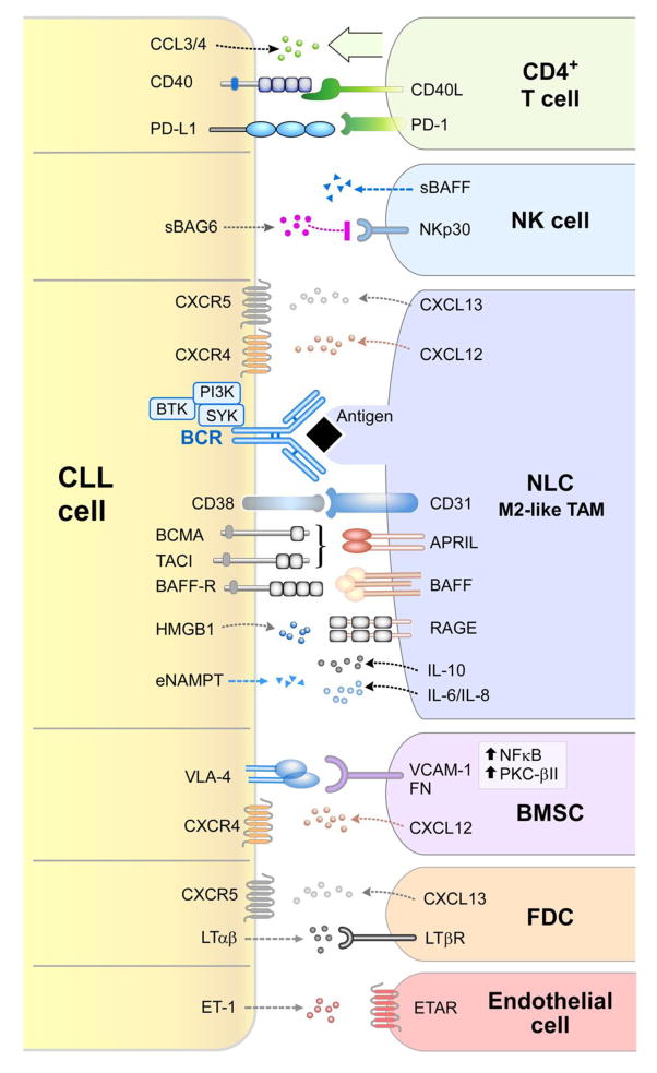
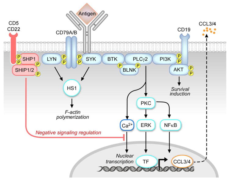
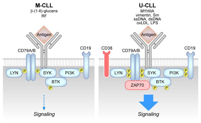
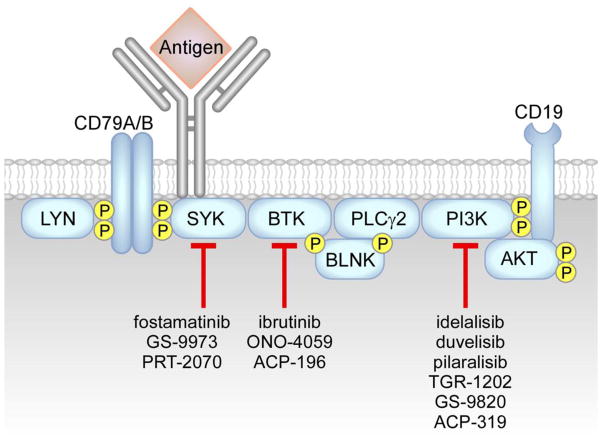
Chronic Lymphocytic Leukemia (CLL) is a malignancy of mature B lymphocytes which are highly dependent on interactions with the tissue microenvironment for their survival and proliferation. Critical components of the microenvironment are monocyte-derived nurselike cells (NLCs), mesenchymal stromal cells, T cells and NK cells, which communicate with CLL cells through a complex network of adhesion molecules, chemokine receptors, tumor necrosis factor (TNF) family members, and soluble factors. (Auto-) antigens and/or autonomous mechanisms activate the B-cell receptor (BCR) and its downstream signaling cascade in secondary lymphatic tissues, playing a central pathogenetic role in CLL. Novel small molecule inhibitors, including the Bruton's tyrosine kinase (BTK) inhibitor ibrutinib and the phosphoinositide-3-kinase delta (PI3Kδ) inhibitor idelalisib, target BCR signaling and have become the most successful new therapeutics in this disease. We here review the cellular and molecular characteristics of CLL cells, and discuss the cellular components and key pathways involved in the cross-talk with their microenvironment. We also highlight the relevant novel treatment strategies, focusing on immunomodulatory agents and BCR signaling inhibitors and how these treatments disrupt CLL-microenvironment interactions. This article is part of a Special Issue entitled: Tumor Microenvironment Regulation of Cancer Cell Survival, Metastasis, Inflammation, and Immune Surveillance edited by Peter Ruvolo and Gregg L. Semenza.

Keywords: BCR; BCR signaling; BCR signaling inhibitors; CLL; Nurselike cells; Stromal cells.


Conflict of interest statement

Conflict of interest disclosure

J. A. Burger has received commercial research grants from Pharmacyclics, Gilead, and Portola Pharmaceuticals, and is a consultant/advisory board member for Noxxon, Boehringer Ingelheim Pharma, Janssen, and Pharmacyclics. E. ten Hacken declares no conflicts of interest.

Figures

Figure 1
Figure 1. Cellular and molecular components of the CLL microenvironment
Contact between CLL cells and nurselike cells (NLCs) is established and maintained by chemokine receptors and adhesion molecules expressed on CLL cells and corresponding ligands on NLCs, which show phenotypic features similar to M2-like TAMs [42, 43]. BCR signaling is activated in CLL cells after co-culture with NLCs [35], possibly by direct recognition of CLL-BCR ligands expressed by NLCs [40]. Pro-survival pathways activated by the NLC-CLL interaction include the CD38-CD31 axis [15, 16] and the TNF family members APRIL and BAFF, which interact with corresponding receptors BCMA, TACI and BAFF-R [37]. Extracellular release of eNAMPT by CLL cells further promotes M2-skewing of TAMs, with associated release of tumor promoting (i.e. IL-6, IL-8) and immunosuppressive (i.e. IL-10) cytokines [44]. Differentiation of NLCs is promoted by HMGB1-RAGE) interactions [41]. NLCs attract CLL cells by secreting CXCL12 [32, 83, 87, 89] and CXCL13 chemokines [34, 66], which interact with their cognate receptors CXCR4 and CXCR5, which are expressed at high levels on CLL cells. BCR stimulation induces CCL3 and CCL4 chemokine secretion [35], which recruit T cells and monocytes to tissue microenvironments. The CD40/CD40L axis favors survival and proliferation of CLL cells [68, 101, 102], and interaction of PD-L1 ligand with PD-1, which is expressed at high levels on the surface of T cells from CLL patients, favors immune evasion of CLL cells from T-cell cytotoxicity [70, 72, 76]. Several factors contribute to reduced NK-cell cytotoxicity, including low expression of NK-cell activating receptors, such as NKp30 [79, 80], soluble BAFF release by NK cells [82], and soluble BAG6 release by CLL cells [77]. Adhesion to bone marrow stromal cells (BMSCs) is mediated by VCAM-1 or FN interaction with VLA-4 integrins [96], and chemotaxis towards BMSCs involves the CXCR4-CXCL12 axis [85]. Cross-talk between CLL cells and follicular dendritic cells (FDCs) through the CXCR5-CXCL13 and LTαβ-LTβR axis is essential for CLL positioning within lymphoid follicles and leukemia progression in vivo [66]. CLL cells additionally secrete ET-1, which interacts with ETAR receptor on endothelial cells and promotes survival and drug resistance [63].
Figure 2
Figure 2. The BCR signaling pathway
BCR triggering by an antigen induces activation of early kinases, including LYN and SYK [199], which then transduce the signal to cytoskeletal activators, including HS1 protein [112, 113], and to other early effectors of the signaling response, including BTK kinase [161]. Through the BLNK adaptor, BTK activates PLCγ2, and subsequent downstream responses, including calcium signaling (Ca2+), PKC, NFκB and ERK kinase [121, 122], and nuclear transcription factors (TF). The positive co-receptor CD19 contributes to the activation of the PI3K-AKT pathway and to survival induction [182]. The signaling response ultimately promotes activation of nuclear transcription, including CCL3 and CCL4 chemokine genes, which are then produced and secreted [35]. The signaling response is tightly modulated by negative coreceptors (e.g. CD22, CD5) and phosphatases, including SHP1 and SHIP1/2.
Figure 3
Figure 3. Differences between M-CLL and U-CLL signaling pathways
M-CLL cells show constitutive phosphorylation of signaling proteins and reduced activation of the signaling response after BCR triggering by external antigens [121, 122], including β-(1,6)-glucans [138] and rheumatoid factors (RF) [–133, 139]. U-CLL cells express BCRs specific for autoantigens, including non-muscle myosin heavy chain IIA (MYHIIA), vimentin, lupus associated ribonuclear protein Smith (Sm), single-stranded DNA (ssDNA), double-stranded DNA (dsDNA), oxidized low-density lipoprotein (oxLDL) as well as microbial antigens, including lipo-polysaccaride (LPS) [, –137]. U-CLL are generally highly responsive to antigenic stimulation [10, 120], as well as those expressing high levels of CD38 [10, 117] and ZAP70 [119].
Figure 4
Figure 4. BCR signaling inhibitors
Interference with the BCR signaling axis can be obtained with inhibitors of SYK kinase, including fostamatinib [200], GS-9973 [202], and PRT-2070, of BTK kinase, including ibrutinib [–180], ACP-196 and ONO-4059 [181], and of PI3K kinases, including idelalisib (δ inhibitor) [165, 183], duvelisib (also called IPI-145, γ,δ inhibitor) [192], pilaralisib (also called SAR245408, pan-PI3K inhibitor) [196], GS-9820 (β,δ inhibitor), TGR-1202 (δ inhibitor) [197], and ACP-319 (δ inhibitor).

References

    1. Chiorazzi N, Rai KR, Ferrarini M. Chronic lymphocytic leukemia. The New England journal of medicine. 2005;352:804–815. - PubMed
    1. Damle RN, Ghiotto F, Valetto A, Albesiano E, Fais F, Yan XJ, Sison CP, Allen SL, Kolitz J, Schulman P, Vinciguerra VP, Budde P, Frey J, Rai KR, Ferrarini M, Chiorazzi N. B-cell chronic lymphocytic leukemia cells express a surface membrane phenotype of activated, antigen-experienced B lymphocytes. Blood. 2002;99:4087–4093. - PubMed
    1. Klein U, Tu Y, Stolovitzky GA, Mattioli M, Cattoretti G, Husson H, Freedman A, Inghirami G, Cro L, Baldini L, Neri A, Califano A, Dalla-Favera R. Gene expression profiling of B cell chronic lymphocytic leukemia reveals a homogeneous phenotype related to memory B cells. The Journal of experimental medicine. 2001;194:1625–1638. - PMC - PubMed
    1. Seifert M, Sellmann L, Bloehdorn J, Wein F, Stilgenbauer S, Durig J, Kuppers R. Cellular origin and pathophysiology of chronic lymphocytic leukemia. The Journal of experimental medicine. 2012;209:2183–2198. - PMC - PubMed
    1. Rai KR, Sawitsky A, Cronkite EP, Chanana AD, Levy RN, Pasternack BS. Clinical staging of chronic lymphocytic leukemia. Blood. 1975;46:219–234. - PubMed

Publication types

MeSH terms