Review
. 2015 Jul 16;7(7):3863-90.
doi: 10.3390/v7072802.

Human Papillomaviruses; Epithelial Tropisms, and the Development of Neoplasia

Affiliations
Review

Human Papillomaviruses; Epithelial Tropisms, and the Development of Neoplasia

Nagayasu Egawa et al. Viruses. .

Abstract

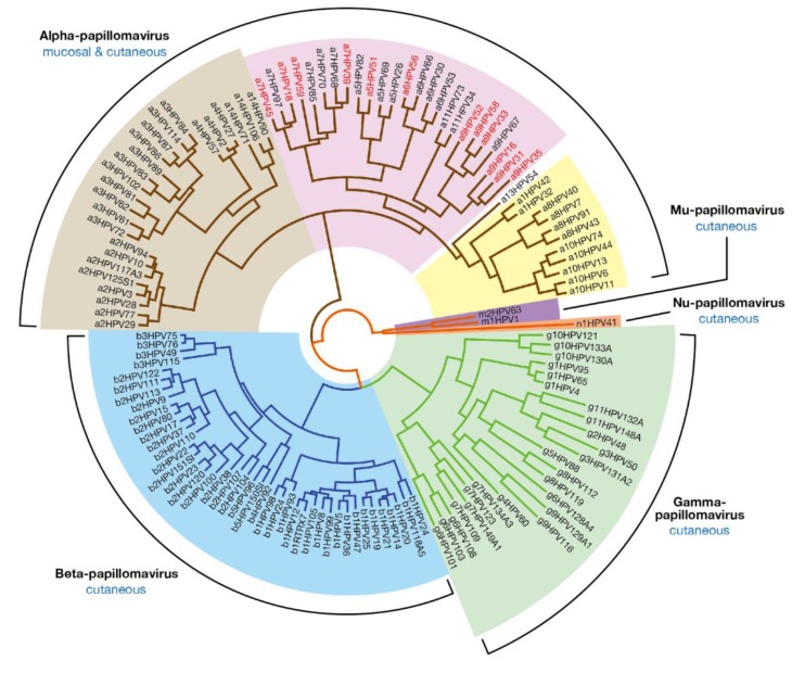
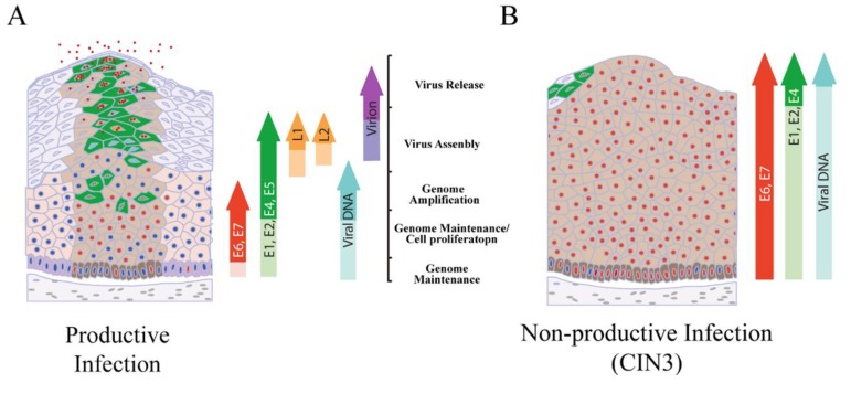
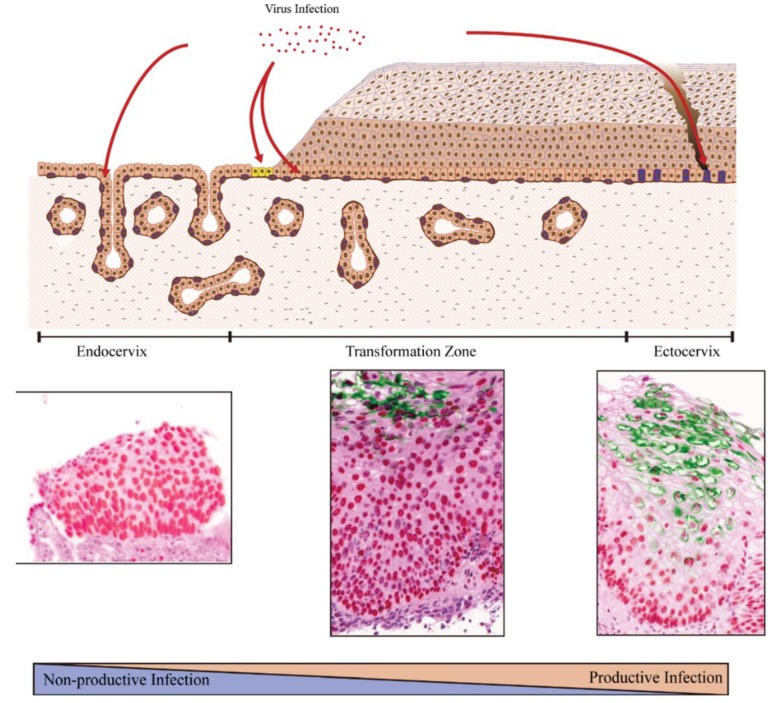
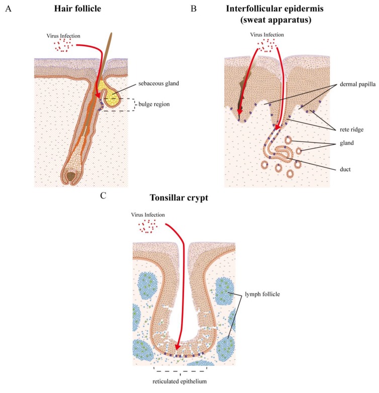
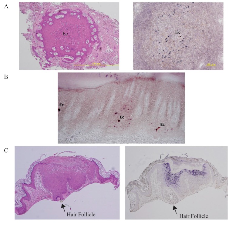
Papillomaviruses have evolved over many millions of years to propagate themselves at specific epithelial niches in a range of different host species. This has led to the great diversity of papillomaviruses that now exist, and to the appearance of distinct strategies for epithelial persistence. Many papillomaviruses minimise the risk of immune clearance by causing chronic asymptomatic infections, accompanied by long-term virion-production with only limited viral gene expression. Such lesions are typical of those caused by Beta HPV types in the general population, with viral activity being suppressed by host immunity. A second strategy requires the evolution of sophisticated immune evasion mechanisms, and allows some HPV types to cause prominent and persistent papillomas, even in immune competent individuals. Some Alphapapillomavirus types have evolved this strategy, including those that cause genital warts in young adults or common warts in children. These strategies reflect broad differences in virus protein function as well as differences in patterns of viral gene expression, with genotype-specific associations underlying the recent introduction of DNA testing, and also the introduction of vaccines to protect against cervical cancer. Interestingly, it appears that cellular environment and the site of infection affect viral pathogenicity by modulating viral gene expression. With the high-risk HPV gene products, changes in E6 and E7 expression are thought to account for the development of neoplasias at the endocervix, the anal and cervical transformation zones, and the tonsilar crypts and other oropharyngeal sites. A detailed analysis of site-specific patterns of gene expression and gene function is now prompted.

Keywords: carcinogenesis; diversity; evolution; human papillomavirus; niche; tissue stem cells; tropism.


Figures

Figure 1
Figure 1
Evolutionary Relationship between Human Papillomaviruses. The human papillomaviruses types found in humans fall into five genera, with the Alpha-, Beta- (blue) and Gammapapillomavirus (green) representing the largest groups; Human papillomaviruses types from the Alphapapillomavirus genus are often classified as low-risk cutaneous (light brown); low-risk mucosal (yellow); or high-risk (pink) according to their association with the development of cancer. The high-risk types highlighted with red text are confirmed as “human carcinogens” on the basis of epidemiological data. The remaining high-risk types are “probable” or “possible” carcinogens. Although the predominant tissue associations of each genus are listed as either cutaneous or mucosal, these designations do not necessarily hold true for every member of the genus. The evolutionary tree is based on alignment of the E1, E2, L1, and L2 genes [26]. HPV sequence data was be obtained from PaVE [1].
Figure 2
Figure 2
Viral gene-expression in adjacent Betapapillomavirus lesions. Immunostaining for the Betapapillomavirus E4 protein (green) reveals distinct patterns of expression in different lesions in an immunosuppressed individual. Lesion (a) and (a`) are atypical, and show marked basal and suprabasal staining; This pattern of E4 expression is distinct from typical E4 pattern of expression, which is usually restricted to the mid/upper epithelial layers as seen in region (c); Lesions (b) and (b`) show an intermediate pattern of E4 staining with expression close to the basal layer, as well as in the suprabasal cell layers (see also review [11]).
Figure 3
Figure 3
Alphapapillomavirus genome organization and the function of HPV proteins. (A) Genome organization typical of the high-risk Alphapapillomavirus types is illustrated by the genome of HPV16. The early (p97) and late (p670) promoters are marked by arrows. The six early ORFs (E1, E2, E4 and E5 (in green) and E6 and E7 (in red)) are expressed from the different promoters at different stages during epithelial cell differentiation. The late ORFs (L1 and L2 (in yellow)) are expressed from the p670 promoter in the upper epithelial layers as result of changes in splicing. The LCR/URR also contains the replication origin as well as post-transcriptional control sequences that contribute to viral gene expression. (B) The function of viral proteins. All known papillomavirus encodes a group of “core” proteins that were present early on during papillomavirus evolution, and which are conserved in sequence and in function between PV types. These include E1, E2, L2 and L1. The E4 protein may also be a core protein that has evolved to meet papillomaviruses epithelial specialization. The accessory proteins have evolved in each papillomavirus type during adaptation to different epithelial niches. The sequence and function of these genes are divergent between types. In general, these proteins are involved in modifying the cellular environment to facilitate virus life cycle completion, contributing to the virulence and pathogenicity. Knowledge of accessory protein function comes primarily from the study of Alphapapillomavirus types.
Figure 4
Figure 4
Regulation and deregulation of the high-risk Alphapapillomavirus life cycle. (A) The papillomavirus life cycle is regulated during epithelial cell differentiation and is shown diagrammatically. Cells that are driven through the cell cycle as a result of E6 and E7 expression are marked with red nuclei. The up-regulation of viral proteins necessary for genome amplification (i.e., E1 and E2) requires activation of the viral late promoter in the upper epithelial layers (cells shown in green with red nuclei), with virus particles subsequently being released from the epithelial surface; (B) In HPV-associated neoplasia, late gene expression is retarded, and although the order of events remains the same, the production of infectious virions is restricted to smaller and smaller areas close to the epithelial surface. This situation is thought to be accompanied elevated E6/E7 expression, and represents a non-productive or poorly productive abortive infection. Integration of HPV DNA into the host cell genome is facilitated by deregulated E6/E7 expression. If integration disrupts the E1/E2 region this can allow the persistent high-level expression of E6 and E7 and the accumulation of genetic errors in the host genome. Eventually, the productive virus life cycle is no longer supported and viral episomes are lost (reviewed in [83]).
Figure 5
Figure 5
The difference of host cells (the site of infection) affects viral pathogenicity. Most cervical cancers arise at the cervical transformation zone. The transformation zone is maintained by a specialized type of tissue stem cell known as the reserve cell (shown in purple in the transformation zone), and possibly also by a cluster of cuboidal cells (yellow) localized more precisely at the squamo-columnar junction. These cells can maintain either the columnar epithelium of the endocervix or the stratified epithelium of transformation zone depending on their extracellular environment. In the ectocervix, the epithelium is populated by conventional epithelial tissue stem cells (purple in the ectocervix). The different characteristics of the various tissue stem cells that HPV infects are thought to influence the pattern of viral gene expression differently [41]. Current thinking suggests that productive infection is favoured at the ectocervix, while a non-productive or abortive infection is more likely at the endocervix. In the immunostains, MCM expression (red) indicates the expression of viral E7 protein. E4 expression (green) indicates the productive infection [59].
Figure 6
Figure 6
Epithelial tissue sites and tissue stem cells as targets for HPV infection. HPVs infect a variety of epithelial tissue sites, and can cause lesions in the vicinity of hair follicles, eccrine and apocrine sweat apparatus), nails, and also the inter-appendageal epidermis. Specialized epithelial sites contain other appendages, such as the salivary glands of the oral cavity and the tonsillar crypts of the oropharynx, where oropharyngeal cancers arise. The transformation zone regions, where stratified epithelium abuts columnar epithelium such as the cervical (Figure 5) or anal transformation zones, are other target sites where infection is thought to be facilitated. (A) The bulge region of the hair follicle is a well characterized region where the stem cells that populate cutaneous epithelial sites reside. HPV virions are thought to gain access to the epithelial stem cells (coloured purple), either through a wound or possibly through the hair follicle; (B) Between the hair follicles, the tissue stem cells are thought to reside in both rete ridge and over the dermal papilla, and are not thought to be clustered at any specific location in the basal component [109]. With the sweat apparatus, at least two distinct stem cell populations have been identified that may be accessible for infection, either in the gland or the duct. These are able to repair damaged epidermis. HPV virions are thought to gain access to these stem cells (coloured purple), either through a wound or maybe through the eccrine duct; (C) The tonsillar crypts are a highly specialized lymphoepithelial tissue. A dense lymphocyte infiltrate generally obscures the junction between the lymphoid and epithelial components and splinters the epithelial sheath into irregular nests and cords. This reticulated epithelium may facilitate viral access to tissue stem cells at an immune-privileged site, which can inhibit virus-specific T cell activity and thereby facilitate immune evasion during initial HPV infection and subsequent virus-induced malignant transformation.
Figure 7
Figure 7
Examples for HPV targeting cutaneous appendages. (A) Haemotoxylin & eosin stain (left) of a horizontal section of a tiny wart. An eccrine (Ec)-centered distribution of histological changes is observed. HPV1 DNAs are identified within the pathology changes following DNA in situ hybridization (right); (B) HPV63 DNA is identified in resident keratinocytes in the vicinity of eccrine ducts (Ec) in a ridge of the plantar skin following DNA in situ hybridization; (C) HPV6/11 histopathological changes are identified in the resident keratinocytes in and around the hair follicle (arrow) by haemotoxylin & eosin staining (left) and DNA in situ hybridization (right).

References

    1. PaVE: Papillomavirus Episteme. [(accessed on 11 July 2015)]; Available online: http://pave.niaid.nih.gov/
    1. Doorbar J., Egawa N., Griffin H., Kranjec C., Murakami I. Human papillomavirus molecular biology and disease association. Rev. Med. Virol. 2015;25(Suppl. 1):2–23. doi: 10.1002/rmv.1822. - DOI - PMC - PubMed
    1. Bottalico D., Chen Z., Dunne A., Ostoloza J., McKinney S., Sun C., Schlecht N.F., Herrero R., Fatahzadeh M., Schiffman M., et al. The oral cavity contains abundant known and novel human papillomaviruses from the betapapillomavirus and gammapapillomavirus genera. J. Infect. Dis. 2011;204:787–792. doi: 10.1093/infdis/jir383. - DOI - PMC - PubMed
    1. De Koning M.N., Quint K.D., Bruggink S.C., Gussekloo J., Bouwes Bavinck J.N., Quint W.G., Feltkamp M.C., Eekhof J.A. High prevalence of cutaneous warts in elementary school children and the ubiquitous presence of wart-associated human papillomavirus on clinically normal skin. Br. J. Dermatol. 2015;172:196–201. doi: 10.1111/bjd.13216. - DOI - PubMed
    1. Poljak M., Kocjan B.J., Potocnik M., Seme K. Anogenital hairs are an important reservoir of α-papillomaviruses in patients with genital warts. J. Infect. Dis. 2009;199:1270–1274. doi: 10.1086/597619. - DOI - PubMed

Publication types